






Abstract
Rapidly progressive Alzheimer’s disease (rpAD) has recently been recognized as a clinical disease subtype characterized by rapidly progressive cognitive decline and/or short disease duration, and the possible occurrence of early focal neurological signs. Consistently, rpAD represents a relatively frequent alternative diagnosis among cases referred as possible or probable Creutzfeldt-Jakob disease (CJD) to surveillance centers for prion disease worldwide. Indeed, the early clinical differential diagnosis between the two disorders can be challenging given the partial overlap in clinical features and cerebrospinal fluid (CSF) levels of the protein surrogate markers 14-3-3 and total-tau. Although typical AD and rpAD seem to share the neuropathological core features, recent evidence suggests that a distinctive molecular signature involving the structure of amyloid-β aggregates and the proteomic landscape of amyloid plaques may distinguish rpAD from typical AD. Here we review clinical, neuropathological, and molecular features and diagnostic findings, including CSF biomarker data, reported to date in rpAD. Furthermore, we summarize the main clinical, pathological and laboratory features of 27 autopsy confirmed cases of rpAD referred to our center. The results of this retrospective analysis, while largely confirming previously published genetic, clinical, and neuropathological data, suggest a higher prevalence of moderate to severe cerebral amyloid angiopathy in rpAD compared to typical AD, a finding to explore further and validate in a larger patient group.
Keywords
INTRODUCTION
Alzheimer’s disease (AD), the most common form of dementia in the elderly, is typically (typical AD) characterized by a slowly progressive cognitive decline with prominent memory loss and an approximate 8-year survival after clinical onset [1]. However, increasing evidence documents a significant variability in both AD clinical phenotype and progression rate, raising questions about the possible biological variables and pathogenic mechanisms underlyingthis heterogeneity.
The term rapidly progressive AD (rpAD) has recently been proposed to identify AD patientsexperiencing a rapid progression of dementia and/or a reduced survival [2–9]. However, no International consensus exists regarding the clinical definition of rpAD. Furthermore, whether rpAD defines a molecularly and pathologically distinct disease subtype or mainly reflects co-morbidities or other factors remains to be fully explored. Notably, preliminary evidence from recent studies supports the idea of rpAD being a specific AD clinical phenotype associated with distinctive molecular features [3, 10]. In apparent contrast to the latter findings, however, no significant differences in the core neuropathological features including their regional distribution, have been detected to date between typical AD and rpAD [2, 6].
In Creutzfeldt-Jakob disease (CJD) surveillance centers worldwide dealing with CJD differential diagnosis, AD is the most frequent non-CJD diagnosis at neuropathological examination, accounting for 14–50% of all non-prion submitted cases [4, 11–13]. Given the diagnostic difficulties caused by the partial clinical overlap between rpAD and CJD, a number of recent studies explored the cerebrospinal fluid (CSF) profile of several biomarkers to differentiate rpAD from prion disease. The very same CSF proteins or other biomarkers may also significantly contribute to the early discrimination of rpAD from typical AD, which is of importance for the clinical and diagnostic management of patients and, possibly, for the development of customized and focusedtherapies.
Here we review clinical, diagnostic, neuropathological, and molecular aspects of this atypical AD phenotype and describe clinical, laboratory, and neuropathological findings in a series of 27 autopsy confirmed rpAD cases.
CLINICAL FEATURES AND DIAGNOSTIC FINDINGS
The existence of a specific clinical phenotype of rpAD is still a matter of debate. The steady report from surveillance centers for prion disease worldwide of a significant proportion of AD postmortem diagnosis in patients referred with the clinical suspicion of CJD [2–5, 11–13] is in apparent contrast with the lack of recognition of rpAD as a distinct disease subtype by the mainstream AD literature. Moreover, the definition of rpAD varied significantly among studies [2, 14–21]. While most authors adopted the clinical definition of rpAD based on the decrease of at least 6 points/year in the score of the Mini-Mental State Examination (MMSE) [3, 18–20], others considered as further selection criterion the disease duration of less than three years [6, 10]. Finally, given that rpAD often mimics CJD because of the rapid course and the frequent occurrence of early focal neurological signs, an operational definition of rpAD based on clinical or laboratory features, suggesting a possible CJD diagnosis independently from the stringent criteria mentioned above, has also been used [7–9] (Table 1).
Selection criteria for rpAD in the main cohorts reported in the literature
C, clinical; B, based on CSF biomarker profile (Aβ42, t-tau, and p-tau); P, pathological; *in a subgroup of cases; μ the availability of CSF biomarker and/or pathological data is not specified; +obligatory selection criterion; -not included as selection criterion; ¥at least one of the marked criteria; y, years; mo, months.
Regarding demographic characteristics, many studies reported a younger mean age at onset (68–74 years) and the lack of a female predominance in comparison with typical AD [2, 17]. An autosomal dominant history of dementia or a significant comorbidity was typically absent [3, 6], whereas the median disease duration varied from 7 to 26 months [2, 10]. Clinically, 56% of rpAD cases were referred as possible or probable sporadic CJD (sCJD) [2] due to the presence of rapidly cognitive decline and/or focal neurological signs at the time of diagnosis. Specifically, early impairment in executive functions or language, motor disturbances such as myoclonus (66–75%), gait impairment (66–87%), pyramidal (53–66%) or extrapyramidal (54%) signs, visual signs (e.g., hallucinations) (44–62%), or psychiatric symptoms frequently occurred in rpAD, either alone or in combination [2, 15]. All these features seem to be predictive of poor outcome in AD patients [22, 23]. Notably, the diagnosis of AD in such cases could be challenging, as the International Working Group-2 criteria contemplate the presence of focal neurological signs as an exclusion criteria for the diagnosis of AD [24]. On the other hand, the real prevalence of rpAD cases with rapidly progressive cognitive decline not associated with early neurological signs remains undetermined given the potential bias of the preferential selection of cases mimicking CJD due to existence of surveillance centers for prion disease in many countries.
Regarding diagnostic investigations, 6% of reported rpAD cases showed periodic sharp-wave complexes (PSWC) at electroencephalographic (EEG) examination, although none of them displayed the typical hyperintensities at diffusion weighted or fluid attenuated inversion recovery magnetic resonance imaging (MRI) that are often seen in typical CJD [2]. One study based on a short-term follow-up period showed no differences in baseline hippocampus volume, white matter hyperintensity (brain MRI) and cortical amyloid deposition (Florbetapir-positron emission tomography (PET)-AV45) between rpAD and typical AD [17]. In the same study, however, a lower and region-specific baseline hypometabolism at FDG-PET was detected in rpAD, with the left angular and left temporal cortices hypometabolism demonstrating the highest accuracy and predictive value in distinguishing rpAD from typical AD at receiver-operating characteristic analysis [17].
As a major center for prion research and diagnosis in Italy, we have collected several cases of autopsy-confirmed AD referred with the suspicion of CJD because of (1) a rapidly progressive cognitive decline, (2) a disease duration≤2 years, and the variable association with other neurological signs. Demographics, clinical features and results of diagnostic investigations of our autopsy-confirmed rpAD population (N = 27) are summarized in Table 2.
Demographics and results of clinical and diagnostic evaluations (N = 27)
*not confirmed as PSWC after an expert revision. IQR, interquartile range; N, number of cases.
Our patients showed an older mean age at onset compared to other reported rpAD cases, but similar disease duration and gender distribution [2, 17]. None of the patients had a history consistent with familial AD or carried autosomal dominant, AD-related, genetic mutations. The clinical phenotype included prominent psychiatric symptoms and motor signs as previously described [2, 5]. Brain computed tomography and/or MRI did not show any distinctive feature in comparison to typical AD. Interestingly, EEG recording showed PSWC in 16.7% of cases, confirming their low accuracy in the differential diagnosis of CJD. Overall, 74% of rpAD patients met the criteria for possible or probablesCJD [25].
CSF BIOMARKERS STUDIES
CSF AD core biomarkers
CSF AD core biomarkers reflect three distinct patho-biological processes that occur in the AD brain: total-tau (t-tau) and phosphorylated-tau (p-tau) are, respectively, biomarkers of downstream non-specific neurodegeneration and intracellular neurofibrillary tangle (NFT) formation, while CSF concentration of amyloid-β peptide 1-42 (Aβ42) reflects the extracellular deposition of Aβ [26]. When used in combination, these biomarkers showed high diagnostic accuracy for AD, reaching 85–90% sensitivity and specificity [27].
Tau is a neuronal cytoplasmatic protein involved in microtubule assembly and stabilization [28]. CSF t-tau levels nonspecifically reflect the rate of neuronal degeneration leading to the release of the protein in the extracellular fluids and CSF [29]. Thus, t-tau is variably increased both in AD and several other diseases, reaching the highest levels in CJD [13, 29].
At variance with t-tau, p-tau more specifically reflects the neurofibrillary degeneration, thus high levels of p-tau are primarily detected in AD [29]. Although the phosphorylation of tau can occur at multiple sites, most of current assays only detect the phosphorylation at one specific amino acid (e.g., in the position 181) [27].
Aβ42 peptide results from the cleavage of transmembrane amyloid-β protein precursor (AβPP) by specific enzymes named secretases. The deposition of Aβ42 insoluble aggregates, such as Aβ plaques, in extracellular spaces, by decreasing the spillage of the protein in the CSF, is responsible for the reduced CSF levels in AD patients [26, 28].
According to the amyloid-beta cascade hypothesis, the formation of Aβ plaques represents the first pathological event in AD, prompting NFT formation, synaptic damage, inflammatory changes and ultimately neuronal loss [26]. Accordingly, CSF Aβ42 levels decrease early in the disease course, even in the pre-symptomatic phase, while the increase in CSF t-tau and p-tau levels would occur later [26, 30–32].
Several studies addressing the role of CSF biomarkers in the differential diagnosis of rapidly progressive dementia found that approximately 8–16% of rpAD cases have relatively high t-tau levels (e.g.,>1250 pg/ml), fitting the inclusion criteria for sCJD, whereas the same t-tau profile is rarely seen in typical AD [12, 33]. In our rpAD cohort, which has the limit of the relatively small sample size, the number of cases showing t-tau levels above 1250 pg/ml was even higher (30.8%, Table 2). Furthermore, in three recent studies, atypical/rapidly progressive AD patients showed higher p-tau levels than typical AD cases [7–9]. Finally, a recent study found that patients with rpAD show a significantly lower p-tau/t-tau ratio than those with typicalAD [17].
The available data on Aβ42 levels in rpAD and typical AD are, to date, more discordant than those on t-tau and p-tau. Specifically, one study reported lower levels in atypical/rpAD compared to typical ones, while three additional studies showed no significant differences between the two groups [7, 16]. CSF t-tau, p-tau and Aβ42 levels in our rpAD cases are reported in Table 2.
Results of other studies addressing the prognostic significance of AD core biomarkers demonstrated a positive correlation between t-tau levels and disease duration. Indeed, both a profile with elevated t-tau level without a proportionally elevated p-tau level [34] and a profile with high t-tau level, high p-tau level, and/or low levels of Aβ42 at time of diagnosis was found to predict a more rapid cognitive decline and a higher mortality rates in AD subjects [35–37]. Other studies, however, found no differences in t-tau, p-tau and Aβ42 CSF levels between rpAD and typical AD [5, 38]. Specifically, Llorens et al. claimed that neither a single biomarker nor any combination of them may discriminate typical AD from rpAD and predict the progression rate in clinically diagnosed AD patients [16]. According to these authors, the findings are consistent with the lack of specific pathological features differentiating the two forms at the time of diagnosis [19].
In conclusion, while there seems to be a trend for t-tau and, to a lesser extent, p-tau to correlate positively with disease progression in AD, the significant heterogeneity in the results among studies, likely reflecting differences in patient selection criteria or pre-analytical and analytical parameters, does not allow a definite conclusion on the prognostic significance of these biomarkers in AD.
Based on the notion that AD cases show lower t-tau but higher p-tau CSF levels than CJD patients, several studies exploited the diagnostic value of the t-tau/p-tau (or p-tau/t-tau) ratio in the differential diagnosis between AD, especially rpAD, and CJD [7–9, 29]. In this clinical scenario (AD versus CJD), sensitivity and specificity of t-tau/p-tau (or p-tau/t-tau) ratio were reported to be 96.3–99.0% and 94.0–95.5%, respectively [8, 16]. Furthermore, the diagnostic accuracy of the same biomarker ratio was excellent (area under the curve (AUC) 0.982–0.990) [8, 16]. Finally, Aβ42/t-tau and Aβ42/p-tau ratios also showed a very good diagnostic performance in the differentiation between rpAD and CJD subjects (AUC = 0.926 and AUC = 0.921 respectively) [8, 16].
Other CSF biomarkers
Amyloid-beta peptide 1–40 (Aβ40) is a small peptide derived from AβPP cleavage which is much more abundant and less prone to aggregation than Aβ42 [28]. According to recent studies, CSF Aβ42/Aβ40 ratio demonstrates a higher concordance with amyloid-PET findings and a better diagnostic performance for AD than CSF Aβ42 alone [39–42]. Since CSF Aβ40 levels seem to reflect the inter-patient variability in Aβ metabolism, such as high or low Aβ production and/or clearance, the improved diagnostic accuracy of Aβ42/Aβ40 ratio may primarily reflect the reduced inter-individual variability in Aβ metabolism that is independent from AD pathophysiology [43, 44]. In the single study completed to date, rpAD patients showed a lower Aβ42/Aβ40 ratio than controls, as previously reported in typical AD [16]. Furthermore, the discrimination power of Aβ42/Aβ40 ratio in the comparison between either typical AD or rpAD and controls was similar. Finally, the Aβ40/t-tau ratio yielded an excellent diagnostic accuracy in the differentiation between rpAD and sCJD (AUC = 0.979) [16].
CSF 14-3-3 protein assay is recommended for the clinical diagnosis of CJD and included in the diagnostic criteria for probable CJD [25]. However, its specificity for CJD is far from being optimal, as reported in several studies [7, 13]. The increase of 14-3-3 in CSF is related to neuronal damage. Thus, any pathological process associated with a severe neuronal damage may yield a positive test. The positivity of 14-3-3 assay, in studies specifically defining a rpAD group, varied from 15.2% to 42.0% according to the different studies [2, 16], whereas in other cohorts of dementia cases submitted for the 14-3-3 assay, the positivity among AD cases ranged from 5.8% to 28.1% [12, 45]. In our group, 29.4% of rpAD cases showed a positive 14-3-3 assay (Table 2). This variability, especially in the latter studies in which the criteria of inclusion of AD cases were not specified, could be related to the relative proportion of typical AD and rpAD included in the different cohorts. Recently we have shown that, when applied to CJD diagnosis, the 14-3-3 western blot assay is associated with a higher percentage of false positive results in comparison to the t-tau enzyme-linked immunosorbent assay (ELISA). However, when the differential diagnosis is limited to CJD and AD, protein 14-3-3 detection is more accurate [13].
The measurement of the prion protein in the CSF is generally referred to as total PrP (t-PrP) assay since it includes the measurement of both cellular (PrPC) and pathological prion protein (PrPSc) levels due to the lack of specificity for one of the two forms of the available antibodies [7]. However, given that PrPSc is virtually undetectable in the CSF due to the extremely low levels, the t-PrP measurement actually corresponds to a PrPC assay. Recently, one study has shown that t-PrP levels are higher in clinical and neuropathological confirmed AD cases, than in controls, with rpAD having higher values than typical AD cases [7]. The rapid clinical course of atypical AD, possibly associated with an upregulation of PrPC has been proposed as a possible cause of these CSF findings [7]. In contrast, we found no significant difference regarding CSF t-PrP values between AD and controls and between typical AD and rpAD cases [8]. However, in both studies the reduction of t-PrP levels in CJD patients has been associated with the extent of PrPC conversion into PrPSc and the burden of PrPSc deposition [7, 8]. Several combinations of t-PrP with t-tau, p-tau and Aβ42 (e.g., Creutzfeldt-Jakob (CJ) factor: t-tau/p-tau×t-PrP; t-tau×Aβ42/(p-tau×t-PrP)) significantly improve the differential diagnosis between AD and CJD. In detail, the CJ factor distinguished CJD from rpAD with 100% sensitivity and 95.7% specificity [7]. Moreover, in the most difficult clinical scenario, namely the clinical distinction between atypical CJD and rpAD, the ratio t-tau×Aβ42/(p-tau×t-PrP) yielded 96.2% sensitivity and 95.5% specificity [8].
Alpha-synuclein (α-syn) represents the main component of Lewy bodies, the neuropathological hallmark of synucleinopathies, namely Parkinson disease, multiple systemic atrophy and dementia with Lewy bodies [18, 46]. Recently, α-syn has been exploited in ELISA or real time quacking induced conversion (RT-QuIC) assays for the CSF biomarker-based diagnosis of synucleinopathies [18, 46]. In a study by Llorens et al. [18] AD cases had slightly higher α-syn CSF levels than controls, although the difference was not statistically significant. In addition, comparable levels were detected in typical AD and rpAD cases. Taken together, these findings prompted the claim that α-syn is not a marker for tissue damage in AD. Furthermore, since sCJD is associated with elevated CSF levels of α-syn, the biomarker could be used in the discrimination between AD and sCJD, especially in cases with inconclusive CSF findings with other assays (e.g., overlapping t-tau levels) [18].
Chitinase-3-like protein 1 (YKL-40) is a marker of inflammation and endothelial dysfunction, which has been found upregulated in the astrocytes of patients with several neurological diseases, including AD [20]. In one study, YKL-40 astrocytic expression was detected in close vicinity to amyloid plaques and NFTs [20]. YKL-40 seems to be not influenced by rate of cognitive decline and/or disease duration, since no differences in CSF YKL-40 levels were reported between typical AD and rpAD [20].
The immune system has also been investigated as a possible modifier of AD pathogenesis and progression rate. According to one study, rpAD patients are characterized by a distinctive pro-inflammatory cytokine profile (IL-6, IL-13, TNF-α, G-CSF) in CSF and blood with respect to either typical AD and CJD patients. Thus, a specific systemic immune response, which is not only the consequence of the rapid disease progression, may be responsible for the rapid clinical course in rpAD [47].
Neurofilament light chain protein (NfL) is a component of neurofilaments, which, together with the medium and heavy chains, contribute in the maintenance of axonal structure [27]. In AD the occurring neuroaxonal degeneration leads to increased CSF and blood NfL levels, although to a lesser extent than in frontotemporal dementia [48–50]. In a recent study [9], we showed no significant differences regarding CSF NfL levels between typical AD and rpAD cases. We suggested that, as for CSF α-syn, the pathophysiological mechanisms leading to a more rapid course in AD could be relatively independent from those affecting NfL CSF levels [9, 18]. Furthermore, we demonstrated that CSF NfL performs better that t-tau in the discrimination between prion disease and AD (AUC 0.981 versus 0.901), as well as between rapidly progressive/atypical AD and atypical prion disease (AUC 0.946 versus 0.654), with NfL/p-tau ratio yielding the highest diagnostic accuracy in this comparison (AUC 0.989) [9]. A comparative summary of CSF biomarker changes reported to date in rpAD, typical AD and CJD is reported in Fig. 1.
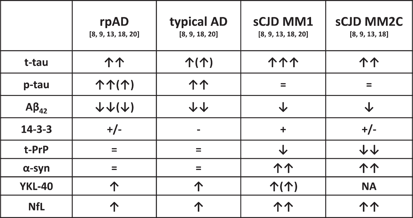
The figure summarizes the CSF biomarker changes reported to date in rpAD, typical AD, and two representative sCJD subtypes (MM1 and MM2-Cortical or MM2C). The latter have been selected because of their clinical and laboratory feature overlap with, respectively, rpAD and typical AD. ↑: increased values in comparison to controls; ↓: decreased values in comparison to controls; the number of arrows indicates the degree of change from the normal range, the bracket indicates some variability among cases; =: values within the control range; +: positive assay result in the large majority of cases; – : negative assay result in the majority of cases; +/– : a significant number of either positive or negative assay results; NA: data not available.
Finally, prion RT-QuIC demonstrated virtually full specificity in the discrimination of CJD from other rapidly progressive dementias, including rpAD [13]. Indeed, RT-QuiC resulted negative in all examined cases (n = 12) of our rpAD cohort, confirming previous findings [13]. However, the strict dependence of this technique on both availability and performance of the used substrate, currently limits its use as routine diagnostic assay. Moreover, RT-QuIC has only moderate sensitivity for sCJD MM2C, an atypical sCJD subtype clinically overlapping with AD [13]. As a consequence, ELISA assay-based biomarkers (e.g., t-tau and NfL) still play a significant role as fast screening procedures for the differential diagnosis of rapidly progressive neurodegenerativedementias.
NEUROPATHOLOGICAL, MOLECULAR, AND GENETIC FEATURES
The extracellular accumulation of Aβ as diffuse or focal deposits, forming diffuse or neuritic plaques, and the intracellular deposition of p-tau, forming NFTs, neuropil threads and the dystrophic neuritis of neuritic plaques, represent the hallmarks of AD neuropathology. Additional findings include a variable extent of non-specific neuronal loss, glial activation and cerebral amyloid angiopathy (CAA) [51]. Among these lesions, neurofibrillary degeneration and neuronal loss correlate best with the levels of cognitive impairment in AD patients [26], whereas the relationship between Aβ load and clinical disease severity is less consistent [52].
Previous studies on the phenotypic spectrum of prion disease revealed a significant heterogeneity in both lesion profiles and type and morphology of lesions correlating with clinical features, especially at onset, and disease duration [53]. In contrast, the studies conducted so far showed no significant differences in the morphology and anatomical distribution of amyloid plaques and neurofibrillary tangles between typical and rpAD [2, 6]. As the only possible exception, a trend toward a milder pathology according to National Institute on Aging–Alzheimer’s Association guidelines and higher levels of Aβ42 in the posterior cingulate cortex in rpAD cases, were observed in one study [6]. In addition, there was no significant comorbid neurodegenerative pathology, such as synuclein or TDP-43 proteinopathies, to explain the faster progression rates in rpAD [6]. Finally, diffuse and glial deposition of Aβ were comparable and rather inconsistent in both typical and rpADpatients [6].
Our retrospective analysis of 27 autopsy confirmed rpAD cases, submitted to our laboratory with thesuspicion of CJD and in which no clinical and pathological evidence of significant neurodegenerative, vascular or inflammatory co-morbidity was found, gave substantially analogous results (Table 3). Most significantly, neuropathological analysis failed to reveal an obvious divergence from the core features of AD pathology seen in typical AD [51]. Moreover, only 37% of the cases showed a high degree of AD pathological changes, which seems to confirm the observed trend toward a lower mean stage of parenchymal degenerative pathology in rpAD in comparison to typical AD [6]. At variance with previous studies, however, we found a relatively high incidence of CAA in our series. Indeed, 59.3% (16 out of 27) of cases had moderate to severe CAA, in contrast with the previously reported 25.6% of typical AD cases [58]. Furthermore in 19 cases, the CAA has progressed from the cerebral cortex to subcortical areas (e.g., phases II and III according to Thal et. al [56]) (Table 3). Thus, future studies on larger case series should address this issue further.
Neuropathological and genetic features
Irrespective of type and distribution of histopathological lesions, biological factors may directly influence the rapid clinical course in rpAD. Recent studies explored this possibility in analogy to what shown in CJD, in which distinct clinicopathological phenotypes have been related to distinct strains of prions that are specified by PrPSc properties [6, 59]. Indeed, preliminary evidence indicates that the molecular structure of Aβ42 or Aβ40 fibrils may vary significantly in vitro and that different amyloid-beta conformers are associated with distinct biochemical properties (e.g., size distributions, resistance to chemical denaturation) and levels of toxicity [6, 60] in vivo. Using advanced conformation-sensitive techniques, Cohen et al. studied Aβ42 and Aβ40 peptides extracted from the brain of AD cases with variable disease progression rates. Notably, rpAD samples showed an expanded conformational heterogeneity with significantly higher levels of Aβ42 conformers composed of 30-300 monomers, which are less stable to denaturation and easily dissociate in vivo leading to fragmentation and replication. Thus, as shown for PrPSc, distinct Aβ42 conformers may affect toxicity, spread of amyloid pathology and, ultimately, disease progression rate [6].
Further proteomic study of amyloid plaques in the same cohort of patients demonstrated a distinct pattern of protein expression in rpAD, with significantly higher levels of neuronal proteins and significantly lower levels of astrocytic proteins in comparison with typical AD [10]. Synaptic proteins, especially those involved in synaptic vesicle release, were particularly abundant in rpAD plaques, suggesting that synaptic dysfunction may have a role in accelerating the spread of amyloid pathology in rpAD [10]. More specifically, the accumulation of Aβ42 oligomers in synapses and their bond with synaptic proteins may lead to synaptic dysfunction and cognitive impairment. This process may occur to a greater extent in rpAD because of a higher order Aβ42 oligomers. On the other side, the decreased number of plaque-associated astrocytes seen in rpAD, could be linked to the impairment of neuroprotective astrogliosis, allowing a faster spread of amyloid pathology [10].
In another study, exploiting a solid-state nuclear magnetic resonance approach, brain extracts from rpAD cases exhibited a significantly greater proportion of Aβ40 fibril structures in comparison to cases with typical AD and posterior cortical atrophy [21]. Thus, one or more fibril structures that are prevalent in rpAD and not in typical AD, may present enhanced neurotoxicity, through either direct or indirect mechanisms. Alternatively, the presence of fibrils with a greater range of thermodynamic stabilities or resistance to degradation may be the consequence of the shorter disease duration rather than the cause of the faster progression of rpAD [21]. Moreover, a structural heterogeneity of Aβ42 fibrils, with at least two predominant structures, was demonstrated in most samples from all AD variants [21].
Apolipoprotein E (ApoE) is an Aβ-associated protein, which plays a central role in AD pathogenesis by modulating Aβ aggregation and clearance in an isoform specific manner [10]. Moreover, ApoE allele ɛ4 is a well-established risk factor for developing typical AD [5], even if its role as a predictor of clinical progression is still debated. Intriguingly, several studies highlighted the significantly lower frequency of ApoE ɛ4 carriers, including ɛ4 homozygotes, in the rpAD population compared to typical AD cases [2, 6]. At variance, a single recent study reported a similar ApoE genotype distribution in the two groups [17]. In our cohort of rpAD cases, the ApoE ɛ4 allele frequency was 23.1%, which is in line with the results obtained in the two largest case series [5, 6](Table 3).
The influence of codon 129 polymorphism on AD susceptibility gave contradicting results [2]. In rpAD cohorts, a slight tendency towards more methionine homozygotes was noted [2, 5]. In our population we demonstrated a prevalence of methionine heterozygotes compared to homozygotes, reflecting the general population distribution (Table 3).
Finally, additional molecular factors that have been linked to rpAD pathogenesis are late ensodomal compartment proteins, such as Rab7a [61] and PrPC [62]. Specifically, these authors characterized prion protein expression, localization and interactome in rpAD cases, and demonstrated a significant decrease in diglycosylated PrP isoforms and the existence of specific proteins with potential interaction with PrP in rpAD cases [62].
CONCLUSIONS
Increasing evidence suggests that rpAD may represent a subtype of AD characterized by distinctive molecular features of Aβ deposits. Mean CSF levels of surrogate markers of neurodegeneration such as t-tau, p-tau, and 14-3-3 are often significantly higher in rpAD than in typical AD patients up to levels sometimes challenging the differential diagnosis against CJD and other rapidly progressive dementias. While standard neuropathological examination does not explain the more rapid clinical course of the former, in absence of a significant co-morbidity, the recent finding of differences in Aβ conformers and protein composition of plaques between rpAD and typical AD may suggest the existence of distinct “prion-like” strains of Aβ underlying the different clinical progression rate between the two ADsubtypes.
Despite these promising advances, several important issues remain unsolved. Firstly, a more reliable and universally accepted clinical definition of rpAD should be achieved. The definition based only on the MMSE score appears rather gross. Moreover, since the majority of studies regarding rpAD was conducted by CJD surveillance centers on autopsy-confirmed AD cases referred as possible or probable CJD, prospective studies analyzing a more homogeneous AD population are needed.
After reaching a consensus on the clinical definition of rpAD, allowing a better comparison among studies, neuropathological and molecular aspects, such as neurofibrillary degeneration, Aβ properties, CAA, astrogliosis, immune response, synaptic dysfunction and vesicle turnover, in vivo and in vitro transmission properties, as well as biomarker profiles should be further compared between typical AD and rpAD before the latter could be definitely established as a distinct variant of AD.
