






Abstract
Because of the failure of all amyloid-β directed treatment strategies for Alzheimer’s disease (AD), the concept of mitochondrial dysfunction as a major pathomechanism of the cognitive decline in aging and AD has received substantial support. Accordingly, improving mitochondrial function as an alternative strategy for new drug development became of increasing interest and many different compounds have been identified which improve mitochondrial function in preclinical in vitro and in vivo experiments. However, very few if any have been investigated in clinical trials, representing a major drawback of the mitochondria directed drug development. To overcome these problems, we used a top-down approach by investigating several older antidementia drugs with clinical evidence of therapeutic efficacy. These include EGb761® (standardized ginkgo biloba extract), piracetam, and Dimebon. All improve experimentally many aspects of mitochondrial dysfunction including mitochondrial dynamics and also improve cognition and impaired neuronal plasticity, the functionally most relevant consequences of mitochondrial dysfunction. All partially inhibit opening events of the mitochondrial permeability transition pore (mPTP) which previously has mainly been discussed as a mechanism relevant for the induction of apoptosis. However, as more recent work suggests the mPTP as a master regulator of many mitochondrial functions, our data suggest the mPTP as a possible relevant drug target within the love triangle between mPTP regulation, mitochondrial dynamics, and mitochondrial function including regulation of neuronal plasticity. Drugs interfering with mPTP function will improve not only mitochondrial impairment in aging and AD but also will have beneficial effects on impaired neuronal plasticity, the pathomechanism which correlates best with functional deficits (cognition, behavior) in aging and AD.
Keywords
MITOCHONDRIAL DYSFUNCTION IN AGING AND DEMENTIA, A UNIFYING CONCEPT
Alzheimer’s disease (AD) is characterized by neurodegeneration (synaptic deficits and finally neuronal loss) and the presence of histopathological alterations (extracellular amyloid-containing plaques and intracellular tangles of hyperphosphorylated tau protein) as well as by severe cognitive deficits clinically often accompanied by neuropsychiatric symptoms. If or if not one or both of the two histopathological hallmarks play a causative role remains unclear for many decades. The discovery of homocygotic risk genes in most of the very rare (probably less than 1%) cases of early onset Alzheimer’s disease (EOAD) which share increased production of amyloid-β (Aβ) as one (but probably not the only one) common property led to the hypothesis of Aβ as the major causative factor not only for EOAD but also for late onset AD (LOAD). These findings were supported by a large number of mainly preclinical data using transgenic cell and animal models finally leading to the amyloid cascade hypothesis [1] suggesting the slow accumulation of Aβ containing plaques as the major causative pathomechanism of AD, even if neurotoxic low molecular weight Aβ aggregates (oligomeric Aβ) were also seen to be relevant in the later years [2]. This hypothesis was strongly driven by the many transgenic animal models of AD which all show a substantial Aβ plaque load, although cognitive deficits and signs of neurodegeneration were often only remote at best and cognitive deficits did not correlate with Aβ levels [3–5]. Based on this hypothesis, many drug treatment strategies were developed to remove amyloid plaques (inhibitors of aggregation, inhibitors of the secretases producing Aβ from its precursor protein, antibodies to remove Aβ, or the increased production of antibodies by vaccination). Even if all seemed to remove Aβ to some extent, all strategies failed to improve the symptoms of dementia, some of the treatments made dementia even worse [6–9].
Accordingly, other aspects of AD pathology, more closely related to the clinical symptoms of the disease are currently investigated as targets for therapeutic improvement like the already mentioned synaptic deficits and impairment of synaptic plasticity [10–13]. Synaptic plasticity, the dynamic regulation of synaptic mechanisms like LTP (long-term potentiation), spine density and form, number and length of dendrites and axons (neuritogenesis), and the number of neurons (neurogenesis and apoptosis) represents a major mechanism by which our brain can adapt to periods of pathologically enhanced or reduced function or to save information at the synaptic level. Mitochondria play an important role as they provide the cellular energy (ATP) for these adaptive responses or initiate apoptosis in case of neuronal damage beyond the possibility of repair [14–16]. Changes of synaptic function and plasticity play a major role for cognitive deficits in aging and dementia [2, 17–20]. Synaptic deficits always showed the best correlations with clinical symptoms of AD patients already in one of the first studies published [21] and also correlated with functional impairment in AD mouse models [5].
Detectable long before the clinical manifestation of AD, impaired cerebral glucose metabolism in several brain regions, most likely due to impaired mitochondrial function, represents a very early pathomechanism of AD [22]. This parallels many other observations of mitochondrial deficits in AD brains like reduced activities of mitochondrial enzymes and of complexes of the respiratory chain and increased oxidative stress due to elevated free radical (ROS) damage [23]. Mitochondrial dysfunction is also a common feature of all AD mouse models [3, 23–25]. Mitochondria are abundant in synaptic terminals since ATP production by mitochondria is crucial for synaptic function. Consequently, impaired mitochondrial function associated with reduced ATP supply leads to synaptic dysfunction, reduced neuronal and synaptic outgrowth, and finally apoptosis [8, 23]. Drugs which improve mitochondrial function enhance neuronal survival and improve neurite outgrowth and neuronal proliferation [27–30].
Both histopathological alternations of early and late onset AD (EOAD and LOAD) like elevated Aβ levels as well as the presence of neurofibrillary tangles and most other relevant risk factors like brain aging, microvascular dysfunction, APOE4 genotype, mtDNA polymorphisms, and gender converge at the level of impaired mitochondrial function [3, 31]. As synaptic function and synaptic plasticity strongly depend on energy (ATP) mainly provided by the mitochondria, mitochondrial dysfunction is closely associated with synaptic deficits in aging and AD [14, 32–36]. These observations led to the hypothesis that impaired mitochondrial function, associated with reduced energy metabolism and enhanced oxidative stress as well as synaptic dysfunction represents a common final pathway of all specific (genetic) and non-specific risk factors for the development of AD [23, 37]. This concept has been put forward in the “mitochondrial cascade hypothesis” first proposed more than 10 years ago by Swerdlow and coworkers [8, 39]. This concept suggests mitochondrial dysfunction not only as the major pathomechanism of AD which slowly develops by aging but also as major driving force for the slow decline from aging to AD. Initially caused by the combined effect of oxidative stress due to aging and slightly elevated Aβ levels caused by individual risk factors, mitochondrial impairment starts to develop long before Aβ deposits begin to form. Further driven by genetic, environmental, and individual factors, mitochondrial dysfunction associated with elevated free radical (ROS) production cumulates in susceptible patients over many years. This process is self-accelerating as ROS will further damage mitochondria which respond with further elevation of ROS. At some point, elevated ROS production will reach a level where Aβ production increases due to β-secretase and γ-secretase activation [37, 40]. Aβ in turn will further impair mitochondrial function and will aggregate to fibrils and finally to plaques. This scenario suggests that Aβ still has a causative role but it is not necessarily the major player. It also seems to be a side product once aggregated to plaques without major functional relevance. This could easily explain that Aβ deposits themselves do not correlate with early signs of neurodegeneration or impaired cognition [41, 42].
The major aspect of this concept relates to mitochondrial dysfunction as the major pathomechanism directly driving neurodegeneration and psychopathology independently of Aβ deposits, from the initial phase of the disease, long before a clinical diagnosis becomes possible, to the later phases of mild to moderate dementia. Accordingly, mitochondrial dysfunction can lead to early signs of neurodegeneration or synaptic deficits as well as distinct cognitive deficits without Aβ deposits being present [43–47]. Additional proof for the mitochondrial cascade hypothesis of dementia may come from studies with mitochondria targeted drugs which should be able to improve cognitive impairment over the whole aging spectrum.
PHARMACOLOGICAL STRATEGIES TO IMPROVE MITOCHONDRIAL FUNCTION
While the concept of mitochondrial dysfunction as a major pathomechanism for the cognitive decline in aging and AD has received substantial support over the last decade, improving mitochondrial function as a strategy for new drug development has not. Preclinical data about improvement of mitochondrial dysfunction and associated deficits of synaptic function and neuronal plasticity as well as cognitive deficits have been reported for several antioxidants, for many polyphenols and other natural compounds, and for some newly developed synthetic drugs. This research was mainly driven by the concept to identify possible targets and/or to investigate effects on individual aspects of mitochondrial function. These studies addressed many aspects of the mitochondrial machinery and investigated known or newly developed compounds [25, 48–53]. For only few of the investigated compounds effects with possible clinical relevance have been reports in animal models, very few if any have been investigated in clinical trials. The lack of clinical evidence or even proof represents a major drawback of the mitochondrial directed drug development. With the limited data available it appears that radical scavenging activity alone (vitamins C and E) is not sufficient for clinical improvement [49]. Compounds which show some clinical benefit seem to act directly at the mitochondrial level [49, 50] and improve one or more mechanisms of impaired mitochondrial function (ATP production, Oxphos activity, synaptic plasticity, mitochondrial dynamics, mitophagy) but a clear common final target mechanism has not yet been identified. A typical example is curcumin which improves many aspects of mitochondrial function in vitro but shows mixed results in men probably due to the low bioavailability of the preparations used so far [49, 50].
To overcome these problems, we used a different “top-down” approach by investigating several older antidementia drugs with clinical evidence of therapeutic efficacy in aging and dementia although not always in line with our todays diagnostic standards for clinical studies. These include EGb761® (standardized ginkgo biloba extract), piracetam, and Dimebon [30, 54–56]. All of them improve specific aspects of mitochondrial function and mechanisms of mitochondrial quality control relevant for mitochondrial dysfunction as present in aging and dementia. Moreover, they seem to affect the mitochondrial permeability transition pore (mPTP) as common target.
Piracetam and ginkgo extract have a long history as so-called “nootropic drugs” which improve cognitive functions in a variety of conditions related to elevated oxidative stress according to our previous concepts including AD and vascular dementia (VaD), aging, and brain injuries) [30, 57]. Pharmacologically both drugs improve energy production (ATP) and glucose metabolism leading to the alternative term “metabolic enhancer” [30, 56]. Even if both drugs showed efficacy in early clinical trial using the dementia concepts of those times, both were seen subsequently rather critically because of the lack of a disease related mechanism of action and of limited therapeutic efficacy. However, when it became known that oxidative stress and impaired mitochondrial function might be a common pathomechanism underlying the various conditions of cognitive deficits mentioned above, the pharmacology of both drugs needed to be reconsidered. Moreover, recent comparisons suggest that clinical efficacy is rather comparable to the acetylcholinesterase inhibitors as the standard treatments for AD [30]. Moreover, with increasing knowledge that all “disease-modifying” Aβ directed AD therapeutic concepts failed, the acceptance of a non-specific mitochondria directed treatment concept increased substantially in the last years.
Clinical efficacy in aging and dementia
EGb 761® has been widely used since its introduction into the market to improve deficits of cognition over a large range of conditions from aging to dementia. However, scientific proof for its use was always seen very critically because of the large range of cognitive disturbances investigated (it is not yet long ago that mild cognitive dysfunction in aging was considered to be completely different from mild stages of AD) and the different and sometimes poor design of some of the older studies. However, when adequate methods were used for the individual conditions, EGb761® sets an example that a mitochondrial-directed drug not only shows substantial clinical benefit in AD but also shows clinical efficacy in patients with mild age-related cognitive deficits and in patients with VaD [30, 56]. This concept is completely different from the concept for drugs related to the amyloid hypothesis which assumes that AD drugs must work via Aβ and therefore cannot be efficacious in VaD. Thus, the broad preclinical and clinical activity of EGb761® might be representative for all future drugs improving mitochondrial dysfunction and cognitive impairment over the whole spectrum of age-related memory disorders [30].
Another example is the metabolic enhancer piracetam, the prototype of the so-called “nootropic” drugs [58]. Piracetam has been shown to improve impaired cognitive functions in various conditions in men from aging, dementia, and brain injuries [54, 59]. Even if its clinical usefulness is seen controversially, piracetam is still used in many countries to treat cognitive impairment in aging and dementia, following brain injuries and stroke, as well as after coronary surgery. A meta-analysis of all available (published and not published) clinical studies provided substantial evidence for a global efficacy in a diverse group of older subjects with cognitive impairment [57]. As it was the case for ginkgo, this broad efficacy was seen very skeptically, but in our days appears to be typical for a mitochondria targeted drug. Contrary to EGb761®, recent clinical data in AD patients are not available. Older placebo-controlled double-blind studies where substantial improvement was seen also used clinical dementia concepts which included patients with AD and VaD [60, 61]. However as outlined above, this drawback gets less relevant in view of our recent concepts of mitochondria targeted drugs as typically seen in case of EGb761® where similar clinical efficacy has been reported for AD and VaD [30, 56]. For both drugs, there is a large range of clinical response (good, moderate, no response) explaining that the clinical data also include negative studies as discussed in the meta-analyses [30, 61]. It will be a major challenge for the future to identify conditions for good clinical response like the presence of neuropsychiatric symptoms in the case of EGb 761® [30] and probably for Dimebon (see below).
Dimebon (latrepirdine) represents an old antihistaminic drug (first generation H1-antagonist) originally developed and clinically used in Russia as an anti-allergic drug [62]. Based on some preclinical studies including improvement of mitochondrial function and findings about robust cognition enhancing properties in a small group of AD patients, a large placebo controlled phase II trial was carried out in nearly 200 AD patients indicating substantial therapeutic benefit over placebo after 24 weeks not only for cognitive symptoms and for activities of daily living but also for neuropsychiatric (mainly affective) symptoms [64]. Dimebon’s large effect was also driven by an improvement over baseline and much more by a reduction of the typical deterioration of AD symptoms as shown in the placebo group. The substantial therapeutic effects of Dimebon remained stable in a continuation phase over additional 6 months. However, a larger consecutive trial in AD patients failed to show positive effects of Dimebon over a similar study time (6 month) and for a similar Dimebon dose (20 mg tid) [65]. Contrary to the initial trial [64] where the placebo group got worse over 6 months (a reduction on the ADAS-cog scale by about 2.0 points), the placebo group in the second trial improved over 6 months by 1.2 ADAS-cog points [66]. However, because of major differences of design and patient characteristics of both clinical studies, because of the rather atypical patients selected for the second trial (no deterioration over time), and regarding the extensive data about effects of Dimebon at the mitochondrial level as reviewed next, it appears that clinical efficacy was mainly associated with a slowing down of the progression of the disease [55, 66]. Quite interestingly, deterioration over time seems to be much more pronounced in patients with high levels of neuropsychiatric symptoms [30, 55]. This pattern seems to be typical for mitochondria targeted drugs as discussed recently for ginkgo extract [30]. Quite interestingly, the patients of the second trial had rather low levels of neuropsychiatric symptoms which, however, also improved with Dimebon treatment [66].
Effects on cognition
EGb 761® is a special dry extract of ginkgo leaves made with acetone 60% (w/w) as extraction solvent developed by the companies Schwabe (Germany) and Ipsen (France) more than 40 years ago. Relative to the original composition of the leaves, pharmacologically active components (flavonoids, terpene lactones) are enriched and possibly toxic components (ginkgolic acids) are downgraded. Nearly all of the clinical studies and the majority of preclinical studies published for ginkgo over the last decades used this standardized extract [30, 68]. After early studies with EGb 761® in patients with cerebral vascular disease reported positive effects on cognition [67], many experimental investigations in mice or rats confirmed improvement of cognitive functions (see the summary about older studies by Müller and Chatterjee [68]). These effects include improvements in many different cognitive domains like learning, short term memory, and aspects of working memory. Most of these older studies already reported better effects on cognitive performance in aged than in young or adult animals [68]. These initial observations have been confirmed in many subsequent studies, extending the better improvement of cognition from aging to overexpression of human Aβ (AD mice), to hypoxia, and cerebral vascular impairment, situations typical for the aging continuum of the mitochondrial cascade hypothesis [30, 56]. With respect to the mitochondrial cascade hypothesis of dementia, it is important to note that EGb 761® also improves mitochondrial dysfunction in aging or other situations of impaired brain function [24, 80].
Piracetam also improves impaired cognitive functions in various experimental conditions in men and in many animal models of impaired brain function as it seems to be typical for mitochondria targeted drugs reviewed in the present communication [54, 74]. Similar to ginkgo, improvement was usually only seen when cognition was impaired by conditions associated with increased oxidative stress. Young animals or humans usually do not benefit from piracetam treatment.
Because of the complete failure to show any pro-cognitive effect in the second AD trial as reported above, it is important to review several animal studies reporting improved cognition after Dimebon administration. Giorgetti et al. [72] reported improved object recognition behavior at single oral doses leading to brain concentrations between 1.7 and 170 nmol/L, where maximal effect was already seen at 5 nmol/l. Cognition improving effects were also seen after 31 days of treatment in a transgenic mouse model expressing high Aβ levels but not in the non-transgenic littermates [75]. Dimebon also enhanced cognition in rats after lesions of the cholinergic forebrain system [76]. Improved cognition in a hippocampus-dependent learning task was also found in mice after acute or repeated dosing with Dimebon [57]. Similarly, Dimebon improved working memory in adult and aged monkeys at rather low doses and also in adult animals after impairment with scopolamine [78].
In a mouse model for depression, aged but not young animals showed anhedonic like behavior (reduction of sucrose preference) [79]. In possible analogy to the beneficial effects of Dimebon on neuropsychiatric symptoms in both AD trials [3, 6], treatment of aged (18 months) but not of the young (3 months) mice with Dimebon for 4 weeks reduced the anhedonic profile [79]. Plasma levels measured in some of the studies correlated quite well with plasma levels seen in AD patients [64, 66].
Effects on mitochondrial function
EGb761®
EGb 761® directly scavenges free oxygen species (ROS) as it can be expected from its flavonoid fraction [81]. This property is not shared by piracetam and Dimebon. Moreover, many experimental studies have clearly shown that EGb 761® additionally reduces mitochondrial ROS production and protects mitochondria and the complexes of the mitochondrial respiratory chain from further damage by ROS, improves the reduced mitochondrial membrane potential, and enhances glucose metabolism and the availability of ATP. Bilobalide and the different ginkgolides are important for these properties [80, 83]. As consequence, neuronal function improves especially following previous impairment (aging, hypoxia, hypoglycemia, elevated Aβ, cerebrovascular pathology) [80, 84–86]. Positive effects of EGb 761® on neuroinflammation may also be secondary to its effects on mitochondrial function [87, 88]. In line with these positive effects of EGb 761® on mitochondrial function outlined above, EGb 761® improves synaptic function and plasticity in a large number of cell and animal models [69]. Its effect is usually mainly seen when these parameters are impaired due to experimental conditions of reduced energy supply by aging, Aβ overexpression, hypoglycemia, or hypoxia, all having in common enhanced oxidative stress and impaired mitochondrial function. All aspects of synaptic plasticity have been shown to benefit from EGb 761® treatment including neuritogenesis, spine density, LTP, and neurogenesis [69, 90].
Piracetam
As a very sensitive indicator for improvement of mitochondrial function, piracetam increased MMP in various cell models in vitro after impairment following many conditions related to aging, oxidative stress, hypoxia, Aβ exposure and also in animal models for brain aging and dementia in vivo and ex vivo [71, 74]. This is parallel with observations of enhanced glucose metabolism and ATP production by piracetam [73, 74]. Ours and others studies indicate substantial neurotrophic properties of piracetam following impairment by oxidative stress like neuritogenesis [28, 74] and neurogenesis [91, 92]. Initial findings suggested that these effects might also be associated with effects on mitochondrial dynamics [93]. In a subsequent communication, we confirmed effects of piracetam on neuritogenesis in a human cell model of LOAD [94] related to the mitochondrial fission and fusion balance (dynamics) and the inhibition of the mPTP opening [28]. Similar findings were obtained for the piracetam analogue levetiracetam [27].
Dimebon
Similar to ginkgo and piracetam, Dimebon also shows substantial positive effects on impaired mitochondrial function [55, 96]. After treating mouse primary neurons or SY5Y neuroblastoma cells with Dimebon at low concentrations (1–10 nmol/l), enhanced mitochondrial membrane potential and ATP production can be measured. Under stress situations (elevated intracellular calcium, serum depravation), Dimebon also protected the cells against the decrease of mitochondrial membrane potential and led to better survival (reduced apoptosis) [97]. Impaired glucose utilization associated with aging has not only been demonstrated in human brains but also in the cortex, hippocampus, and somewhat less the cerebellum of mice [98]. Treatment of aged (20 months) but not of young (3 months) mice with Dimebon 75 min before measuring of glucose uptake with the PET tracer 18-fluoro-deoxyglucose showed significantly enhanced glucose uptake as indicator for a restoration of impaired glucose metabolism [98]. These findings fit nicely into our findings about effects of Dimebon on oxidative phosphorylation activity in HEK cells [99, 100]. Treating HEK control cells with 100 nmol/l Dimebon had no effects on OXPHOS activity as measured by high resolution respirometry. The same treatment significantly enhanced OXPHOS activity in HEK cells where OXPHOS was reduced by the overexpression of Aβ or by rotenone treatment as a model for the impairment of complex I function during aging [99, 100].
Similarly to ginkgo and piracetam, mitochondrial improvement by Dimebon (up to 100 nmol/) has been associated with enhanced neurite outgrowth in several cell systems [101–103]. Dimebon also enhances neuronal cell proliferation and neurogenesis [75, 104].
Improvement of mitochondrial quality control
Mitochondrial dysfunction as it occurs in aging and many neurodegenerative diseases like AD usually takes years or even decades before symptoms arise, since it only gets functionally relevant when the rate of damage exceeds the rate of continual repair by the mitochondrial quality control system. Mitochondrial dynamics, meaning the ability of mitochondria to undergo changes in size and form [105], are gaining more and more attention as an important factor regulating mitochondrial function and as a mechanism of mitochondrial quality control and seems to be substantially impaired in AD [106, 107]. Even if reports are sometimes controversial, in most cases mitochondrial fragmentation is accompanied by reduced mitochondrial function and vice versa [108–110]. Accordingly, shorter mitochondria seem to be energetically unfavorable. We have previously used confocal microscopy of fixed mitochondria as a very reliable method to analyze mitochondrial dynamics in many situations of impaired mitochondrial function, where as a common feature the fission and fusion balance is shifted to the energetically less favorable fission site [24, 99].
We initially used HEK cells overexpressing Aβ with a pronounced shift of fission and fusion balance to the smaller size mitochondria. Treating these cells shifted back fission and fusion balance to the fusion site as shown for Dimebon [99], piracetam [28, 93], and ginkgo (Müller et al., unpublished findings). Similar effects have also been reported for the ginkgo ingredient ginkgolide K [111]. We also used human SY5Y cells slightly overexpressing Aβ as a model for LOAD and were able to confirm this effect for piracetam and levetiracetam [27, 28].
Mitochondrial fission is regulated by the interaction of mainly two proteins: the cytosolic GTPase dynamin-related protein 1 (Drp1) and an outer mitochondrial membrane anchored protein, mitochondrial fission protein 1 (Fis1). Fusion processes are chiefly regulated by the two GTP-ase isoforms: mitofusin 1 and 2 (Mfn1 and Mfn2), as well as optic atrophy type 1 (OPA1) protein. Parallel to the effect on mitochondrial dynamics, Dimebon and ginkgo reduced the elevated levels of the fission protein Drp1 [55, 111].
THE MITOCHONDRIAL PERMEABILITY TRANSITION PORE AS COMMON TARGET
The mPTP represents a dynamic multiprotein complex, which spans the inner and outer mitochondrial membranes at special contact sites. Although, the structure of the mPTP is not yet fully elucidated, there are several identified components or modulators of the mPTP. The most common proposed structure of mPTP includes the voltage-dependent anion channel (VDAC) and the 18 kDa translocator protein (formerly known as the peripheral benzodiazepine receptor) in the outer membrane, the adenine nucleotide translocator (ANT) in the inner membrane, cyclophilin D from the matrix, and possibly other proteins such as creatine kinase from the intermembrane space, and hexokinase at the outer surface of the outer membrane. Opening of mPTP plays a causative role not only in apoptosis by releasing cytochrome c but also in mitochondrial fragmentation. Inhibition of mPTP showed both reduction in expression of fission proteins and increase in expression of fusion proteins and an impaired fission and fusion balance [27, 114].
Numerous effectors can open the mPTP, in particular calcium ions, ROS, Aβ, and atractyloside as experimental compounds. On the other hand, many endogenous and exogenous inhibitors of mPTP have been described including high negative potential, low matrix pH, ADP, magnesium and strontium, and the immunosuppressive drug cyclosporine A, which was used in our experiments as a control where it inhibited mPTP opening induced by calcium ions [27, 99]. Induction of mPTP leads to a nonspecific high permeability for different agents, to a collapse of MMP and loss of ATP. Mitochondria become permeable to all solutes up to a molecular mass of about 1500 Da and undergo a dramatic swelling. This finally ends in the rupture of the OMM and release of proapoptotic intermembrane proteins into the cytosol like cytochrome c [117, 118]. Cyclosporine A inhibits mPTP trough interaction with cyclophilin D [117]. Similar to cyclosporine A, all three antidementia drugs investigated function as inhibitors of mPTP opening by different agents like calcium, atrytyloside, and oxidative stress as reported for Dimebon [99, 120], piracetam [28, 121], and ginkgo extract as well as some of its ingredients [111, 123].
FINAL CONCLUSIONS AND OUTLOOK
Even though there are still multiple models and viewpoints regarding mPTP and its components, the prevention of mPTP opening has been shown to provide neuroprotection in different paradigms by inhibiting the induction of apoptosis [115, 125]. However, more and more data suggest that beside its role in the regulation of apoptosis, the mPTP functions as a master regulator of all mitochondrial functions including OXPHOS activity, ATP production, MMP, and dynamics which are typically affected in aging and dementia and which benefit from the three antidementia drugs investigated (Fig. 1). Moreover, reducing mPTP function by reducing the concentration of one of its individual components improves mitochondrial function, synaptic deficits, and cognition as shown for cyclophilin D deficiency [132] and reduced VDAC1 levels [133]. Vice versa, elevating cyclophilin D levels impairs mitochondrial function, synaptic plasticity, and cognitive performance [137]. Thus, it appears quite plausible that interfering with mPTP opening events represents a major mechanism by which the three antidementia drugs improve disturbed mitochondrial function and finally enhance neuronal plasticity. The mPTP has already been suggested by several authors as promising target to treat age-related neurodegenerative disorders [116, 130]. However, up to now, a relationship between inhibition of mPTP opening and therapeutic improvement in age-related memory impairment was missing. Our data summarized in the present communication can fill this gap and span a bridge from mitochondrial improvement to therapeutic outcome in dementia and might be very important for the future development of mitochondria targeted antidementia drugs. On the other hand, one should be quite careful in over-interpreting this concept as the therapeutic benefit for the three drugs is limited as summarized above. Even for EGb761®, which has the best data according to today’s standards, effect sizes and percentages of patients responding to treatment are modest and are within the range reported for acetylcholinesterase inhibitors as our present standard treatment of AD [30, 129]. On the other hand, this moderate response is obtained without major side effects for of all three drugs.
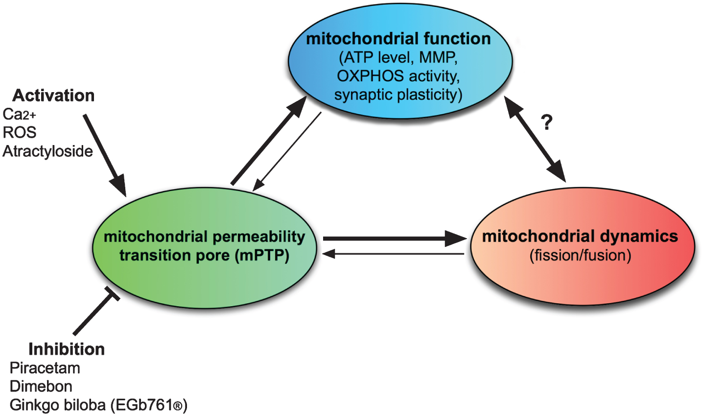
The love triangle of mitochondrial function, dynamics, and mPTP function. Beside its well-known function as regulator of programmed cell death, there is increasing knowledge that the mPTP works as a master regulator of different mitochondrial mechanisms including MMP and ATP production and mitochondrial quality control, especially fission and fusion balance [28, 135]. Inhibition of PTP function has been shown to improve many aspects of mitochondrial function and impaired synaptic plasticity and to shift fission and fusion balance to the fusion side. For example, in a cell model of LOAD, piracetam shifted back mitochondrial fission and fusion balance to larger mitochondria accompanied by reduced mPTP opening events and improved neuritogenesis [28]. Moreover, mPTP opening by atractyloside was accompanied by enhanced fission which also was reduced by piracetam [28]. Consequentially, altering mitochondrial dynamics by down-regulation OPA1 was leading to larger mitochondria and reduced mPTP function [135]. Similarly, enhancing mitochondrial fusion by upregulating Mfs2 reduced the sensitivity of mPTP to opening by ROS [136]. Moreover, impairing mPTP function by downregulating individual mPTP compounds improves mitochondrial function synaptic deficits, and cognition as shown for cyclophilin D 132] and VDAC1 [133]. Vice versa, elevating cyclophylin D levels impair mitochondrial function, synaptic plasticity, and cognitive performance [134].
This will give rise to several important questions: Up to now, therapeutic benefit for mitochondria targeted drugs is modest [30, 139]. Is this already the best possible or do we have the chance to develop mitochondria targeted drugs/drug combinations with better efficacy? Based on the data available we think that other mitochondria targeted drugs can show better efficacy but it seems rather unlikely that a magic bullet will be found. This has to be seen against the background of the many disappointments in the field and the perspective that no other approach to treat age-related memory disorders can be expected for the time coming. Which models to test mitochondrial function should we use as there is some discrepancy between the sometimes substantial mitochondria improving effects of the three drugs in preclinical settings and the modest clinical efficacy? Adequate animal models seem to be mandatory. The three drugs described in the present communication are quite different. All three only show modest impairment of mPTP function but not complete inhibition. How much inhibition is possible without blocking beneficial effects of mPTP opening like regulating programmed cell death [141]? The mPTP represents a very complex system. Is it suitable as a specific molecular drug target? Which of the individual components should be targeted [139]? The three drugs described in the present communication are chemically and pharmacologically quite different and very likely interfere with different parts of this large supramelucular structure. Should we continue to develop mitochondria targeted drugs or should we still wait for the right Aβ-directed drug even if all developments investigated so far failed to show relevant clinical benefit? It appears very unlikely that any other compound following this line will be better. Moreover, there is nothing on the horizon giving hope for the magic bullet within the next decade. Thus, it seems plausible to follow the mitochondrial concept which could result at least in reasonable efficacious drugs in a not too long time.
DISCLOSURE STATEMENT
Authors’ disclosures available online (https://www.j-alz.com/manuscript-disclosures/17-9915).