






Abstract
Background:
Trials of supplementation with omega-3 fatty acids (ω3-FAs) in patients with mild cognitive impairment or Alzheimer’s disease (AD) have produced inconsistent effects on cognitive decline. There is evidence of an interaction between B vitamin status and ω3-FAs in relation to brain atrophy and cognitive decline.
Objective:
We investigated whether baseline levels of plasma total homocysteine (tHcy), a marker of B vitamin status, modify the effects of ω3-FAs supplementation on cognitive performance in moderate AD.
Methods:
This post hoc analysis of the OmegAD trial included 171 community-based patients with AD (MMSE≥15): 88 patients received daily doses of 1.7 g docosahexaenoic acid and 0.6 g eicosapentaenoic acid for 6 months. Treatment outcome on cognition was analyzed according to baseline levels of tHcy using a general linear model and ANCOVA.
Results:
We found significant interactions between ω3-FA supplementation and tHcy on cognition and clinical stage assessed by MMSE (p = 0.040), global CDR (p = 0.059), and CDRsob (p = 0.023), but not on ADAS-cog (p = 0.649). In patients with tHcy levels <11.7μmol/L, ω3-FA supplementation improved cognitive performance as measured by MMSE (+7.1%, 95% CI: 0.59 to 13.7%, p = 0.033) and clinical status as measured by CDRsob (–22.3%, 95% CI: –5.8 to –38.7%, p = 0.009) compared with placebo.
Conclusion:
The effect of ω3-FA supplementation on MMSE and CDR appears to be influenced by baseline tHcy, suggesting that adequate B vitamin status is required to obtain beneficial effects of ω3-FA on cognition.
INTRODUCTION
Nearly 50 million people worldwide suffer from dementia at an annual cost of over $800 billion, and it is regarded as a public health priority by the World Health Organization [1]. Alzheimer’s disease (AD) is the leading cause of dementia, for which there currently is no effective drug treatment. A number of dietary factors have been suggested to have disease-modifying effect on the progression of cognitive decline to dementia. These include long-chain polyunsaturated omega-3 fatty acids (ω3-FAs) and also B vitamins.
The ω3-FAs of main interest are docosahexaenoic acid (DHA) and eicosapentaenoic acid (EPA). DHA has been shown to pass the blood-brain barrier and can be measured both in blood and in the cerebrospinal fluid [2]. DHA is heavily enriched in the brain, and forms a major component of neuronal membranes, particularly in synapses [3, 4]. Vitamins B6, B12, and folate (B9) are vital for one-carbon metabolism and methylation reactions, necessary for the production of monoamine neurotransmitters, phospholipids, and nucleotides. Intake of B vitamins lower levels of the sulphur-containing amino acid homocysteine (Hcy), a recognized risk factor for cognitive impairment and AD (as reviewed in [5]). Despite epidemiological support for beneficial effects of B vitamins and ω3-FAs on cognitive decline and dementia, clinical trials on both nutrients have demonstrated conflicting results [6–11].
B vitamins are necessary for the methylation of phosphatidylethanolamine to form phosphatidylcholine (PC), an important carrier molecule of DHA and EPA in blood and for their transport over the blood-brain barrier [12]. Adequate blood levels of B vitamins could therefore be of importance for the delivery of ω3-FAs to the brain. VITACOG is a randomized, double-blind, placebo-controlled study (RCT) evaluating the effect of high dose B vitamin treatment (folic acid, 0.8 mg; vitamin B6, 20 mg; vitamin B12, 0.5 mg) in elderly with mild cognitive impairment (MCI) [11]. A post-hoc analysis of VITACOG results showed that the protective effect of the B vitamin treatment on brain atrophy rates was dependent on the plasma concentrations of DHA and EPA [13]. In subjects with high concentration of ω3-FA at baseline (plasma DHA+EPA >590μM), B vitamin treatment slowed the brain atrophy rate by 40% compared with placebo. Similarly, significant interaction effects were also observed in relation to cognitive functions [14]. These findings could suggest that the protective effect of ω3-FA supplementation may be dependent on sufficient B vitamin levels.
OmegAD is an RCT investigating the effect of supplemental ω3-FAs in patients with mild to moderate AD on cognitive outcomes, primarily the Mini-Mental State Examination (MMSE) and the modified cognitive part of the Alzheimer’s Disease Assessment Scale (ADAS-cog) [15]. In the main study, we observed no significant effects on delay of cognitive decline, but in a small patient group (n = 32) with very mild AD (MMSE of >27), there were indications of significant beneficial cognitive effects. The main objective of this post hoc study is to investigate interactions between baseline levels of total Hcy (tHcy), as a marker of B vitamin status, and supplementation with ω3-FAs on cognitive and functional assessments in patients with mild to moderate AD, with the hypothesis that the potential positive effects of supplementation with ω3-FA on the cognitive and functional outcome is related to the baseline levels of tHcy.
MATERIALS AND METHODS
Participants
This post hoc analysis was performed as a part of the OmegAD RCT (registered at Clinicaltrials.gov as NCT00211159), conducted between December 2000 and March 2004 in Stockholm, Sweden. The study was carried out according to the principles of the Declaration of Helsinki and was approved by the local Ethics Committee of Karolinska Institutet, Stockholm, Sweden. Details on study design, participants, inclusion and exclusion criteria and the randomization procedure can be found elsewhere [15]. In short, the study enrolled 204 patients with diagnosed AD (Fig. 1), according to the criteria in Diagnostic and Statistical Manual of Mental Disorders, Fourth Edition (DSM-IV). Further inclusion criteria were an MMSE score ≥15, and that the subjects had received treatment with acetylcholinesterase inhibitors for at least three months before the start of the study, and that the treatment was planned to continue throughout the study. Group sizes were calculated in order to detect significant effects on cognition after 6 months with 80% statistical power. Allocation was generated by randomization in blocks of four. Patients treated with ω3-FA preparations, anticoagulants, or non-steroidal anti-inflammatory drugs other than low-dose acetylsalicylic acid, were excluded along with patients suffering from other serious diseases or with alcohol abuse. At the time of this post hoc analysis, no information regarding B vitamin supplementation was available.
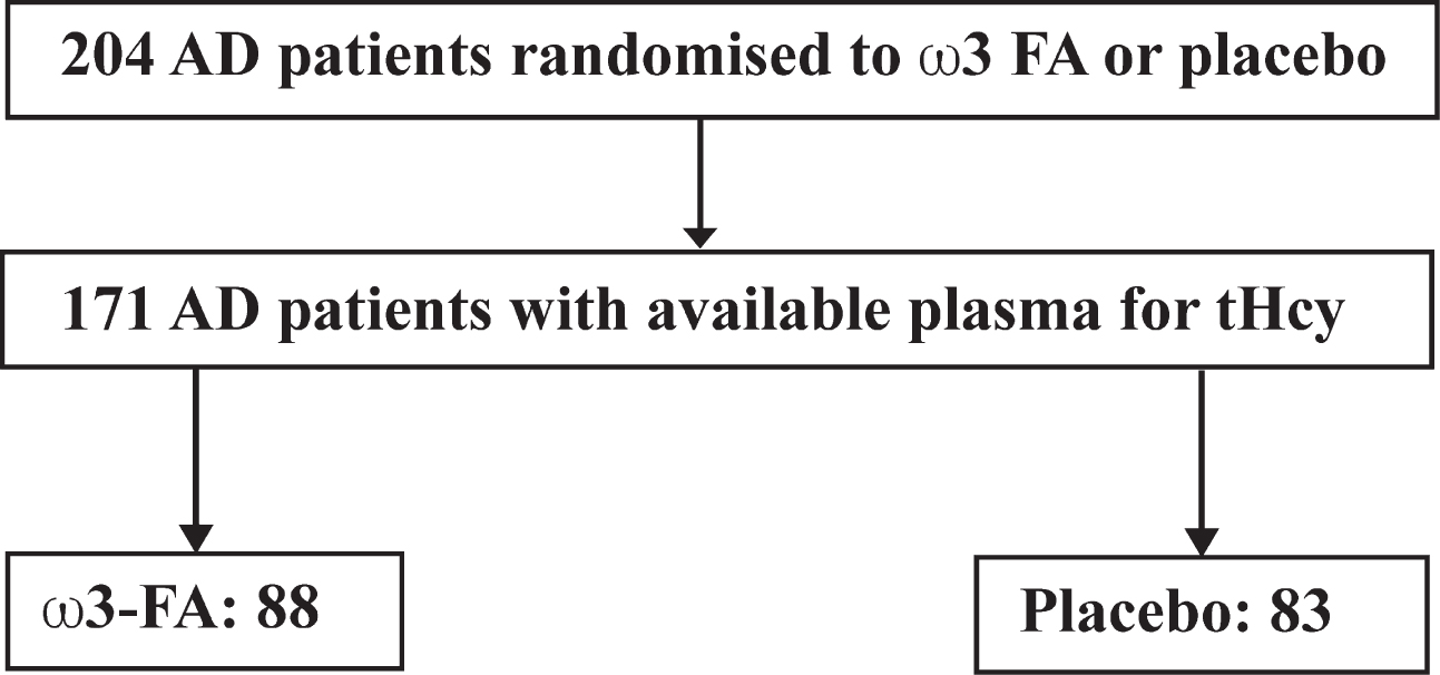
Design of the present post hoc study. 204 patients completed the baseline assessment, of which 174 completed the OmegAD study. At the time of this post-hoc analysis, baseline plasma was available from 171 patients for analysis of tHcy, 88 in the ω3-FA group, and 83 in the placebo group.
Design and data collection
Patients were randomized to receive four 1 g capsules daily, each containing either 430 mg of DHA and 150 mg of EPA, i.e., EPAX1050TG (Pronova Biocare A/S, Lysaker, Norway), or isocaloric placebo oil (containing 1 g of corn oil, including 0.6 g of linoleic acid) for 6 months. Both preparations included 4 mg of vitamin E (α-tocopherol) as an antioxidant. This was followed by an additional 6 months of open treatment with ω3-FA supplementation in all patients. Altogether, 174 patients completed the protocol. Fasting plasma samples were collected at baseline and at follow-up (6 months). Data on the primary endpoints (MMSE and ADAS-cog) were available from 171 patients with blood for amino acid analysis, 88 of which had received ω3-FAs. This study used the outcomes after the initial 6 months of treatment, to enable comparisons between the ω3-FA and placebo groups.
The primary outcome measure was defined as cognitive functions assessed by MMSE and ADAS-cog [15]. As secondary outcomes, global function assessed by the Clinical Dementia Rating scale (global, 0–3, CDR-gl) and sum-of-boxes (CDR-sob) was used.
Biochemical assays
Plasma FA levels were analyzed by gas chromatography (TR-Fame column, 30 m×0.32 mm inner diameter, 0.25μm film; Thermo Electron Corp, Waltham, MA, USA) coupled to a flame ionization detector, as described elsewhere [16]. Measurements of FA composition of red blood cells may have provided a more stable assessment of long term ω3-FA intake, but was not available at the time of this post hoc analysis. Here, measurements of plasma levels of DHA, EPA, docosapentaenoic acid (DPA), and arachidonic acid were expressed as the abundance of individual FAs relative to total FA.
Plasma sulphur amino acids, including tHcy, were measured by liquid chromatography–tandem mass spectrometry (LC-MS/MS) using a modified version of a previously described method [17]. Briefly, deuterium-labelled isotopes were added to plasma as internal standards, followed by reduction of disulphides using dithioerythritol [100 mM] and then protein precipitation using 5-sulfosalicyclic acid [10%]. The extracts were diluted with an aqueous solution of formic acid [0.5%] and heptafluorobutyric acid (HFBA) [0.3%] prior to analysis. LC-MS/MS was carried out using a Shimadzu LC-20ADXR Prominence LC system (Kyoto, Japan) coupled to a Sciex QTRAP5500 mass spectrometer with a Turbo V ion source (Framingham, MA, USA). Chromatographic separation was achieved on a Phenomenex Kinetex Core Shell C18 (100×4.6 mm, 2.6μm) LC column (Torrance, CA, USA) with an aqueous solution of formic acid [0.5%] and HFBA [0.3%] and acetonitrile gradient mobile phase. Positive mode multiple reaction monitoring was used for detection. Linear calibration curves of the peak area ratios of analyte and internal standard in water were used for quantification. The coefficient of variations for the amino acids were 3.1–7.8%. The method for tHcy was validated using spiked plasma quality assurance (QA) samples from an external QA scheme ERNDIM (http://www.erndim.org).
Statistical analysis
A general linear model and analysis of (co)variance was used to examine associations between plasma tHcy, treatment category and primary (MMSE and ADAS-cog) and secondary outcome measures (CDRgl and CDRsob), and the presence of an interaction effect. These models were adjusted for age, sex, ApoE status, baseline levels of total ω3-FA (DHA + EPA + DPA), baseline concentration of creatinine, and initial score of the relevant outcome measure. Total ω3-FA and creatinine data were transformed to their natural logarithms and checked for normality using the Kolmogorov-Smirnov and Shapiro-Wilks tests. Levene’s test was used to test homogeneity of variances. Baseline tHcy concentrations, categorized into tertiles, were used in all statistical analyses unless otherwise stated. Pairwise comparisons following ANCOVA, adjusted for variables specified above, were performed to investigate differences between the placebo group and the ω3-FA treated group for each tHcy tertile.
Pearson correlations were used to examine the relationships between sulphur amino acids, ω3-FAs, and outcome measures at baseline. p values (two-tailed) <0.05 (p < 0.1 for interaction term) were considered statistically significant. As this is a hypothesis testing explorative study that was conducted post hoc, no corrections for multiple comparisons were made. Data are given as mean and 95% confidence interval unless otherwise stated. All statistical analyses were performed using IBM SPSS Statistics for Mac, version 25 (IBM Corp., Armonk, NY, USA).
RESULTS
Participants
Baseline characteristics of the 171 subjects who were included in the present study are presented in Table 1. As reported in the original publication, baseline characteristics were similar in the placebo group and ω3-FA treatment group, and the treatment was well tolerated [15]. In addition, the new analyses show that baseline levels of tHcy were similar in both groups, with geometric means of 13.6μmol/L in both the placebo and treatment group. The upper reference limit without folate supplementation is 15μmol/L in adults between 15–65 years of age, and 20μmol/L in elderly (>65 years) [18].
Baseline characteristics of participants in the OmegAD study1
1OmegAD subjects with available plasma at baseline and data on the primary endpoints (MMSE and ADAS-cog) at baseline and after six months. ApoE, apolipoprotein E; BMI, body mass index; BP, blood pressure; DHA, docosahexaenoic acid; DPA, docosapentaenoic acid; EPA, eicosapentaenoic acid; tHcy, total homocysteine. ω3-FAs denotes the sum of the amounts of EPA + DHA + DPA. 2Means; 95% CI in parentheses (all such values). 3Data only from a limited number of participants, n = 143, 130, and 150 for diabetes, smoking, and family history of AD, respectively. 4Geometric means; 95% CI of geometric means in parentheses (all such values).
To examine the relationships between sulphur amino acids (tHcy, total cysteine, methionine, and total glutathione) and cognitive performance at baseline, simple Pearson’s correlation analyses were performed. None of the sulphur amino acids showed significant correlations with any of the cognitive scores at baseline (Table 2).
Pearson’s correlation between sulphur-containing amino acids and cognitive and neuropsychiatric performances at baseline1
1Pearson’s correlation coefficients along with their two-sided p values. All amino acid variables were entered as the natural log of the baseline concentrations in μmol/L, to ensure normal distribution. ADAS-cog, cognitive portion of the Alzheimer’s Disease Assessment Scale; CDR, Clinical Dementia Score; CDRsob, CDR sum-of-boxes; MMSE, Mini-Mental State Examination.
Correlation analyses were made to examine the relationship between sulphur amino acids and selected plasma FA levels at baseline. tHcy showed significant inverse relationships with a number of plasma polyunsaturated FAs, including the ω6 polyunsaturated FA arachidonic acid (r = –0.153, p = 0.045), and the ω3-FAs EPA (r = –0.190, p = 0.013), DPA (r = –0.270, p < 0.001), and DHA (r = –0.290, p < 0.001), indicating a possible biochemical link between tHcy and polyunsaturated FA levels.
Relations of ω3-FA treatment to MMSE and ADAS-cog scores at 6 months according to baseline tHcy levels
The main objective of this study was to examine the possibility of interaction effects between ω3-FA treatment and baseline levels of tHcy on MMSE and ADAS-cog at 6 months. Cognitive scores after 6 months of ω3-FA or placebo treatment according to baseline tHcy are shown in Fig. 2A and B, adjusted for age, sex, ApoE status, baseline concentration of ω3-FAs and creatinine. Using a general linear model, we found significant interactions between treatment and tHcy tertiles for MMSE (p = 0.040), but not for ADAS-cog (p = 0.649).
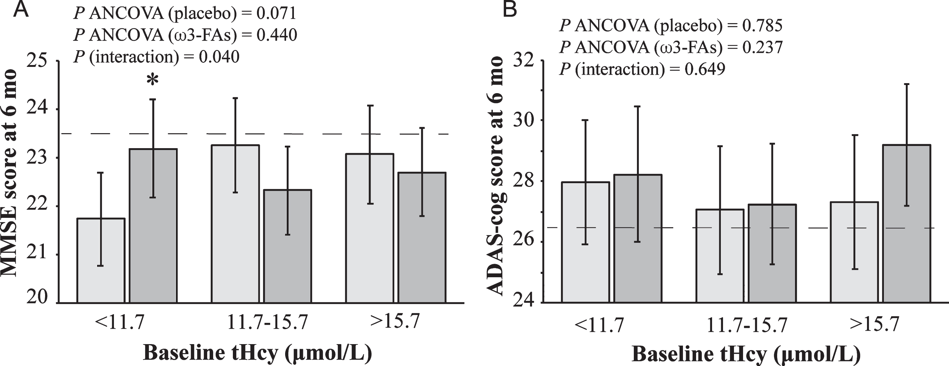
Cognitive test scores at six months among subjects receiving placebo (light grey) and ω3-FA treatment (dark grey) (mean with 95% CI) according to tertiles of plasma baseline tHcy. All models were adjusted for age, sex, ApoE status, baseline concentration of ω3-FAs (ln), baseline concentration of creatinine (ln), and baseline score on the test investigated. A) Mini-Mental State Examination Scores (MMSE), where higher scores indicate better cognitive function. Baseline MMSE appearing in this model is indicated by a horizontal dashed line at 23.5. B) The cognitive portion of the Alzheimer’s Disease Assessment Scale (ADAS-cog), where lower scores indicate better cognitive function. Baseline ADAS-cog appearing in this model is indicated by a horizontal dashed line at 26.4. Group sizes varied between 26–31 among the placebo and treatment groups among the tHcy tertiles. *p < 0.05 compared with the corresponding placebo tertile.
Comparisons by ANCOVA of MMSE and ADAS-cog scores in the placebo and treatment groups separately showed no statistically significant differences over tHcy tertiles in either group (Fig. 2A, B). However, when comparing MMSE scores between the ω3-FAs treatment and placebo groups in individual tHcy tertiles, adjusted pairwise comparisons show significant effects of ω3-FA treatment on MMSE (7.1% higher, 95% CI: 0.59 to 13.7%, p = 0.033) in subjects with low baseline tHcy (<11.7μmol/L). Thus, we found that the effect of ω3-FA treatment on MMSE was modified by baseline tHcy levels. Patients with lower tHcy at baseline (<11.7μmol/L), consistent with better B vitamin status, seemed to benefit more from the treatment (Fig. 2A, B).
Relations of ω3-FA concentrations and CDR according to baseline tHcy levels
General linear models were used to evaluate modifying effects of baseline tHcy on CDR, adjusted for age, sex, ApoE status, baseline concentration of ω3-FAs and creatinine. CDR is a clinical assessment test, where higher scores indicate a worse clinical status (Fig. 3A, B). We found significant interaction effects on CDRgl (p = 0.059) and CDRsob (p = 0.023). ANCOVAs performed in the placebo and treatment groups separately (Fig. 3A, B) revealed a significant difference in CDRsob between tHcy tertiles in subjects treated with ω3-FAs (p = 0.022). Post-hoc pairwise comparisons showed differences between the 1st and the other tHcy tertiles, with scores being 30.7% higher in the 2nd tertile (95% CI: 8.43 to 52.9%, p = 0.007), and 25.4% higher in the 3rd tertile (95% CI: 1.58 to 49.3%, p = 0.037). When comparing scores in those with low baseline tHcy (<11.7μmol/L), CDRsob was 22.3% lower among patients receiving ω3-FAs compared with the placebo group (95% CI: 5.8 to 38.7%, p = 0.009). In short, the effect of the ω3-FA treatment on CDR was modified by tHcy, with a lower tHcy concentration at baseline being beneficial for the effect of ω3-FA supplementation (Fig. 3A, B).
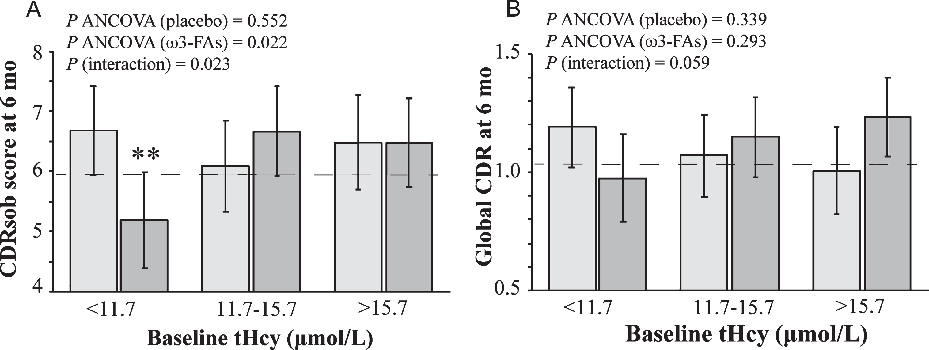
Clinical status as assessed by the Clinical Dementia Rating (CDR) scale at six motnhs among subjects receiving placebo (light grey) and ω3-FA treatment (dark grey) (mean with 95% CI) according to tertiles of plasma baseline tHcy. All models were adjusted for age, sex, ApoE status, baseline concentration of ω3-FAs (ln), baseline concentration of creatinine (ln), and baseline score on the test investigated. A) CDR sum-of boxes (CDRsob), where lower values indicate a better clinical status. Baseline CDRsob appearing in this model is indicated by a horizontal dashed line at 5.90. B) Global CDR score, where lower values indicate a better clinical status. Baseline global CDR appearing in this model is indicated by a horizontal dashed line at 1.03. Group sizes varied between 26–31 among the placebo and treatment groups among the tHcy tertiles. **p < 0.01 compared with the corresponding placebo tertile.
DISCUSSION
In this post hoc analysis of the OmegAD study, an RCT investigating the effects of ω3-FA supplementation on cognitive measurements in patients with mild to moderate AD, we demonstrate significant interactions between ω3-FA treatment and baseline levels of tHcy on MMSE and CDR score (global and sum of boxes) after six months of treatment. Lower tHcy concentration (<11.7μmol/L), indicating a better B vitamin status, was associated with a beneficial effect of ω3-FA supplementation on both cognitive and clinical outcomes. Compared with the placebo group, MMSE scores were 7.1% higher and CDRsob scores 22.3% lower in the ω3-FA group, indicating better cognitive performance and improved clinical status. No significant effects were evident when assessing ADAS-cog, a possible reason being that change scores in this scale may be of limited reliability [19].
Apart from VITACOG, available clinical studies demonstrating an interaction effect between ω3-FAs and B vitamins on cognitive impairment or dementia are few. The studies usually use relatively low doses of vitamins B12, B6, and B9 (folate) and include multiple nutrients and/or vitamins. Plasma tHcy is rarely measured in trials involving ω3-FAs, making any assessment of the effect of the dose on one-carbon metabolism difficult. Furthermore, most ω3-FA trials are not designed specifically to detect effects on cognitive performance and are not sufficiently powered. The SU.FOL.OM3 study investigated the effects of B-vitamins and ω3-FA supplementation on cardiovascular diseases. An ancillary analysis showed an apparent benefit of the combination on temporal orientation in a subgroup with prior stroke [20]. A study on 86 healthy elderly found no beneficial effect of ω3-FA in combination with a multinutrient supplement including vitamins B9 and B12, after 6 months of treatment, during which no cognitive decline was evident in the placebo treated group [21]. From the multicenter study LipiDiDiet, where patients with prodromal AD received medical food containing DHA, EPA, folic acid, vitamins B12 and B6 and multiple other nutrients, there was no significant effect on the neuropsychological test battery outcome over a 2-year period [22]. However, cognitive decline over this period was much lower than expected, rendering the primary endpoint inadequately powered. Nevertheless, there were beneficial effects on some secondary endpoints of disease progression such as CDRsob and hippocampal atrophy.
DHA is enriched in the brain but cannot itself be synthesized in brain tissue. PC is quantitatively the most important carrier of DHA in plasma, and is depleted in blood and brain tissue of AD patients [23–26]. B vitamins are crucial for efficient synthesis of PC-DHA through their role in methylation. Without sufficient intake of B vitamins, formation of the relevant forms of PC could be impaired, and the brain may not obtain enough ω3-FAs. This hypothesis is yet to be investigated experimentally in humans, but a cross-generational study on rats suggest that ω3-FAs together with B vitamins may result in higher DHA levels in the hippocampus of their offspring, compared with ω3-FAs alone [27]. Also, exposure to elevated levels of Hcy in chick embryos caused reduced PC and elevated PE levels in chick brains, and reduced DHA levels in chick brain membranes [28]. The effect of B vitamins on ω3-FA distribution has been investigated in RCTs. Fish oil plus a multivitamin supplementation that included B vitamin, increased the incorporation of ω3-FAs into erythrocyte membranes [29]. This effect was not achieved with the fish oil alone. A larger study on B vitamin supplementation (1 mg folate, 0.5 mg vitamin B12, and 10 mg vitamin B6 over 2 years failed to increase the proportion of ω3-FAs in plasma PC [30]; however, only relative concentrations were reported.
There are some limitations to our study. First, this is an exploratory analysis and was not designed to be adequately powered for the detection of interaction effects or subgroup analyses. Second, this study included patients with mild to moderate AD. Third, the duration of the ω3-FA intervention was only 6 months. Fourth, B vitamin status was only assessed indirectly by measuring tHcy. OmegAD was the first randomized trial on ω3-FA treatment in AD to be published. Since its publication, it has become apparent that subjects with established AD may not be the ideal target population for nutritional supplements. It is now clear that the underlying processes behind AD may start several decades before the onset of cognitive symptoms. Therefore, more focus is now on the prevention of age-related cognitive decline and treatment of preclinical stages of AD, e.g., MCI, with disease-modifying interventions like ω3-FAs. Since no immediate relief of symptoms may be expected, such studies generally require durations of several years before significant differences between treatment groups become significant. In fact, in this study neither the placebo nor the ω3-FA group showed any significant cognitive or clinical decline over 6 months, as judged by MMSE, ADAS-cog, and CDR scores [15]. Considering these shortcomings, it is encouraging that significant interaction effects on cognition could be detected, highlighting the need for further studies specifically designed to address this topic.
In conclusion, our results indicate that the effect of ω3-FA supplementation on cognitive and clinical outcomes is related to baseline tHcy levels, which implies that functional B-vitamin status is a potentially important factor to consider when evaluating the effect of ω3-FA supplementation. Future randomized clinical trials with ω3-FAs therapies should be conducted to provide further elucidation of the relationships between ω3-FAs, Hcy, and B vitamins on cognitive as well as neuropsychiatric symptoms.
Footnotes
ACKNOWLEDGMENTS
We are grateful to Cynthia Prendergast for valuable help with sulphur amino acid analyses, Ann-Christine Tysén-Bäckström and Andreas Svensson for patient data management, Siv Tengblad for analyzing plasma fatty acids, and Jonas Selling for statistical support.
This work was supported by the Regional Agreement on Medical Training and Clinical Research (ALF) between Stockholm County Council and the Karolinska Institutet, Funds of Capio, Demensförbundet, Gamla Tjänarinnor, Swedish Alzheimer Foundation, Norwegian Research Council, Odd Fellows, Swedish Brain Foundation, Swedish Nutrition Foundation, Gun and Bertil Stohnes Stiftelse, Swedish Society of Physicians, Lion’s Sweden, and from Åke Wibergs foundation (#M16-0251). The OmegAD study was partly funded by Pronova Biocare A/S, Lysaker, Norway. The company was represented in the trial steering committee with regard to study design, and provided the EPAX1050TG and placebo preparations; the company has not been involved in the data and patient collection, analyses or interpretations of scientific data.
