






Abstract
Alzheimer’s disease (AD) and age-related macular degeneration (AMD) are two complex incurable neurodegenerative disorders the common pathogenesis of which is actively discussed. There are overlapping risk factors and molecular mechanisms of the two diseases; at the same time, there are arguments in favor of the notion that susceptibility to each of these diseases is associated with a distinct genetic background. Here we identified single-nucleotide polymorphisms (SNPs) that are specific for senescence-accelerated OXYS rats, which simulate key characteristics of both sporadic AD and AMD. Transcriptomes of the hippocampus, prefrontal cortex, and retina (data of RNA-Seq) were analyzed. We detected SNPs in genes Rims2, AABR07072639.2, Lemd2, and AABR07045405.1, which thus can express significantly truncated proteins lacking functionally important domains. Additionally, 33 mutations in genes—which are related to various metabolic and signaling pathways—cause nonsynonymous amino acid substitutions presumably leading to disturbances in protein structure or functions. Some of the genes carrying these SNPs are associated with aging, neurodegenerative, and mental diseases. Thus, we revealed the SNPs can lead to abnormalities in protein structure or functions and affect the development of the senescence-accelerated phenotype of OXYS rats. Our data are consistent with the latest results of genome-wide association studies that highlight the importance of multiple pathways for the pathogenesis of AD and AMD. Identified SNPs can serve as promising research objects for further studies on the molecular mechanisms underlying this particular rat model as well as for the prediction of potential biomarkers of AD and AMD.
INTRODUCTION
Aging is the main risk factor of late-onset Alzheimer’s disease (AD) and age-related macular degeneration (AMD). These are two complex incurable neurodegenerative diseases, the common pathogenesis of which is currently actively discussed. According to the estimates on population aging by 2050, the number of patients with AD will reach 152 million [1], and the expected number of individuals affected by AMD in 2020 is 196 million (288 million in 2040) [2]. These age-related neurodegenerative diseases primarily affect various parts of the central nervous system (CNS) but are substantially similar in terms of abnormal extracellular amyloid-β (Aβ) deposits: senile plaques in the brain of patients with AD and druses in the eyes of patients with AMD, microvascular abnormalities, neuroinflammation, and metabolic and oxidative stress [3–5]. Population studies have revealed a high incidence of cognitive impairment among patients with AMD and pathological changes in the fundus of most AD patients [5–10]. AD and AMD have complex signs, the initiation of which is controlled by a variety of interacting genetic and environmental factors. There are overlapping risk factors and molecular mechanisms of the two diseases; at the same time, there are arguments in favor of the notion that susceptibility to each of these diseases is associated with a distinct genetic background [11–13]. Investigation of the correlates of AMD and especially AD is worthwhile but has rarely been done [14]. Many genetic and environmental/lifestyle factors not only affect the mechanisms that trigger the transition from normal age-related changes to the diseases but also modulate the age of manifestation of AD and AMD, their progression, and severity. This situation makes it difficult to study these factors and diseases in the human population.
Undoubtedly, none of the available animal models reproduces all the features of human AD and AMD and therefore cannot be considered a representative model of AD or AMD as a complete disease [15, 16]. Nevertheless, animal models can add to our understanding of how various clinical signs or genes can contribute to the etiology and pathogenesis of these diseases. Among such models, monogenic ones predominate: these are transgenic animals, gene knockout animals, or animals with certain mutations [17]. Such models allow for elucidation of a contribution of a specific gene to the development of a symptom but do not reproduce all phenotypic manifestations of polygenic diseases, such as AD and AMD.
In recent years, convincing arguments were made confirming that the strain of senescence-accelerated OXYS rats meets the requirements for both the AD models [18–22] and AMD models [23–29] owing to a good match between the accelerated brain/retina aging and the development of key “clinical” signs of these diseases. The current concepts of AD pathogenesis in humans correspond well to the rather long list of pathological changes in OXYS rats: dysfunction of mitochondria, tau protein hyperphosphorylation, an aberration of long-term post-tetanic potentiation, synaptic insufficiency, destructive changes in neurons, behavioral disorders, and a decrease in cognitive functions at the early stages and aggravation of these functions during an increase in the amyloid-β protein precursor (AβPP) level, enhanced accumulation of Aβ, and formation of Aβ plaques in the brain [18–21]. In the genome of OXYS rats, the absence of mutations in genes App, Psen1, and Psen2 [20] (which are specific for the early form of AD) also suggests that this model exactly meets the criteria of the sporadic form of this disease (>95% of cases).
Developing already at a young age, retinopathy in OXYS rats corresponds to the dry atrophic form of AMD in humans. As in humans, neovascularization develops in some (∼10–20%) OXYS rats with age. The clinical signs of AMD-like retinopathy appear in conjunction with a reduction in the transverse area of the retinal pigment epithelium (RPE) and impairment of choroidal microcirculation. Significant pathological changes in the RPE as well as clinical signs of advanced stages of retinopathy in OXYS rats manifest themselves as excessive accumulation of lipofuscin and Aβ in RPE regions and whirling extensions of the basement membrane into the cytoplasm. As in the dry form of human AMD, the initial alterations in the OXYS RPE cells later lead to the atrophy of choriocapillaris and a complete loss of photoreceptor cells in the OXYS retina by age of 24 months.
Emergence of the signs of AMD-like and AD-like pathologies in OXYS rats (ages 3–5 months) is associated with a change in the expression of genes related to the immune system, inflammation, oxidative stress, calcium homeostasis, and apoptosis. Progression of these pathologies (age 3–18 months) takes place during the expression alteration of genes in the retina and cortex; these genes are associated with the metabolic pathway of AD, including the genes related to the processing of AβPP, aggregation and degradation of Aβ, disturbances of synaptic processes, and mitochondrial dysfunction [18–21, 31]. These observations are consistent with RNA-Seq data on changes in the transcriptome of the prefrontal cortex of patients with AD [21]. It is important that in the preclinical period (20 days) in both the retina and cortex of OXYS rats, there are changes in the expression of genes whose products are involved in the development of the CNS, synaptic transmission, and neuronal plasticity [21, 29]. The nature of gene expression changes in the retina, hippocampus, and cerebral cortex of OXYS rats during maturation completion indicates a contribution of delayed maturation of the brain [32] and retina to the development of AMD and AD signs in OXYS rats. Nevertheless, the reason for these changes remains unknown.
Here we identified single-nucleotide polymorphisms (SNPs) that are specific for the transcriptome of OXYS rats and could affect the manifestation of the traits characterizing the senescence-accelerated phenotype of these rats, with a focus on AD-like and AMD-like pathologies.
METHODS
Animals
The OXYS rat strain was developed at the Institute of Cytology and Genetics, Russian Academy of Sciences (Novosibirsk), from Wistar stock via selection for susceptibility to cataractogenic effect of a galactoserich diet and via inbreeding of highly susceptible rats as described earlier [18, 33]. After 5 cycles of inbreeding, feeding of galactose-rich diet and selection, the subsequent generations of rats developed cataracts spontaneously, without the galactose-rich diet. At present, we have the 112th generation of OXYS rats with spontaneously developing cataract and accelerated senescence syndrome, which is characterized by early development of a phenotype similar to human geriatric. This pathological phenotype primarily includes accelerated thymus involution [34], senile osteoporosis [35], retinopathy similar to human AMD [23–29], cardiomyopathy [36], and AD [18–22, 30–32]. Male senescence-accelerated OXYS rats were used at ages 20 days and 3, 5, and 18 months, while age-matched male Wistar rats served as controls (3–5 per group). The animals were kept at the Center for Genetic Resources of Laboratory Animals at the Institute of Cytology and Genetics, the Siberian Branch of the Russian Academy of Sciences. At the age of 4 weeks, the pups were weaned, housed in groups of five animals per cage (57×36×20 cm), and kept under standard laboratory conditions (22±2°C, 60% relative humidity, 12 h light/12 h dark cycle, lights on at 9 a.m.). The food and water were available ad libitum. The protocol of the animal experiment was approved by the Bioethical Committee of the Institute of Cytology and Genetics.
Tissue preparation
After decapitation, the prefrontal cortex, retina, and hippocampus were excised rapidly, placed in RNAlater (Ambion, catalog # AM7020), frozen, and stored at –20°C until analysis. The frozen rat tissues were lysed with the TRIzol Reagent (Invitrogen, cat. # 15596-018), and total RNA was isolated. RNA quality and quantity were evaluated on an Agilent Bioanalyzer (Agilent). The RNA samples were sent to Genoanalytica Inc. (http://www.genoanalytica.ru, Moscow, Russia) where the RNA sequencing was performed.
RNA sequencing
Over 40 million single-end sequenced reads of 50 bp length were obtained for each sample of retinal, prefrontal-cortex, and hippocampal RNA by Illumina nonstranded sequencing (on an Illumina GA IIx at Genoanalytica) in accordance with standard Illumina protocols (mRNA-Seq Sample Prep Kit) as described previously [21, 25]. All the samples were analyzed as biological replicates for each rat group.
SNP detection
After barcode trimming, the sequencing data were tested for quality using the FastQC software and mapped to Rattus norvegicus reference genome assembly RGSC 6.0 (Ensemble release 75) in Bowtie 2 or TopHat software v2.0.4. Quality assessment of the mapped data was carried out using the CollectRnaSeqMetrics module in the Picard software package (https://www. broadinstitute.github.io/picard).
SNP positions within the aligned reads relative to the reference genome were identified via the pileup function in SAMtools (v. 0.1.17). By means of various filter commands, SNPs were predicted for various positions with minimum mapping quality (Q) of 100. These parameters ensure high-quality, reliable mapping of the reads, which is important for variant calling. Using custom-designed Perl scripts, the VCF files were converted into MySQL tables. The obtained polymorphic variants in the tissues of OXYS rats were further filtered by the following criteria: only those positions were selected whose genotype was found to be in a homozygous state for at least three animals in one experimental group. Coverage (DP) had to be ≥10 in at least one animal.
The list of SNPs of OXYS rats was next compared with RGSC 6.0 data for genome sequences of 32 rat strains and substrains: 10 of these strains/substrains are commonly employed as a normotensive control (FHL/EurMcwi, LN/MavRrrc, LL/MavRrrc, MNS/Gib, SBN/Ygl, SR/Jr, WKY/N, WKY/Gla, WKY/NCrl, and WKY/NHsd), and 22 rat strains/substrains serve as control or experimental animals in the studies on various pathological conditions that have no relation to hypertension or aging [ACI/N, ACI/EurMcwi, BBDP/Wor, BN-Lx/Cub, BN-Lx/CubPrin, BN/SsN, BUF/N, DA/BklArbNsi, F334/N, F344/NHsd, F344/NCrl, SUO_F344, GK/Ox, LE/Stm (SOLiD), LEW/Crl, LEW/NCrlBR, LE/Stm (Illumina), M520/N, MR/N, WAG/Rij, WN/N, and Wistar [37]. A comparison of SNPs of OXYS rats with genomes of the 32 rat strains and substrains was performed only at genomic loci sequenced during this transcriptome analysis of OXYS rats.
Prediction of SNP phenotypes
The influence of each amino acid substitution on protein function was predicted using a Web-based tool called Variant Effect Predictor (https://www.ensembl.org/Multi/Tools/VEP); the consequence type, SIFT score, and prediction were obtained for each variant. Generally, SIFT scores of 0–0.05 were classified as “deleterious” and 0.05–1.00 as “tolerated.”
Functional annotation
The gene list with SNPs was subjected to functional enrichment analyses by means of the DAVID tool (https://www.david-d.ncifcrf.gov/). Gene pathway analysis was conducted in the KEGG pathway database (http://www.genome.jp/kegg/). An atlas of combinatorial transcriptional regulation in mice and humans was utilized to determine the transcription factor genes carrying SNPs [38]. The RGD (https://rgd.mcw.edu/) was used to identify the association of SNP-containing genes with aging and diseases associated with aging: hypertension, premature aging, cerebrovascular disorders, dementia, AD (as part of dementia), macular degeneration, demyelinating diseases, mental disorders (anxiety and neurocognitive disorders), and neurodegenerative diseases.
RESULTS
SNP profiles of the prefrontal cortex, hippocampus, and retina of OXYS rats
A comparison of transcriptome sequences of the prefrontal cortex, hippocampus, and retina of OXYS rats with the reference genome of BN/NHsdMcwi rats revealed 42,478 SNPs in 9,903 genes. The total numbers of SNPs in each tissue are given in Table 1. The numbers of tissue-specific and common SNPs detected in the prefrontal cortex, hippocampus, and retina of OXYS rats are shown in Fig. 1A, and the numbers of genes with tissue-specific and common SNPs are presented in Fig. 1B. The functional and structural consequences and the effects of the identified SNPs are listed in Table 2. Altogether, 178 SNPs were found to possibly have major consequences for protein function or structure.
The numbers of SNPs detected in the transcriptomes of the prefrontal cortex, hippocampus, and retina of OXYS rats as compared with the reference genome of BN/NHsdMcwi rats
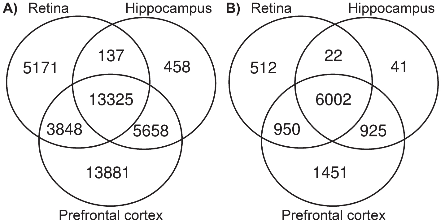
Venn diagrams depicting the numbers of tissue-specific and common SNPs (A) and genes (B) with tissue-specific and common SNPs detected in OXYS rats.
Classification of effects of the SNPs that can influence the senescence-accelerated phenotype of OXYS rats
*Number of SNPs in OXYS rats as compared with the reference genome of BN/NHsdMcwi rats. UTR, untranslated region; NMD, nonsense-mediated mRNA decay.
SNPs related to the development of the senescence-accelerated phenotype of OXYS rats
Among the 42,478 SNPs found in OXYS rats, 40,373 SNPs in 9,699 genes have also been detected in the genomes of rat strains (and control strains) in studies on various pathological conditions that have no relation to aging or age-related diseases. The group of the remaining 2,105 SNPs was of interest in terms of the search for genetic variants that may influence manifestation of the specific phenotype (accelerated senescence) in OXYS rats. The functional and structural consequences and other effects of these SNPs located in the sequences of 1,211 genes (for which 1,809 transcripts are known) are summarized in Table 2.
The classification of SNPs possibly influencing the senescence-accelerated phenotype in OXYS rats indicated that there were seven SNPs characterized by a high impact on transcript structure (Table 3). One of them deserves a special mention because it is located in the mRNA sequence of the Lhx2 gene encoding a transcription factor (Table 3). Two SNPs classified as exerting a high impact on transcript structure were found in the mRNA sequence of Csnk1e, which is annotated in the Rat Genome Database (RGD) as a gene associated with aging, neurodegenerative diseases (including AD), and mental disorders. The SNP in the Rims2 gene was found in three transcripts of different lengths.
SNPs that are specific to OXYS rats and can lead to functionally significant structural rearrangements of transcripts
Genes associated with: *aging, #neurodegenerative diseases (including AD), or Δ mental disorders (including neurocognitive disorders) according to the Rat Genome Database (https://rgd.mcw.edu/). H, hippocampus; R, retina; PC, prefrontal cortex.
The SNPs’ effects on the transcript structure of the genes listed in Table 3 are depicted in Fig. 2, and the expected effects of the structural rearrangements of these transcripts on protein structure and functions are summarized in Table 4. The most substantially truncated protein structures are expected for RIMS2, LEMD2, AABR07072639.2, and AABR07045405.1; moreover, in three of them, functionally significant domains are expected to get lost. The SNP in Lemd2 was found in the retina but not in the hippocampus and prefrontal cortex, whereas the SNPs in AABR07072639.2 and Rims2 were detected only in the prefrontal cortex, and the SNP in AABR07045405.1 was detected in the retina and prefrontal cortex. In addition, in OXYS rats the levels of mRNA of genes Lemd2, Csnk1e, and Lhx2 were increased at some ages in one or more of the studied tissues relative to control animals (Supplementary Table 1).
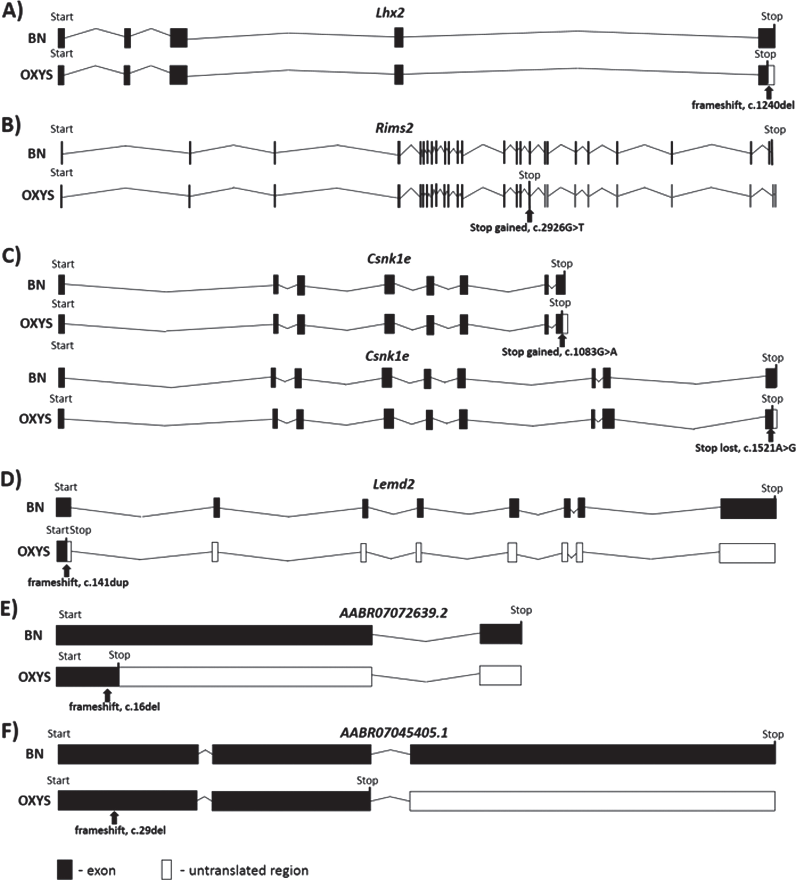
The SNPs’ influence on the transcript structure of genes Lhx2 (A), Rims2 (B), Csnk1e (C), Lemd2 (D), AABR07072639.2 (E), and AABR07045405.1 (F). The black arrow indicates the location of an SNP in a transcript. BN: BN/NHsdMcwi rats.
The effects of the SNPs on the protein length and function in OXYS rats
Domain information was obtained from UniProt (http://www.uniprot.org/uniprot) and Ensembl (https://www.ensembl.org/Rattus_norvegicus). BN: BN/NHsdMcwi rats.
According to the SIFT software algorithm, 33 SNPs having presumably a significant negative effect on the structure and/or function of a protein were detected (Table 5). Among them according to the RNA-Seq analysis we identified 26 genes in OXYS rats whose mRNA level was different from the Wistar rat mRNA level (Supplementary Table 1). Two genes (Acat2 and Ephx1) were associated with aging according to the gene annotation in the RGD. Pla2r1, Zmym6, Trappc9, Nqo2, Ephx1, Ano10, Chrna5, Man2c1, Pcm1, and LOC100364500 are annotated in the RGD as genes associated with neurodegenerative diseases and/or mental disorders. One gene (Ephx1) among those listed in Table 5 is currently known to be associated with hypertension. A number of SNPs designated as novel in Table 5 are described for the first time.
Mutations that have a presumably significant impact on the structure and/or function of protein products according to the SIFT algorithm
Genes associated with: *aging, #neurodegenerative diseases, ∧AD,
Δ
mental disorders, or
The most SNPs listed in Table 5 were detected in transcripts in two or three tissues. Accordingly, these mutations can be regarded as relevant SNPs: those that may be associated with the development of both AD- and AMD-like pathologies.
Functional annotation of genes carrying mutations exerting significant effects on protein or transcript structure and/or functions
Functional annotation of genes carrying mutations that may have an influence on the accelerated aging in OXYS rats (Tables 3–5) were subjected to functional enrichment analysis to identify the affected biological processes and metabolic pathways. The assigned Gene Ontology (GO) terms included antigen processing and presentation (RT1-A1, LOC100364500, and RT1-CE5), developmental cell growth (Lhx2, Akap6, and Rims2), and positive regulation of cellular protein localization (Cnst, Akap6, and Pcm1). Furthermore, several genes were associated with the regulation of binding proteins (Gtpbp4, Csnk1e, Cald1, and Lhx2) and single-multicellular organism process (Gtpbp4, Zmym6, Lemd2, Ephx1, Ostm1, Rims2, Acat2, Pcm1, Rt1-A1, Trappc9, Csnk1e, Lhx2, Chrna5, Akap6, Pla2r1, and Nqo2).
According to Kyoto Encyclopedia of Genes and Genomes (KEGG) pathway analysis, several SNPs were found in the genes associated with cellular senescence (RT1-A1, RT1-CE5, and RT1-CE7), with major histocompatibility complex I (MHC1) protein complex (RT1-A1, LOC100364500, RT1-CE7, and RT1-CE5), and phagosomes (Pla2r1, RT1-CE7, RT1-A1, and RT1-CE5).
DISCUSSION
The aim of this study was to identify SNPs that can potentially contribute to the accelerated senescence and simultaneous development of AD-like and AMD-like pathologies in OXYS rats. As the candidate mutations most likely contributing to the accelerated senescence, we detected seven SNPs leading to functionally significant structural rearrangements of transcripts and 33 SNPs causing the nonsynonymous amino acid substitutions characterized by a deleterious effect on protein structure or function in OXYS rats. As expected, the list of identified genes carrying SNPs in OXYS rats included genes associated with aging and neurodegenerative diseases. All these genes belong to different metabolic signaling pathways.
Age is the strongest risk factor of AD and AMD. Four SNPs specific for OXYS rats were found in the mRNA of genes associated with aging: Csnk1e, Ephx1, and Acat2. CSNK1E (casein kinase 1, epsilon) as an integral component of the circadian clock, takes part in the regulation of degradation and subcellular localization of human period protein 1 (hPER1) through phosphorylation [39, 40]. CSNK1E is also a known component of the WNT signaling pathway, which plays an essential part in neurodevelopment and in the regulation of adult brain function and structure [41, 42]. Alterations in Csnk1e activity and Csnk1e mutations correlate with neurodegenerative diseases and schizophrenia [40]. Here, SNPs in Csnk1e were detected in the transcriptomes of the retina, hippocampus, and prefrontal cortex of OXYS rats. This state of affairs could be one of the reasons for the development of combined pathologies of the retina and brain. Nonetheless, SNPs in the Csnk1e gene in OXYS rats are expected to cause slight changes in protein length and to not affect the known functionally important regions of the protein product.
EPHX1 (epoxide hydrolase 1) encodes an evolutionarily highly conserved enzyme participating in biotransformation of xenobiotic and in general physiological maintenance of some organs [43]. The presence of the EPHX1 protein in neurons and astrocytes may be related to neurotoxicity [44]. A role of EPHX1 in the pathogenesis of neurodegeneration was further supported by the discovery of its differential expression in patients with AD [45]. Indeed, the mRNA expression of Ephx1 is higher in the prefrontal cortex of OXYS rats during the manifestation of AD signs [21], these data may indicate EPHX1 involvement in the AD pathogenesis and make it a promising candidate for further research.
The third gene is ACAT2 (acetyl-CoA acetyltransferase 2) encoding a cholesterol acyltransferase that controls the equilibrium between free and esterified cholesterol [46, 47]. It is well known that lipid metabolism worsens with age, thereby leading to obesity and atherosclerosis. ACAT2 expression may be induced by higher cholesterol concentrations [48]. Excessive free cholesterol and free fatty acids are major risk factors of atherosclerosis and type 2 diabetes mellitus. On the other hand, our studies have revealed that in OXYS rats, cerebral dysfunction [49] and fibrosis of myocardial vessels [36] develop against the background of hypocholesterolemia [50]. These observations are in good agreement with the finding that the mRNA expression of Acat2 is lowered in the retina of OXYS rats [25, 51].
It is noteworthy that we identified a mutation in gene Lemd2, which encodes a nuclear envelope protein called LEM domain–containing protein 2 (LEMD2). The latter is involved in nuclear-structure organization and in cell signaling and differentiation. Disruption of the mouse Lemd2 gene leads to embryonic death [52]. Mutation of LEMD2 is associated with juvenile cataract and inherited dilated cardiomyopathy in humans [53]. Recently, it was demonstrated that a recurrent de novo mutation in LEMD2 results in a progeria-like facial phenotype in humans [54] and leads to chromatin remodeling and accelerated aging of cardiac tissue [55]. In OXYS rats in this study, we found a mutation causing a frameshift in the first exon of Lemd2. The emergence of an additional premature stop codon causes accumulation of a defective protein product in the absence of a full-sized one; however, the position of the mutation spares one of the two transmembrane domains, and a partially preserved protein function may be expected. It should be noted that spontaneous cataract has been the basis for OXYS rat selection. It has sped up senescence, which manifests itself in the simultaneous development of AMD, AD, and hypertrophic cardiomyopathy during moderately high blood pressure [36].
A frameshift mutation was also identified in exon 18 of the Rims2 gene in OXYS rats. We expect the presence of a defective protein product lacking several sites of serine phosphorylation. RIMS2 is known as a regulator of synaptic membrane exocytosis in the CNS [56]. The reported association between mutation in Rims2 and inherited retinal diseases is characterized by progressive pathological changes in retinal cells, which are responsible for absorbing and converting light into electrical signals [57, 58]. In addition, other researchers have demonstrated that RIMS2 is a candidate gene associated with the risk of age-related diseases [56].
A frameshift mutation in the first exon of genes AABR07045405.1 and AABR07072639.2 can also lead to the accumulation of a defective protein product. Both of these genes are poorly characterized, but it is known that the AABR07072639.2 gene contains a domain similar to the mitochondrial carrier superfamily, which participates in the transport of pyrimidine nucleotides for mitochondrial-DNA synthesis. It is noteworthy that the OXYS rat transcriptome contains a mutation with a presumably significant effect on the structure and/or function of the protein product in gene Slc25a32 (mitochondrial folate transporter/carrier, also known as solute carrier family 25 member 32). The product of this gene performs folate transport through the mitochondrial membrane; this transport is necessary for proper nucleotide biosynthesis and methylation reactions. Mitochondrial dysfunctions are believed to be common pathophysiological events between AMD and AD. Already at the preclinical stage, OXYS rats show some characteristic structural and functional changes in hippocampal and cortical mitochondria; these changes are exacerbated with the emergence and progression of the AD-like pathology, including a decrease in the activity of respiratory complexes accompanied by the process of fusion and formation of larger mitochondria [30, 31]. Here, we identified SNPs in genes Slc25a32 and AABR07072639.2, which might be implicated in mitochondrial dysfunctions. Thus, these two genes may be considered promising targets for research into the mechanism of mitochondrial dysfunction during early neurodegeneration.
Behavioral disorders and deterioration of cognitive functions in OXYS rats develop by age 3–5 months and progress with age, thereby leading to brain neurodegeneration. In this regard, an interesting set of SNPs that may have a significant impact on protein structure and/or function was found here in the mRNA of genes associated with neurodegenerative diseases and mental disorders: Trappc9, Man2c1, Pcm1, Zmym6, Ano10, Pla2r1, Nqo2, Ephx1, Chrna5, and LOC100364500. These genes take part in various metabolic and signaling pathways.
The protein encoded by human TRAPPC9 (trafficking protein particle complex 9) has been implicated in human brain development, possibly through its influence on NF-κB activation and protein trafficking in postmitotic neurons of the cerebral cortex [59]. The nucleotide variations of TRAPPC9 have been linked to autosomal recessive mental retardation [60, 61]. The affected patients are characterized by defects in axonal connectivity [59]. In our study, a mutation of gene Trappc9 was detected in all the tissues studied, meaning a high probability that this gene is involved in the combined pathology of the brain and retina.
The manifestation of signs of AMD-like and AD-like pathologies in OXYS rats in this study is related to a change in the expression of genes of calcium homeostasis and apoptosis. Among the genes containing SNPs in OXYS rats, Ano10 and Chrna5 are known to be associated with ion channels. SNPs in these genes cause cognitive disturbances. The ANO10 gene encodes a transmembrane protein (anoctamin 10), which is a member of the family of calcium-activated chloride channels. It may participate in the regulation of intracellular Ca2 + signaling, including a release of Ca2 + from intracellular stores and regulation of mitochondrial function [62]. Mutations in ANO10 cause neurological and immunological defects by disrupting local Ca2 + signaling, e.g., in autosomal recessive cerebellar ataxia [63–65], Purkinje cell dysfunction, and Ca2 +-triggered neurodegeneration [66]. Another gene carrying a mutation in OXYS rats and associated with calcium (Chrna5; cholinergic receptor nicotinic alpha 5 subunit) is a component of neuronal nicotinic acetylcholine receptors, which are ion channels. CHRNA5 is an accessory subunit that can change receptor function, e.g., by increasing calcium permeability [67]. Mice lacking Chrna5 feature impaired attention, increased anxiety, and decreased novelty-induced behavior [68]. We have observed similar changes in behavioral tests of OXYS rats [18].
Among the genes associated with neurodegenerative diseases, MAN2C1 and NQO2 may be indirectly involved in apoptosis alterations. MAN2C1 encodes cytosolic α-mannosidase, which plays an essential part in the catabolism of free oligosaccharides. MAN2C1 downregulation induces mitochondria-dependent apoptosis [69]. Histological analysis of the CNS in Man2c1-deficient mice has uncovered neuronal and glial degeneration with formation of multiple vacuoles in deep neocortical layers and in major telencephalic white matter tracts [70]. NQO2 (N-ribosyl-dihydronicotinamide quinone reductase 2) contributes to vitamin K metabolism and to a reduction in cellular quinone concentration, thus preventing a build-up of reactive oxygen species [71]. Additionally, it binds to and stabilizes the apoptosis regulator p53 [72]. Polymorphism is associated with increased expression of this gene and increased susceptibility to Parkinson’s disease. An increase in hippocampal NQO2 amounts may be a cause of AD or might promote AD progression by raising toxic quinone levels with a consequent loss of cognitive function [73].
An alteration of neuroplasticity is proposed as the basis of behavioral and memory dysfunction under pathological conditions including AD [74, 75]. One of the underlying mechanisms is probably based on impaired development and migration of brain cells. Analysis of our data revealed SNPs in genes Lhx2, Pcm1, and Cald1 associated with these processes. Of note, there is a mutation in the Lhx2 gene in OXYS rats. LHX2 encodes a transcription factor known to be involved in the regulation of cortical neurogenesis [76, 77] and cerebral-cortex development [78, 79]. In addition, LHX2 is a critical regulator of the neuron–glia cell fate switch in the developing murine hippocampus [80, 81]. Recently, we reported that in OXYS rats, there is a delay in brain maturation [32]. LHX2 controls the relative balance between neurogenesis and gliogenesis in the retina [82] and is a key regulator of retinal progenitor cells’ properties contributing to retinal-tissue formation [83–85]. On the basis of known LHX2 functions, one may conclude that the mutations in Lhx2 can determine the timing of both AD and AMD presentation. The protein encoded by PCM1 (pericentriolar material 1) is a component of centriolar satellites. It is necessary for the correct localization of centrosomal proteins and for the attachment of microtubules to the centrosome. Downregulation of PCM1 produces neuronal-migration defects in a developing cerebral cortex [86]. Mutations in this gene are associated with schizophrenia too [87]. The mice with haploinsufficiency at the Pcm1 locus [Pcm1(+/−)] have a smaller whole-brain volume and possibly neuroanatomical and behavioral deficits, suggesting that this locus is implicated in neuropsychiatric disorders [88]. CALD1 (caldesmon 1) is involved in the brain cell migration process too [89–91].
Phospholipase A2 receptor 1 (PLA2R1) encodes a transmembrane receptor that plays a role in the clearance of phospholipase A2 [92]. This protein mediates neuronal homeostatic processes related to neurite outgrowth and differentiation during neurodevelopment [93, 94] and Aβ-induced neuronal apoptosis [95]. Constitutive expression of PLA2R1 in normal human cells induces premature senescence [96, 97]. Moreover, PLA2R1 mediates key premature-aging phenotypes through the p53–FDPS pathway [98]. Downregulation of PLA2R1 has been shown in rd3 mice, which are a model of retinal photoreceptor degeneration [99].
Among the genes containing OXYS-specific SNPs, we identified RT1-A1 (RT1 class Ia, locus A1), LOC100364500, and RT1-CE5 (RT1 class I, locus CE5) associated with major histocompatibility complex I (MHC1). This is a protein complex best known for antigen presentation and immunological surveillance in the adaptive immune system and is important for neuronal homeostasis and for brain development and plasticity [100–103]. On the basis of these data, we suggest that mutations in MHC1 genes found in the current study might be related to impaired neuroplasticity in OXYS rats.
We also detected SNPs in several genes implicated in cognitive impairment (Entpd3, Upf3a, Akap6, and Aldh16a1). A decrease in ENTPD3 (ectonucleoside triphosphate diphosphohydrolase 3) expression has been noted in the late stages of neurodegenerative diseases such as AD and Parkinson’s disease [104]. Other investigators have proved that UPF3A (a regulator of nonsense-mediated mRNA decay) mutations cause several types of mental retardation [105]. Mutations in AKAP6 (A-kinase anchoring protein 6) are associated with problems in episodic memory, vocabulary, and speed of perception [106] and raise the risk of AD [107]. According to the literature, the ALDH16A1 gene is related to the development of gout and hyperuricemia [108] as well as Mast syndrome, which is associated in more complex cases with cognitive impairment, dementia, and other neurological anomalies [109].
Thus, in the present study, numerous SNPs specific for the OXYS rat strain were found. According to the analysis, some of these SNPs can have a significant effect on the structure and functions of encoded proteins. These include SNPs detected in four transcripts (Rims2, AABR07072639.2, Lemd2, and AABR07045405.1), which may yield significantly truncated proteins lacking functionally important domains. AABR07072639.2 participates in the transport of pyrimidine nucleotides for mitochondrial-DNA synthesis and may be associated with disturbances of mitochondrial function. Another 33 mutations in genes related to different metabolic pathways are likely to cause nonsynonymous amino acid substitutions possibly leading to the disturbances in protein structure or functions. Some of the genes carrying these SNPs are associated with aging, neurodegenerative and mental diseases. It should be noted that among them, new SNPs are found in genes Ephx1 and Csnk1e, which are associated with AD according to the RGD. Until now, there has been no information on the relation of polymorphisms in these genes with either AD or AMD. A significant proportion of the identified SNPs are located in genes that are assigned to the main metabolic and signaling pathways involved in the development of these diseases, e.g., Ca2 + signaling, the complement system, and lipid metabolism, thus confirming the polygenicity of AD and AMD.
Generally, our data are consistent with results of the latest genome-wide association studies that demonstrated AD as well as AMD shared risk SNPs with age-related phenotypes and longevity and underscore the importance of the additional pathways in the pathogenesis of these neurodegenerative diseases. SNPs we identified may be implicated in the development of the senescence-accelerated phenotype in OXYS rats and can serve as promising research objects for further studies on the molecular mechanisms underlying this particular rat model as well as for the prediction of potential biomarkers of AD and AMD.
Footnotes
ACKNOWLEDGMENTS
The authors would like to thank Nikita Ershov, who was strongly involved in the analysis of sequencing data. The English language was corrected and certified by shevchuk-editing.com.
The analysis of transcriptome data from OXYS rats was supported by the Russian Science Foundation [grant # 19-15-00044]. The sponsor had no role in study design; in the collection, analysis and interpretation of data; in the writing of the report; and in the decision to submit the article for publication.
