






Abstract
Deemed as incurable, Alzheimer’s disease (AD) research is becoming less convoluted as our understanding of its pathology increases. With current treatments focusing on merely mitigating the symptoms of AD, there have been many attempts to find a molecular culprit to serve as the single underlying cause and therapeutic target for clinical applications to approach the disease from its roots. Indeed, over the course of decades, the endless search for a singular target culprit in AD has uncovered a cascade of pathological defects, adding on to each other throughout the progression of the disease. The developmental patterns of amyloid-β (Aβ) oligomers have been studied as a means to discover the complex molecular interplay between various immune responses, genetic mutations, pathway disturbances, and regulating factors that disturb synapse homeostasis before disease manifestation. This new understanding has shifted the underlying goal of the research community from merely removing Aβ oligomers to finding methods that can predict high risk individuals and resorting to cocktail-drug treatments in an attempt to regulate multiple pathways that cumulatively result in the debilitating symptoms of the disease. By utilizing various assays from immuno-targeting to molecular biomarkers, we then interfere in the molecular cascades in an endeavor to avoid synapse dysfunction before disease maturity. Here, we review the current literature supporting the importance of synapses in AD, our current understanding of the molecular interactions leading up to clinical diagnoses, and the techniques used in targeted therapies.
Keywords
BACKGROUND
Alzheimer’s disease (AD), the most common cause of dementia, is a debilitating degenerative brain disease that often involves a slow and stealthy progression characterized by memory loss and cognition deficit in clinic. According to the Alzheimer’s Association, AD can cause disturbances in brain homeostasis to occur over 20 years before clinical symptoms first appear [1 –4]. As shown in Fig. 1, the appearance of pathological changes precedes onset of cognitive impairment and dementia. The pathophysiology of AD is defined by a preclinical phase, also called mild cognitive impairment (MCI), during which synapse loss occurs, preceding the clinical phase, during which the afflicted individuals begin to show cognitive decline and neuronal death occurs [5 –7]. MCI is common in elderly individuals, but its appearance only signifies at-risk status and does not necessitate later development of dementia [7].
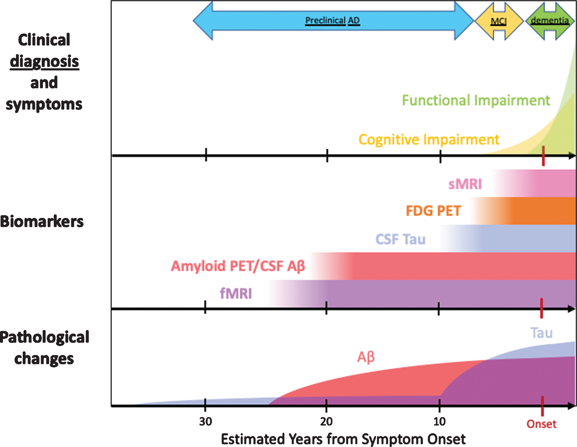
Timeline of AD clinical features, biomarkers, and pathology. Tau pathology correlates most with the onset of clinical symptoms. Aβ accumulation starts ∼20 years prior to dementia onset, even in preclinical AD, where a decrease in CSF Aβ levels or amyloid PET may be detected [7].
Symptoms of AD are subtle at first, instigated by neuronal damage to various areas of the brain and typically emerging over the course of many years [5]. In the early development of AD, this can lead to difficulty in remembering recent conversations or general apathy, followed by disorientation and impaired communication. Eventually, fully developed AD is associated with difficulties in speaking, walking, or swallowing—ultimately proving to be fatal. AD patients at this stage have brain atrophy (i.e., 15–40% loss in hippocampal volume [8]) and inflammation in pathological vulnerable areas of the brain (i.e., the hippocampus and cerebrocortex) [5 , 10].
Afflicting mostly the elderly demographic over 65 years of age, late onset AD makes up for more than 95% of clinical cases. The remaining percentage of cases are early onset AD, characterized by more aggressive cognitive degradation and shorter life expectancy, of which less than 5% are due to sporadic AD that stems from genetic mutation [11].
Postmortem molecular analysis of developed AD reveals two primary pathological indicators: neurofibrillary tangles, made up of aggregated hyperphosphorylated tau proteins within neurons, and senile plaques, composed of insoluble amyloid-β (Aβ) fibrils within the extracellular matrix [12]. Hyperphosphorylation causes tau helical filaments to pair up and produce intracellular neurofibrillary tangles. These abnormal tau protein accumulations block neuronal transport systems and harm synaptic communication, ultimately leading to cell deaths. While tau can also form aggregates in the absence of amyloid pathology [13], there has been evidence to suggest that Aβ promotes tau hyperphosphorylation [14, 15].
There are a variety of Aβ species present in the brain, ranging from soluble monomers and oligomers to insoluble, fibular forms found in amyloid plaques [16, 17]. Because Aβ is an amphipathic peptide, soluble oligomers can act as protein micelles and come in the form of spherical particles (2.7–4.2 nm in diameter) or curvilinear structures [18]. Both neurofibrillary tangles and senile plaques can be influenced by neurotoxic soluble Aβ oligomer presence [19 –21]. Specifically, soluble Aβ oligomers are thought to not only contribute to the aggregation of senile plaques, but also have a precursor effect on the development of AD; they occur up to 10–15 years before the AD diagnosis. The neurotoxicity of soluble Aβ oligomers underlies many deleterious pathologies seen in AD, such as synapse shrinkage and cell death which lead to the impairment of hippocampal long-term potentiation (LTP) and memory function [22, 23]. Additionally, Aβ can trigger cellular stress signaling pathways, which can increase oxidative stress and impair energy metabolism [24].
In familial AD, the presence of the genetic marker, apolipoprotein E (APOE) ɛ4 alleles of the APOE gene, increases the risk of developing the disease 8–12-fold and decreases the age of disease onset [25]. Typically, the APOE gene encodes for the cholesterol transport protein receptors of APOE (APOER2) and the very low-density lipoprotein (VLDLR), which serve as signaling receptors for Reelin, an extracellular protein crucial to synaptic function—enhancing N-methyl-D-aspartate (NMDA) receptor activity through phosphorylation [26]. This LTP stimulation helps to counteract Aβ oligomers. The APOE ɛ4 represents a loss-of-function toxicity that reduces Aβ clearing [27] and depletes APOER2, NMDA, and α-amino-3-hydroxy-5-methyl-4-isoxazoleproopionic acid (AMPA) receptors as well as a harmful gain-of-function toxicity that increases cellular responses to stress or injury [28]. Thus, the risk gene can play a large role in the increased Aβ deposition within the cell [29].
SYNAPSES
Within the human brain, neurons communicate chemically through neurotransmitters across synapses. In fact, there are about 100 billion neurons and 100 trillion synapses in the brain [30]. This chemical stimulation occurs across a synaptic cleft, about 20 to 25 nm long, extending from the presynaptic axon terminals to the postsynaptic membrane which contains neurotransmitter receptors on the receiving neuron’s dendritic spines [31 –33]. This signal transmission creates neuronal circuits that aid in forming memories, thoughts, sensations, emotions, movements, and skills.
Encompassing the small membranous protrusions at the receiving postsynaptic end of a neuron, dendritic spines are made up of various elements such as: glutamate receptors, postsynaptic densities (PSDs), the actin cytoskeleton, membrane-bound organelles, etc. In particular, the PSD is composed of “a multilayered scaffold of densely organized proteins underneath the post-synaptic membrane that anchors cell surface proteins, neurotransmitter receptors, and cell-adhesion molecules” [34]. Visualized as an electron-dense structure in imaging experiments, a visible PSD is often used as a distinguishing factor between excitatory and inhibitory synapses, appearing asymmetric and symmetric, respectively [35].
In addition to performing information transmission, these dynamic synapses change throughout one’s lifetime to facilitate memory, a concept called synaptic plasticity. They can be altered and strengthened through LTP expression, a phenomenon associated with learning, which involves upregulation of AMPA receptors [36] in neural synapses and enlargement of spine heads. In contrast, long-term depression (LTD), typically induced by low-frequency stimulation, is associated with AMPA receptor removal and spine head shrinkage [37, 38]. Upon glutamate binding, both AMPA and NMDA ionotropic glutamate receptors allow for the strong influx of Na+ into and slight efflux of K+ out of the cell, resulting in net depolarization of the membrane. At negative potentials, Mg2+ blocks NMDARs, but slight depolarization dispels Mg2+, making them NMDARs permeable to Ca2+ influx [39]. The rapid and high Ca2+ influx into the synapse through NMDARs facilitates LTP induction, while the low level Ca2+ influx favors LTD [40].
ALZHEIMER’S DISEASE AND SYNAPSES
Research into the pathology of AD suggests that synaptic dysfunction and nerve ending loss strongly correlate with cognitive impairment [41]. There have been several studies suggesting that AD is a synaptopathy, leading to debilitating changes in spine morphology. As a primary pre-cursor to AD development and strongly associated with cognitive decline, synapse loss serves as a morphological reflection of synaptic dysfunction beginning early in the disease [42]. It was reported that patients with mild AD had decreased synapse counts (55%), as measured by transmission electron microscopy, in comparison to groups with either mild or no cognitive impairment; the total synapse number was correlated with performance on cognitive tests examining both immediate and delayed recall [43, 44]. Indeed, infusing soluble Aβ into the brain causes disrupted cognitive function in rodents, as seen through the rapid, potent, and transient learned behavior deficits [22], leading to the synaptic Aβ hypothesis of AD that soluble, non-fibrillar Aβ oligomers underlie neurotoxicity and synapse loss and produce memory and cognition deficits [45 –47]. This is supported by a recent study demonstrating the existence of a regionally specific relationship across posterior cortical regions between episodic memory decline and increasing amyloid accumulation [48]. Interestingly, Aβ neurotoxicity can spread in a prion-linked manner from cell to cell [49]. This causes amyloid deposition by transforming endogenous normal Aβ within the new cell, giving further evidence to its causative role in AD development.
Aβ oligomers also interfere with synaptic interneuron communication and stimulate LTD, leading to cell death. Aβ plaques serve as reservoirs for colocalization of Aβ oligomers, which, at certain concentrations, can determine the aggregation rate of Aβ plaques [50].
Aβ42 overproduction
Several genetic mutations can predispose a patient to familial AD. The amyloid-β protein precursor (AβPP) is a type I transmembrane protein that is cleaved to form a variety of soluble and membrane-bound products [51, 52]. Normally, AβPP protein processing follows two pathways of cleavage: a non-amyloidogenic pathway generating a p3 peptide following sequential α-secretase and γ-secretase cleavage and an amyloidogenic pathway leading to the generation of either Aβ40 or Aβ42 peptides following sequential β-secretase and γ-secretase cleavage. The soluble extracellular peptide Aβ42 is more prone to clump together to create Aβ oligomers [51]. Thus, Aβ42 formation is favored through the amyloidogenic pathway and can be enhanced by mutations to AβPP and γ-secretase protein complex.
Specifically, multiple sites on the APP gene can be targeted to include mutations known to cause AD in humans (i.e., Swedish mutation). This increases the likelihood of AD development in these transgenic animal models through favoring either the amyloidogenic pathway over the non-amyloidogenic pathway or Aβ42 peptide production over Aβ40 peptide production.
One way the amyloidogenic pathway can be disrupted to favor the pro-amyloidogenic pathway is through the altered functionality of α-secretase. ADAM10, one of the ADAM (a disintegrin and metalloproteinase) family members, catalyzes the non-amyloidogenic α-secretase cleavage of AβPP in the brain and prevents amyloid formation. However, a decrease in PKC-dependent phosphorylation of SAP97 molecule prevents its association with and the mediated successful delivery of ADAM10 to synaptic spines, thus reducing α-secretase activity [53]. Alternatively, mutating the presenilin-1 (PSEN 1) and presenilin-2 (PSEN 2) gene regions within the AβPP-catalyzing proteolytic γ-secretase protein complex favors cleavage at the 42nd amino acid, increasing the rate of Aβ42 production and thus the Aβ42/Aβ40 ratio [24].
Ultimately, these autosomal dominant mutations in the APP gene that stimulate familial AD generate the same disease pathologies as non-genetic, sporadic AD. Once Aβ42 recycles to the cell surface after processing, it is released into the brain interstitial fluid and is able to trigger post-synaptic membrane signaling cascades, increasing AMPA receptor endocytosis and dendritic spine loss [54].
Aβ actions
Aβ oligomers have been found to directly cause LTD and indirectly disturb the molecular interactions at the pre- and post- synaptic membranes that lead to LTD (Fig. 2). These oligomers interact with several synaptic proteins, leading to disturbance of neuronal homeostasis and debilitation of downstream mechanisms. The culmination of this causes synaptic dysfunction and neuronal loss [55].
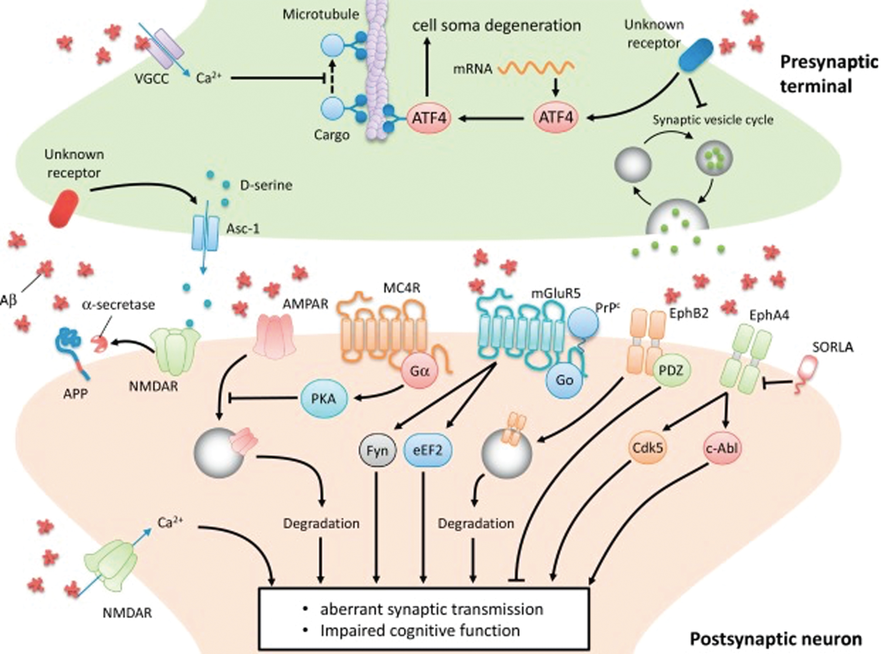
Pre- and post-synaptic interactions with Aβ. Presynaptically, Aβ interacts with VGCC to enhance calcium influx and subsequent disruption of retrograde brain-derived neurotropic factor (BDNF) transport and facilitates ATF4 transport into the soma and thus neurodegeneration. Furthermore, Aβ triggers the presynaptic release of D-serine, which is a co-agonist of post-synaptic NMDARs that mediate synaptotoxicity. Aβ binds to extrasynaptic NMDARs on the postsynaptic neuron to induce excitotoxicity in addition to synaptic NMDARs, which shifts AβPP processing to α-secretase mediated non-amyloidogenic pathway. Aβ also induces the internalization and degradation of EphB2 and AMPAR, the latter leading to synaptic depression. Finally, Aβ interactions with mGLuR5 and EphA4, which leads to its aberrant activation, ultimately leads to synaptic dysfunction through Fyn/eEF2 or Cdk5/c-Abl mediated pathways, respectively (Reprinted from [55] Pharmacol Ther, Vol. 195, Chen Y, Fu AKY, Ip NY, Synaptic dysfunction in Alzheimer’s disease: Mechanisms and therapeutic strategies, pp. 186–198, 2018, with permission from Elsevier.).
In a physiologically normal brain, both Aβ40 and Aβ42 are produced to control and provide a balance between LTP and LTD, maintaining neuronal homeostasis. In this case, regulated LTD stimulation does not cause a synaptic disruption [24]. However, Aβ oligomers initiate LTD independently of synaptic changes, through the various mechanisms of glutamic receptor internalization and dendritic spine shrinkage and loss. Spine loss in this manner is interlinked with mitochondrial apoptosis, which is directly or indirectly stimulated by Aβ through its sub-apoptotic activation of caspase-3, which results in LTD or cell death [24] (Fig. 3).
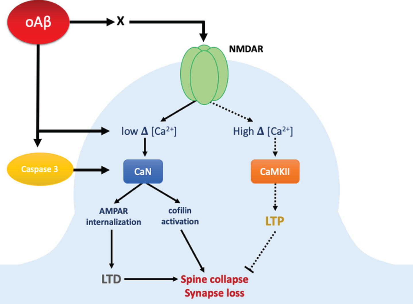
Synaptic dysfunction induced by oligomeric Aβ (oAβ). OAβ can mediate activation of caspase 3 and disrupt calcium homeostasis either directly or through intermediates (X) modulating NMDAR activity. OAβ synaptic interactions favor the activation of NR2B containing NMDARs, which may cause a low increase in calcium levels and a subsequent activation of calcineurin (CaN), leading to dephosphorylation of cofilin. Dephosphorylated cofilin is now active and depolymerizes actin in the dendritic spine, causing dendritic spine collapse and loss of synapses. Activated CaN also dephosphorylates AMPARs at the GluR subunit, leading to the internalization of AMPARs from synapses and thus LTD, finally causing shrinking of dendritic spines.
Pre-synapse
Presynaptic Aβ buildup interferes with axonal transport, synaptic vesicle cycling, and neurotransmitter release. Aβ oligomers disrupt fast axonal transport by co-localizing with axonal voltage-gated calcium channels, thus elevating calcium influx, leading to the impairment of brain-derived neurotropic factor transport [56]. Aβ also uses pathological axonal transport as a retrograde means to spread AD pathology. This is accomplished through the intra-axonal translation of the transcription factor ATF4 that transmits a neurodegenerative signal through alterations in gene expression (i.e., suppression of memory-related genes and activation of proapoptotic genes) [57, 58].
Aβ deregulates synaptic vesicle cycling through the calpain-dependent increases of Cdk5 activity that reduce the vesicle recycling pool while increasing the resting pool [59]. Furthermore, Aβ oligomers exacerbate neurotransmitter release at excitatory synapses. For example: glutamate re-uptake inhibition indirectly triggers aberrant activation of NMDRs [60]. Additionally, Aβ oligomers cause an excess in D-serine production, a co-agonist for NMDAR-mediated synaptotoxicity.
Post-synapse
Post-synaptically, Aβ oligomers interact with a myriad of different receptors. By binding to these cell surface molecules and creating Aβ receptor complexes, Aβ oligomers alter the structure, composition, and functioning of the synapses, some of which include ionotropic glutamate receptors such as NMDARs and AMPARs, leading to receptor internalization and synaptic depression [61, 62]. Aβ oligomers also interact with Eph receptor tyrosine kinases, causing the internalization and degradation of EphB2 as well as the aberrant activation of EphA4, leading to synaptic dysfunction with the help of downstream Cdk5 and c-Abl kinases [55].
Aβ also interacts with GPCRs such as metabotropic glutamate receptors, in particular mGluR5, which plays a critical role in glutamatergic synaptic excitatory transmission and NMDAR-dependent synaptic plasticity regulation. Scaffolding causes the aberrant clustering of mGluR5 receptors and inhibits mGluR5 antagonists that help prevent elevated post-synaptic intracellular Ca2+ and synaptic defects [63, 64]. mGluR5 interacts with the cellular prion protein (PrPc), leading to downstream interactions through Fyn kinase and eukaryotic elongation factor 2 (eEF2), which induces the deregulation of protein translation in dendrites while also facilitating a loss of dendritic spines and subsequent LTD [55 , 66].
As one of its most damaging post-synaptic interactions, Aβ is able to directly bind to NMDA receptors to induce excitotoxity that leads to spine loss and synaptic depression [67]. Aβ-induced Ca2+ influx through NMDAR causes an excess in formation of a reactive oxygen species, activation of calpain, and degradation of critical proteins [68, 69]. In addition, Aβ42 induces clathrin-mediated NMDA receptor endocytosis [69, 70] (Fig. 4). More specifically, Aβ42 enters the synaptic cleft after formation from AβPP and binds to the α7-nicotinic receptor, leading to activation of calcineurin (PP2B) and subsequent dephosphorylation of STEP46. Activated STEP46 gates NMDA receptor activation and LTP and increases dephosphorylation of the NMDA receptor subunit NR2B at Tyr1472, resulting in decreases in NMDA receptor density at synapses, glutamatergic transmission, and LTP [71].
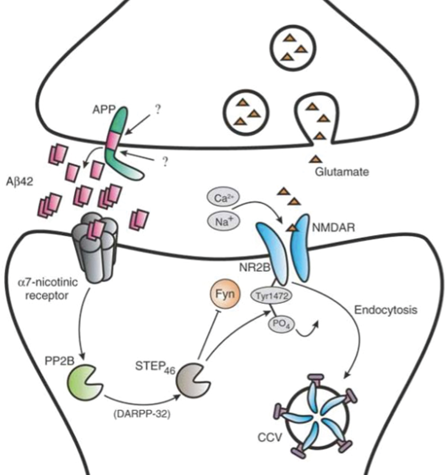
NMDAR internalization induced by Aβ42 at the synapse. Familial AD mutations in APP and PSEN1/2 genes increase production and levels of Aβ42 secretion in the synapse. Synaptic Aβ42 binds to α7-nicotinic receptors, which ultimately leads to the endocytosis of NMDAR via clathrin-coated vesicles (CCVs) (Reprinted from [71] Nat Neurosci, Vol. 8, Tanzi RE, The synaptic Aβ hypothesis of Alzheimer disease, pp. 977-979, 2005, with the permission from Springer Nature.).
Non-Aβ related synapse degradation
Microglia
As part of the immune system, microglia cells are macrophages localized to the central nervous system that help maintain brain homeostasis by performing a multitude of tasks, including engulfing cellular debris, removing excess synaptic connections in circuit formations, and removing protein aggregates. In AD patient brains, microglia eliminate toxic proteins and debris from dead cells. However, overactivity of microglia can lead to chronic neuroinflammation in the brain [72].
In knocking out the gene for TDP-43, a protein regulator of microglial phagocytosis, researchers have found that TDP-43 depletion not only reduces amyloid load in AD models, but also, paradoxically, induces synapse loss [73]. This is due to removal of the TDP-43 negative lysosomal biogenesis control, creating a non-cell-autonomous neurotoxic effect, which stimulates abnormal synapse loss independent of Aβ presence. This pathway exemplifies the complexity of AD, as it shows how targeting one aspect may lead to unintended downstream consequences. This complexity explains many issues faced by failed clinical treatment trials while attempting to improve cognitive function, as it proves the presence of factors other than Aβ involved in synaptic degradation [73].
Presenilin
Outside of being the catalytic component of γ-secretase, presenilins can also act independently to affect synaptic function. In fact, mutations of presenilin account for 40% of familial AD cases, making them more functionally relevant than APP mutations [74]. Presenilins help with cellular Ca2+ handling and ER leakage regulation [75]. It has been found that pre-synaptic presenilin is integral to normal LTP functioning, and its disruption decreases glutamate release resulting in presynaptic dysfunction. Hence, a loss-of-function mutation in presenilin can be an early contributor to synaptic dysfunction and neurotoxicity in AD [76].
Tau
In addition to the intracellular tau aggregations that lead to cellular apoptosis, extracellularly localized tau oligomers can be taken up by neurons and can affect both axonal transport and synaptic transmission (Fig. 5). As with Aβ oligomers, tau disrupts axonal transport in the pre-synapse, but instead of interfering with calcium channels as Aβ does, tau hyperphosphorylation causes microtubule destabilization. Tau also activates PP1, leading to enhancement of GSK-3β-mediated kinesin phosphorylation, which impairs kinesin-mediated axon transport. Tau impairs synaptic vesicle cycling through Syantogyrin-3, a transmembrane vesical protein [55 , 78].
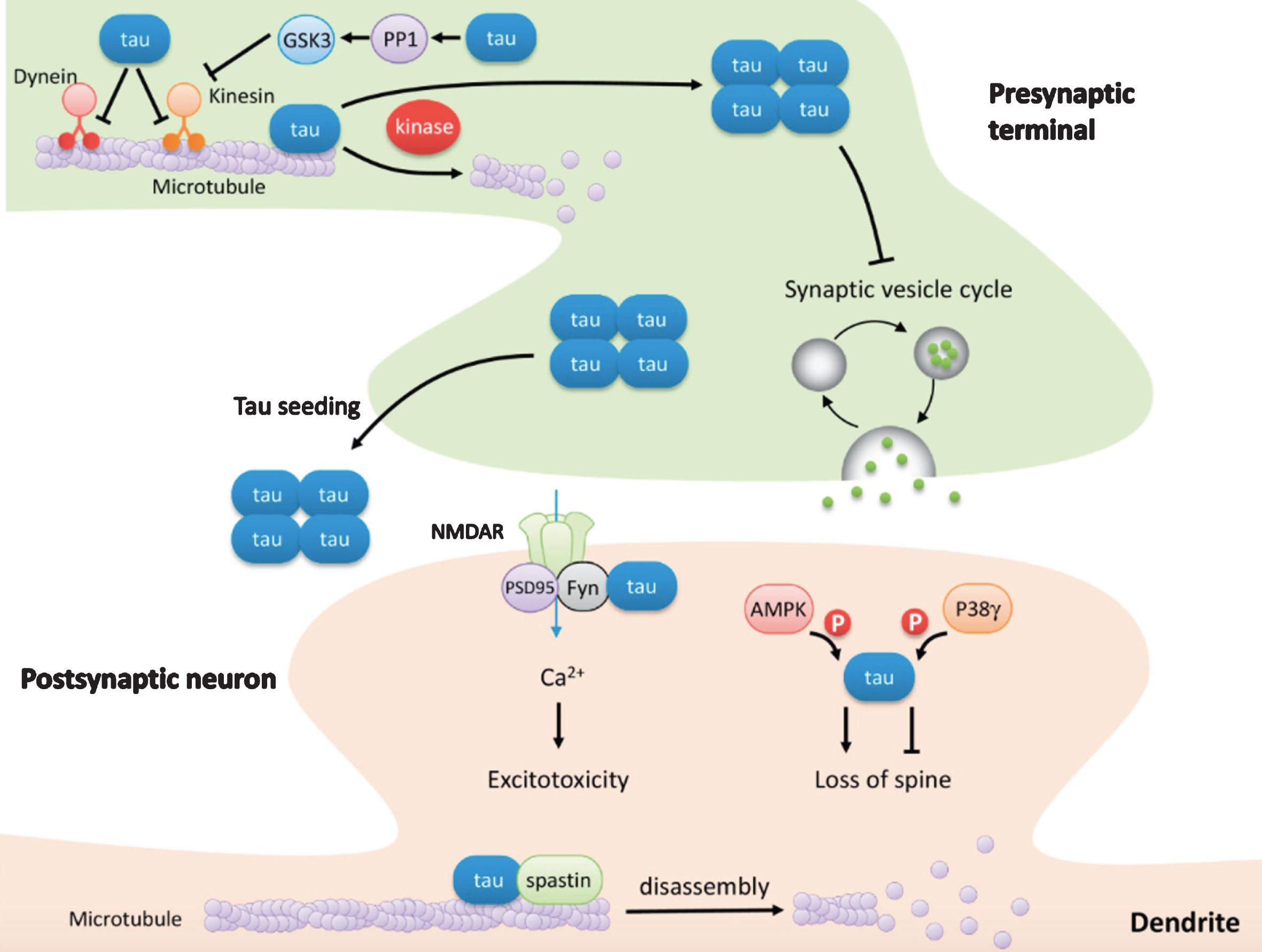
Synaptic dysfunction mediated by tau. Tau hyperphosphorylation by the kinases GSK-3β and Cdk5 leads to its dissociation from microtubules and microtubular destabilization. Tau also competes with dynein and kinesin, disrupting the speed of axonal transport. Furthermore, tau activates PP1, which in turn enhances GSK3 phosphorylation of kinesin, further disrupting axonal transport. Tau interacts with Synaptogyrin-3 to disrupt synaptic vesicle cycling in the presynaptic terminal. In the postsynaptic neuron, missorted tau helps to recruit spastin, a microtubule severing enzyme, which ultimately leads to the disassembly of microtubules. Tau also facilitates Aβ-induced excitotoxicity by recruiting Fyn to the postsynaptic density and binding to NMDARs. Differential phosphorylation by various kinases, such as AMPK or P38γ, leads to opposite effects on spine loss. Tau spreads transynaptically, enhanced by synaptic contact and activity as well as non-synaptic mechanisms, leading to the further propagation of tauopathy throughout the brain (Reprinted from [55] Pharmacol Ther, Vol. 195, Chen Y, Fu AKY, Ip NY, Synaptic dysfunction in Alzheimer’s disease: Mechanisms and therapeutic strategies, pp. 186–198, 2018, with permission from Elsevier.).
In post-synapses of neurons, tau infiltrates dendritic spines to interact with NMDA receptors, recruiting Fyn molecule to the PSD and phosphorylating it, facilitating Aβ-induced excitotoxicity through Ca2+ influx [79, 80]. Tau can be phosphorylated by kinases in the post-synapse, including the AMPK and P38γ, resulting in loss of spine [81]. Finally, tau missorting to neuronal dendrites recruits the microtubule-severing enzyme, spastin, which then proceeds to disassemble microtubules, leading to spine loss [82].
Dkk1
Through a positive feedback mechanism by Aβ to increase its own production, Dickkof-1 (Dkk1) expression has also been shown to play a large role in AD progression. High expression of Dkk1 has been found in the postmortem AD brain. Molecularly, this is due to its involvement in inhibiting the Wnt-β-catenin (canonical Wnt) pathway by blocking the binding of LRP6 and frizzled [83, 84]. In effect, this action activates an opposing non-canonical Wnt-planar cell polarity (Wnt-PCP) pathway that leads to an increase in Aβ production.
Due to its inhibition of the homeostatic balance of Wnt-β-catenin and Wnt-PCP signaling pathways, Dkk1 carries out a substantial portion of the neurotoxic effects seen in Aβ aggregation, favoring both synapse disassembly and synapse loss. Further, the promotion of Wnt-β-catenin signaling reduces Aβ production and enhances synapse stability.
Two primary avenues of the Wnt-PCP pathway exist. First is through the c-Jun N-terminal kinase (JNK1) and c-Jun signaling, which regulates gene expression to increase tau phosphorylation [85]. Second, Wnt-PCP also signals through the RhoA and Rho-associated kinase (ROCK) pathway, both of which act as key regulators in synapse formation and synaptic plasticity [86, 87]. RhoA has an antagonistic effect on dendritic plasticity, whereas ROCK helps to regulate dendritic spines of hippocampal neurons downstream of RhoA [86]. This helps in the disassembly of synaptic actin filaments, which leads to synaptic loss [87].
CURRENT TREATMENTS AND SYNAPTIC PROTEINS
The aforementioned findings regarding the Aβ molecular pathways within AD onset reveal new potential for future treatment directions. It has been found that the inhibition of clathrin-mediated endocytosis reduces AβPP internalization and thus reduces interstitial fluid Aβ levels in vivo [88]. This leads the way for follow-up experiments to explore the treatment possibility of selectively inhibiting individual components of the endocytic machinery to decrease Aβ generation.
There are currently 213 therapeutics that are being or previously have been tested for AD [89]. Of these, a myriad of AD treatment targets exist, ranging from the most common amyloid-related ones, to others targeting cholesterol, cholinergic system, inflammation, neurotransmitter, and tau. These therapies take the form of dietary supplements, DNA- or RNA-based therapies, active and passive immunotherapies, procedural interventions, and finally the most common, small molecule targeting therapies. Of these, the only ones to have made it past phase IV of the U.S. Food and Drug Administration (FDA) clinical trials and been approved are 5 small molecule targeting therapies. Even so, there remain no approved disease modifying drugs available to effectively target the underlying causes behind disease progression or stop the neuronal loss caused by AD.
It should be noted that these drugs were all previously licensed for use in other indications. Due to the extensive formulation, trial, and approval procedure for establishing a new drug, it is easier to repurpose existing drugs that have already passed FDA toxicity studies and go directly into phase II trials. Not having been originally designed toward AD treatment may explain why these untargeted drugs show poor success.
Given the difficulty in directly targeting synapses, many treatments also manipulate pathological proteins, such as Aβ and tau, as doing so helps relieve some downstream consequences of AD. Anti-amyloid therapies try to reduce amyloid load in two ways: either reducing Aβ production and aggregation or enhancing its clearance. Elayta, or CT1812, is an example of drugs that target Aβ. This small molecule antagonist blocks Aβ’s interaction with the sigma2/progesterone receptor membrane component 1 (PGRMC1) receptor, interfering with Aβ-induced synaptic toxicity. Currently, this drug is undergoing phase II of clinical trials.
Approaching through a more indirect method, the drug fasudil has been shown to target the Dkk1-induced synapse loss by antagonizing Wnt-PCP, specifically as a ROCK inhibitor, and thus blocking the effects of Aβ [90]. By inhibiting the Aβ-Dkk1 positive feedback loop, fasudil has been shown not only to prevent dendritic spine withdrawal in vitro but also to reduce Aβ load in vivo in mouse models with advanced Aβ pathology.
Some treatments try to block Aβ42 peptide formation by evading the amyloidogenic pathway entirely. Mutations in the β-secretase cleavage site on AβPP increases the overall production of the Aβ peptide by improving AβPP’s substrate aptness for β-secretase, while mutations in the presenilin-1 (PSEN 1) and presenilin-2 (PSEN 2) gene regions of γ-secretase favor Aβ42 production. Thus, both favor the amyloidogenic pathway, which can be avoided by inhibiting β-secretase and γ-secretase cleavage activity [91].
Another avenue of AD treatment is through active or passive immunotherapies. While active immunotherapy involves inducing an immune response in the patient—utilizing the body’s own adaptive immune responses against Aβ [92]—passive immunotherapy simply administers immune molecules (i.e., antibodies, cytokines) to patients, providing immediate and short-term protection without generating immune memory. Anti-Aβ antibodies have been utilized as prophylactics that can delay or even prevent AD onset in high-risk familial AD mutation carriers.
The U.S. FDA has approved various drugs that target neurotransmitters within the brain for temporary symptom reduction [93]. Rivastigmine, galantamine, and donepezil are cholinesterase inhibitors that assist in preventing acetylcholine breakdown. In contrast, the cognition-enhancing drug, memantine, is a noncompetitive NMDA receptor antagonist that acts to inhibit extrasynaptic Ca2+ influx and prevents the excitotoxic effects through NMDA. Memantine is also combined with donepezil in a drug called Namzaric to create a “cocktail-like” drug that has been shown to have increased effectiveness due to its multi-targeting treatment approach.
It should be noted that some clinical applications have extended away from research-based medicine and into experimental medicine. One example of this is demonstrated by the current industry attempts at testing the therapeutic effects of mesenchymal stem cell-derived exosomes in mitigating neuronal loss [94]. It must be considered that untargeted treatments as these are likely not a cure-all solution but rather merely serve as a bandage-solution providing temporary and limited symptom amelioration.
Synaptic biomarkers
With evidence supporting synaptic loss as a structural correlate of cognitive impairment in AD, the ability to measure synapse-specific biomarkers in vivo not only serves as a useful prognostic for disease progression for at-risk individuals but also as an outcome measurement for potential disease-modifying therapies (i.e., drug efficacy in pharmaceutical trials). When compared to AD pathological proteins, such as Aβ and tau, synaptic proteins serve as a more direct form of synapse monitoring. Thus, assays tracking these biomarkers can be used to quantitatively assess AD progression and assess drug effectiveness during clinical trials.
One example of this is the use of radionuclides for synaptic vesicle glycoprotein 2A (SV2A), using positron emission tomographic (PET) imaging with the radioligand 11C-UCB-J to measure levels of the hippocampal SV2A in vivo. Because SV2A is a vesicle membrane protein expressed at almost all synapses, it serves as a correlate to synaptic density and thus a good molecular target for PET. SV2A is a more synapse-specific alternative to receptor biomarkers (i.e., AMPA, PSD95, NMDA) that are often used in animal research. One study showed a 41% decrease in SV2A specific binding in AD patients [95]. This imaging technique provides a non-invasive, in vivo method of measuring decreases in synaptic density due to AD development in humans.
Cerebrospinal fluid (CSF) assays measure the amounts of certain synaptic proteins in the CSF that serve as biomarkers for AD disease severity before neurodegeneration. Directly connected to the central nervous system, the CSF is continuous with the brain interstitial fluid and is thus a source of synapse-derived proteins. Specifically, levels of six synaptic proteins, Calsyntenin-1, GluR4, Neurexin-2A, Neurexin-3A, Syntaxin-1B, and Thy-1, have been found to be reduced 0.8-fold in preclinical AD [96]. The assay is performed by extracting the CSF fluid using lumbar puncture in living subjects. This is a method of monitoring AD development during pre-clinical stages, after which the protein levels are confounded by neuronal loss. Assays targeting CSF-derived proteins provide a cheaper alternative to those necessitating brain samples.
Difficulties with evidence
Several barriers exist for developing effective clinical treatments. Even with the abundance of conclusions made from accumulated AD research, there is limited applicability to experimental studies performed in animal models. Neurological test results obtained from animal models have occasionally proved to be unreliable in determining effectiveness of human treatment due to pathological differences between human brains and animal brains. At the most fundamental level, human brains are known to have different electrophysiology and contain more neurons and synapses than animal model brains [41].
Additionally, the experiments are limited by the specific disease-mimicking mutations they are founded on. Most experiments exclusively use animals induced with familial AD, overexpressing risk genes such as APP, PS1, PS2, and/or tau as models. However, familial AD is responsible for less than 1% of all actual AD cases seen in human patients. Rather, 95% of sporadic AD arises as a symptom of synaptic signaling dysregulation and synaptic failure developed over the course of many years. Although the pathologies of different disease manifestations seem indistinguishable at the molecular level, we must consider other hidden mental/stress factors underlying sudden and aggressive AD onset verses slow decade-long progression of the disease. This difference in disease inception calls into question the applicability of these experimental results derived from genetically modified models to future clinical treatment of the developed disease in humans.
An example of pathological inconsistency of using animal models is demonstrated in the non-AD-conditional, APP transgenic mouse, Tg2576, which overexpresses AβPP with an Aβ Swedish mutation. In this animal model, only Aβ aggregation pathology is observed without any of the accompanying hyperphosphorylated tau associated neurofibrillary tangles that are typically co-expressed. This indicates an alternative pathological progression than that seen in humans. One explanation for this discrepancy is that mice models might not live long enough for the pathological effects to manifest as they do in humans. But ultimately, we must not undermine the many external factors that come to play in the development of this complex disease, and we must not oversimplify the human system.
The possible untranslatability of experimental results is most pronounced in failed clinical trials. The inability to establish successful phase III trials for immuno-targeting of Aβ-oligomers, despite the great success of immunotherapy in mouse models in preventing AD, is a prime example of this. It seems that when similar treatments were utilized in humans, many unexpected complications including encephalitis in active immunization trials and vasogenic edema or amyloid-related imaging abnormalities in passive immunization trials were presented [97].
Hence, although finding an accurate treatment model is important, it serves as only the first step of the research process. Only by moving on to clinical trials can we work our way toward a viable solution for the populous. However, the clinical study recruitment process is slow and can be risky for treatments that are not adequately developed. There are many issues with patient consent due to the ethical consideration of whether a person with MCI or dementia is able to properly give clinical consent. Clinical trials are very expensive to run, and the best read out is on a cognition and dementia scale. Additionally, due to the slow progression of AD, a lengthy observational period is required to investigate each treatment’s effect on disease progression.
Given the high risk and monetary resources necessary to conduct clinical trials along with the notoriety of unsuccessful clinical trials in the past, AD research has declined in popularity, despite the enormous demographic afflicted by the disease. By creating biomarkers that help both diagnostically and in clinical trials to easily detect the effectiveness of treatments, researchers aim to increase its appeal to the scientific and medical community.
Another variable factor in treatment has been the discovery that the progression of the disease is gender biased. Making up 2/3 of total AD cases, women are not only known to develop AD at an earlier age, but also as a more aggressive form. There exists a strong link between the sudden and significant decline in estrogen and progesterone hormones during menopausal onset and the elevated risk of developing AD [98]. Notably, when the same human mutations were given to transgenic animal models, their disease progression showed similar gender bias [99, 100]. This calls into consideration the population demographics being sampled in experimental studies, as significant differences in synapse dysfunction in men and women may warrant differential AD treatment approaches.
The transition from research-based medical approaches to experimental medicine likely stems from the aforementioned complex molecular interplay between various pathways. As with antibiotic treatments, another approach to facing these overlapping factors is to use the cocktail treatment method to target more than one pathway at once. But with the cascading effects of Aβ oligomers in inducing defects in synaptic homeostasis, it is hard to say if the amplification of these imbalances can be fixed at all after the indistinct and limited preclinical time-frame. Currently, the easiest route to AD treatment is prevention at its early stages of development. Once the homeostasis is disrupted and cognitive decline starts, it may be hard to come back from the accumulated cognitive deficiencies.
CONCLUSION REMARKS
AD has long been considered incurable due to its wide-ranging pathological causes and effects. Current treatment options merely provide temporary relief through symptom mitigation or otherwise seem to only delay AD onset with drugs or behavioral therapies. By gaining a deeper understanding into the synaptic mechanisms behind the disease, we are able to target various signaling components, including some critical proteins, for further treatment. Aβ oligomers may not be the only factor causing synaptic loss, but they appear to be deeply intertwined with many molecular mediators that contribute to AD progression.
During the recent years, the prevalence of AD research has been on the decline due to the unfavorable costs, length of studies, and risk factors involved in previously failed attempts at advancing clinical trials. In response, the FDA has taken active measures to lower its standards for approval to decrease the likelihood of AD clinical trial failure. Given the devastating and wide-ranging effects of this disease, it is crucial that we do not stagnate in our efforts to understand it further. The newfound emphasis on synapse-centered treatments reflects a more updated and targeted approach in AD research using synaptic failure as a metric to assess in disease development. With more precise methods of early detection and more dependable assays that evaluate treatment status, we have never been in a better position to be developing and testing clinical applications of all the knowledge we have acquired and yet to acquire to remedy the disease.
