






Abstract
Background:
Alzheimer’s disease (AD) is the most common cause of dementia in the elderly and is characterized by progressive cognitive decline. Considerable evidence supports an important role of amyloid-β oligomers (AβOs) in the pathogenesis of AD, including the induction of aberrant glial activation and memory impairment.
Objective:
We have investigated the protective actions of a nutritional formulation, denoted AZ formulation, on glial activation and memory deficits induced by intracerebroventricular (i.c.v.) infusion of AβOs in mice.
Methods:
Two-month-old male mice were treated orally with AZ formulation or isocaloric placebo for 30 consecutive days. Microglial and astrocytic activation were analyzed by immunohistochemistry in the hippocampus 10 days after i.c.v. infusion of AβOs (n = 5 mice per experimental condition). Memory loss was assessed by the novel object recognition (NOR) test (n = 6–10 mice per experimental condition).
Results:
Oral treatment with the AZ formulation prevented hippocampal microglial and astrocytic activation induced by i.c.v. infusion of AβOs. The AZ formulation further protected mice from AβO-induced memory impairment.
Conclusion:
Results suggest that administration of the AZ formulation may comprise a promising preventative and non-pharmacological strategy to reduce brain inflammation and attenuate memory impairment in AD.
INTRODUCTION
Alzheimer’s disease (AD) is the most common cause of dementia in the elderly, affecting over 35 million people worldwide [1]. Characterized by progressive cognitive decline, AD is an incapacitating disorder that costs over $305 billion/year in patient care in the United States alone [1]. The underlying mechanisms leading to memory impairment in AD are complex and not yet fully understood, but a considerable body of evidence suggests an important role of amyloid-β oligomers (AβOs) in the pathogenesis of AD [2, 3]. AβOs accumulate in the AD brain and cerebrospinal fluid [4 –6] and have been shown to trigger aberrant glial activation and neuroinflammation [7 –9], synapse failure and loss [10 –12], and memory impairment [7 , 13–16].
Neuroinflammation is a hallmark of many neurodegenerative diseases [17], is thought to play a significant role in the pathogenesis of AD [18, 19], and is correlated with cognitive decline in AD patients [20, 21]. Microglia and astrocytes are the major contributors to inflammation in the central nervous system, and previous reports have shown that activation of glial responses by AβOs contributes to neuronal dysfunction and memory deficits via the release of pro-inflammatory cytokines [9 , 22]. Despite our increased understanding of the neurobiology of AD, no effective treatments are available to cure or stop the progression of the disease. Thus, development of approaches to prevent neurodegeneration and cognitive impairment in AD is an urgent and unmet need. The role of brain inflammation in pathogenesis suggests that targeting glial activation may comprise an effective approach to slow down or prevent AD progression.
Nutritional formulations represent non-pharmacological therapeutic alternatives for several disorders, and a growing body of evidence suggests that combinations of nutraceutical compounds may be more effective to minimize brain damage in AD than the use of single nutrients [23 –25]. Studies using natural dietary or nutraceutical supplementation with neuroimmune modulators have been shown to ameliorate symptoms and improve patient life quality [26, 27]. For example, phosphatidylserine (PS) was shown to reduce hippocampal inflammation and improve memory in AD [28, 29]. PS incorporation into cell membranes is related to the availability of docosahexaenoic acid (DHA) [30], and DHA deficiency is correlated with cognitive decline [31, 32]. In rat models of depression and ischemia, supplementation with DHA was found to be neuroprotective and to attenuate inflammation [33, 34]. Furthermore, impaired brain glucose transport/utilization and insulin resistance are features of AD [22, 35], and metabolic dysregulation is correlated with impaired synaptic plasticity and cognitive dysfunction [9, 22]. Evidence indicates that diets containing medium-chain fatty acids such as capric (C10:0) and caprylic (C8:0) acids increase ketone bodies levels [36, 37], providing an alternative brain fuel source and reducing oxidative stress and inflammation [38].
Several additional nutrients play important roles in proper brain function, and numerous studies have highlighted correlations between AD and low levels of choline, selenium, and vitamins [39 –41]. Vitamins D, C, E, and B-complex have been implicated in modulation of inflammation and cognitive activity. For example, administration of vitamin B-complex was found to decrease pro-inflammatory cytokines in a rat model of peripheral nerve injury [42]. In aged patients with mild cognitive impairment, treatment with B vitamin slowed brain atrophy and cognitive decline, and lowered homocysteine levels [43]. Studies with vitamin D supplementation showed reductions in inflammation and enhanced learning and memory in animal models of AD [44, 45]. Moreover, vitamin E or C were found to mitigate the production of inflammatory mediators and rescue memory impairment in animal models of AD [46 –48].
Although some of these nutrients may be individually important to combat AD pathogenesis, little is known on the potential beneficial action of a nutritional formulation comprising a defined mixture of such components. In the current study, we investigated the effects of a nutritional formulation based on a unique combination of capric and caprylic acids, PS, DHA, vitamins and minerals, henceforth denoted AZ formulation, in a murine model of AD. We hypothesized that treatment with the AZ formulation might prevent neuroinflammation-related glial activation and memory impairment induced by intracerebroventricular (i.c.v) infusion of AβOs in mice. Our findings suggest that the AZ formulation represents a promising non-pharmacological strategy to attenuate brain inflammation and memory deficits in AD.
MATERIALS AND METHODS
Reagents
Alexa Fluor-labeled secondary antibodies (A-11008) and ProLong antifade reagent (P36935) were from Invitrogen. Rabbit polyclonal antibody against mouse Iba-1 was from Wako (019-19741); rabbit polyclonal antibody against mouse GFAP was from Abcam (ab7260). Synthetic Aβ1–42 was from American Peptide (641-15). Dimethyl sulfoxide (DMSO) was from Sigma-Aldrich (D2650-5X5ML).
Preparation and characterization of AβOs
AβOs were prepared weekly from synthetic Aβ1–42 following the original procedure described by Lambert et al. [49] and were routinely characterized by size-exclusion chromatography and, occasionally, by western immunoblots, as previously described [13 , 50–52]. Oligomers were kept at 4°C and were used within 48 h of preparation.
Animals, treatment by oral gavage and intracerebroventricular infusion of AβOs
Male Swiss mice were obtained from ANILAB Ltd. (São Paulo, Brazil) and were 2 months old at the beginning of experiments. Animals were housed in groups of five per cage with free access to food and water, with controlled room temperature and humidity, and under a 12-h light/12-h dark cycle. All procedures followed the Principles of Laboratory Animal Care from the National Institutes of Health and were approved by the Institutional Animal Care and Use Committee of the Federal University of Rio de of Rio de Janeiro (protocol 010/21).
The composition of the AZ formulation (Prodiet Medical Nutrition, Curitiba, Brazil) is presented in Table 1. In the control group, we used an isocaloric placebo diet composed of soybean oil and maltodextrin. The AZ formulation has a significant energy content coming from medium-chain fatty acids (capric and caprylic acids). Use of soybean oil in the placebo diet provides an effective approach to obtain the same calories in the same volume of diet for administration to mice. In addition, soybean oil is rich in long-chain triglycerides, and thus unrelated to the formation of ketone bodies derived from capric and caprylic acids in the AZ formulation. Maltodextrin was used to balance the additional calories coming from other components in the AZ formulation. AZ formulation or isocaloric placebo were suspended in deionized water and administered by oral gavage (1 g/kg/d) for 30 consecutive days in a final volume of 55 microliters.
Nutrient composition per 100 g of AZ formulation composition
For intracerebroventricular (i.c.v.) infusion of AβOs, animals were anesthetized for 7 min with 2.5%isoflurane (Cristália Ltd., São Paulo, Brazil) using a vaporizer system and were gently restrained only during the injection procedure. A 2.5 mm-long needle was unilaterally inserted 1 mm to the right of the midline point equidistant from each eye and 1 mm posterior to a line drawn through the anterior base of the eyes, as described [13]. AβOs (10 pmol) or vehicle (2%DMSO in PBS) were infused in a final volume of 3 microliters, and the needle was kept in place for 30 s to avoid backflow. Accurate placement of the needle into the right lateral ventricle was confirmed by macroscopic examination of dissected brains.
Novel object recognition (NOR) test
An open field arena measuring 30×30×45 cm (W x L x H) was used. Test objects had different shapes, colors, sizes, and textures and were fixed to the box to prevent displacement caused by exploratory activity of the animals during sessions. Previous tests showed that none of the objects used evoked innate preference. Mice (12 per group) were initially submitted to a habituation session to freely explore the empty arena for 5 min. The training session consisted in placing the animals in the same arena with 2 identical objects for 5 min. The arena and objects were cleaned thoroughly between trials to eliminate olfactory cues. Two hours after the training session, animals performed the test session in which one of the two objects in the arena had been replaced by a new one. The amount of time spent exploring each object was recorded. Sniffing and touching the object were considered exploratory behavior. Results were expressed as percentage of time exploring each object (novel or familiar) during the test session. Animals that spent less than 5 seconds in total exploring both objects were excluded from the test.
Immunohistochemistry
Ten days after i.c.v. infusion of AβOs, animals were anesthetized and perfused with saline, followed by 4%paraformaldehyde. Fixed brains were removed and cryoprotected in 20%sucrose for 7 days. Frozen 40-micrometer coronal brain sections were obtained on a cryostat (Leica Microsystems) and stored in PBS containing 0.01%sodium azide at 4°C. Five animals were used per experimental condition and 4–6 sections were obtained per animal. Free-floating immunohistochemistry was performed after washing sections with PBS tree times. Sections were incubated for 1 h with 5%bovine serum albumin (BSA) and 0.15%Triton X-100 in PBS, followed by overnight incubation at 4°C with primary antibodies (anti-Iba-1, 1:400; or anti-GFAP, 1:400) diluted in PBS. After washing, sections were incubated for 2 h at room temperature with anti-rabbit IgG Alexa Fluor 488-conjugated secondary antibody (1:1,000) and mounted with ProLong antifade reagent with DAPI. Images were acquired on a Zeiss Axio Observer Z1 microscope and mosaic representative images were acquired on a Nikon C2 confocal microscope.
Image analysis
Four to six z-stack images (10–20 planes each) of each hippocampal subregion (CA1, CA3, dentate gyrus) were acquired from each animal to account for possible variability across the antero-posterior axis. Microglia and astrocyte morphology were analyzed by measuring the average of total area, normalized area per cell, total fluorescence integrated density and normalized integrated density per cell from multiple sections of each animal. Quantification of area, integrated density and total positive cell number was performed using ImageJ analysis software (NIH) and results are expressed as percentage of control, vehicle-infused mice. Total hippocampal quantification was obtained by averaging results from all three hippocampal regions (CA1, CA3, and DG) from each animal. Mosaic images were obtained by merging individual images with the aid of Adobe Photoshop. Some distortion may be present at the edges of individual images. However, analysis and quantification were performed exclusively on Zeiss microscope raw images.
Statistical analysis
All analyses were performed with GraphPad Prism. Datasets were assessed for normality using the D’Agostino-Pearson test and analysis of the residuals was performed to verify the equality of variances in the samples (homogeneity of variance). Values are expressed as means±SEM. For statistical analysis of immunohistochemistry data, we used two-way ANOVA followed by the Sidak post hoc test, as indicated in the figure legends. Data from the novel object recognition task were analyzed by one-sample t-test, comparing the percentage of time of exploration of the novel object against the fixed value of 50%(chance level). Statistical confidence levels are indicated in each figure. A p value < 0.05 was considered significant. Choices regarding animal exclusion criteria and statistical tests employed were made beforehand, to minimize potential bias in the results of analysis.
RESULTS
Treatment with the AZ formulation prevents AβO-induced hippocampal gliosis
Activated microglia and astrocytes are major players in inflammatory response in the CNS. To determine whether treatment with AZ might attenuate glial activation in AβO-infused mice, we pre-treated mice for 30 days by oral gavage with AZ or an isocaloric placebo formulation and examined glial morphology in hippocampal sections obtained 10 days after i.c.v. infusion of AβOs (or vehicle).
Consistent with our previous findings [7, 9], AβO infusion in placebo-treated mice caused an increase in hippocampal immunoreactivity to Iba-1 (microglia marker; Fig. 1A), indicated by both total area and integrated fluorescence density analysis (Fig. 1B, C) as well as when these parameters were normalized by the number of Iba-1-positive cells (Fig. 1D, E). A more detailed analysis of hippocampal subregions revealed a statistically significant increase in Iba-1 immunoreactivity in CA1, but not in CA3 or dentate gyrus (Supplementary Figure 1). Interestingly, no increase in hippocampal Iba-1 immunoreactivity was verified in AβO-infused mice that had been pretreated with the AZ formulation (Fig. 1). No changes in total numbers of Iba-1-positive cells were detected in any of the experimental groups investigated.
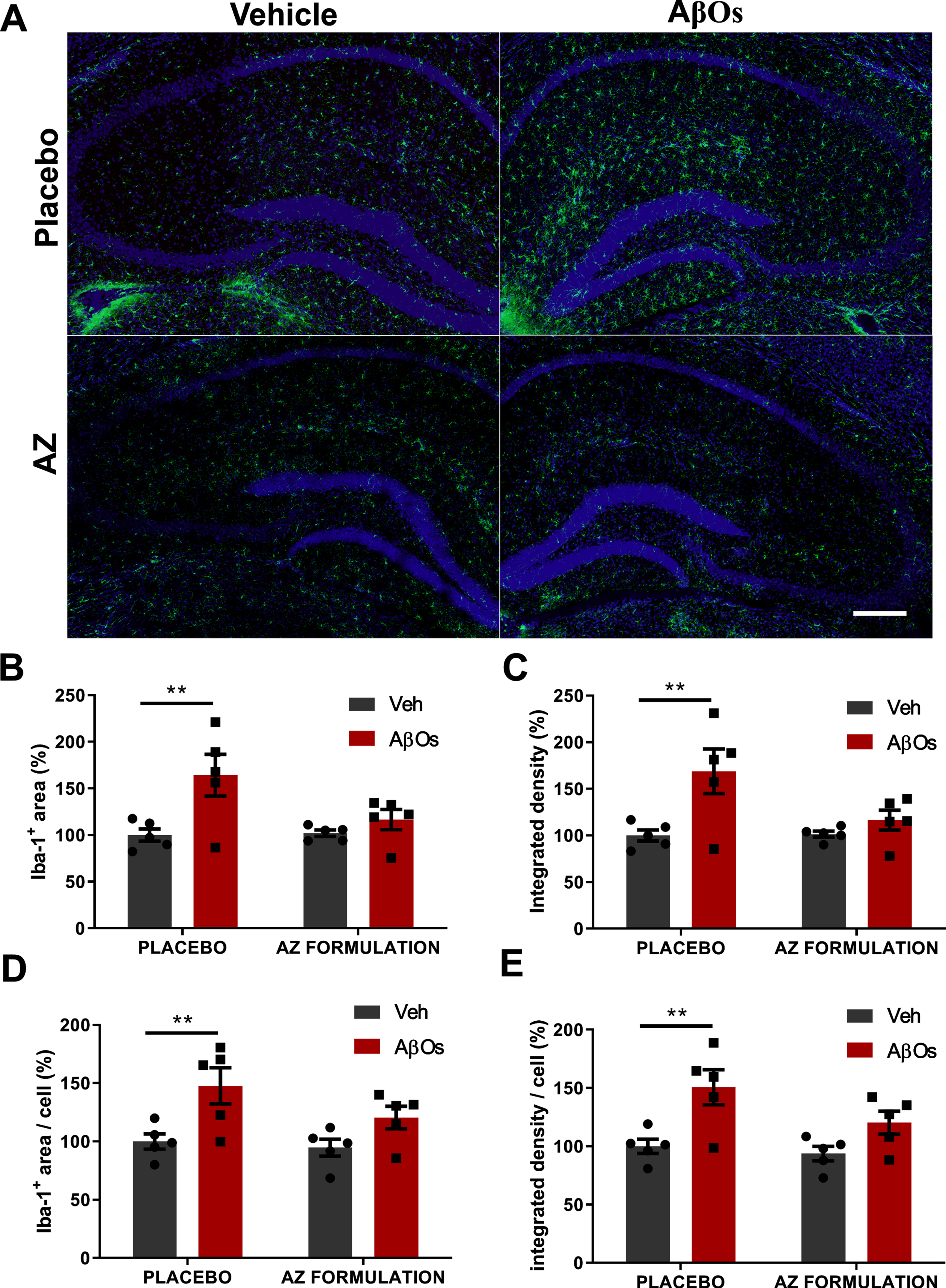
Treatment with the AZ formulation prevents hippocampal microglial activation induced by i.c.v. infusion of AβOs. A) Representative mosaic hippocampal immunohistochemistry image for Iba-1 (green) and DAPI (blue) from vehicle- or AβO-infused mice treated with placebo or AZ formulation (as indicated). Scale bars, 200μm. B-E) Total hippocampal Iba-1 quantification (average of CA1, CA3 and DG hippocampal regions) for each animal. Results were obtained by averaging 4–6 hippocampal sections per animal and using five animals/group (symbols represent individual mice). Panels show total Iba-1+ area (B), total Iba-1+ integrated fluorescence density (C), cell-normalized Iba-1+ area (D), and cell-normalized Iba-1+ integrated fluorescence density (E).
We further analyzed astrocyte activation by immunohistochemistry for glial fibrillary acidic protein (GFAP). Ten days after infusion of AβOs, mice that had been pretreated with placebo exhibited increased GFAP immunoreactivity (Fig. 2A) in the hippocampus, but no differences in numbers of GFAP-positive cells. Increased GFAP immunoreactivity was detected by analysis of both total and cell-normalized area (Fig. 2B, D) and by integrated fluorescence density (Fig. 2C, E). Astrocyte activation was more prominent in the dentate gyrus of the hippocampus (Supplementary Figure 2). Similar to our findings with microglia, pretreatment with the AZ formulation prevented astrocyte activation in the hippocampi of AβO-infused mice. These findings demonstrate that AβOs induce hippocampal glial activation that persists for at least 10 days after i.c.v. infusion, and that this is prevented by pretreatment with the AZ formulation.
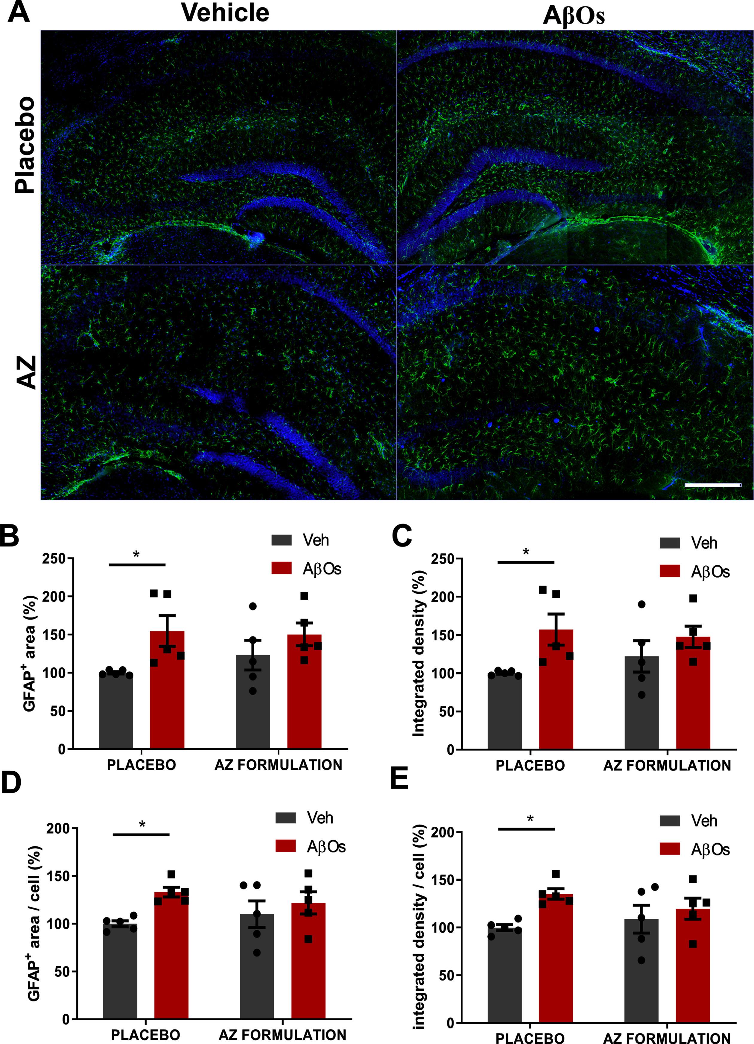
Treatment with the AZ formulation prevents hippocampal astrocyte activation induced by infusion of AβOs. A) Representative mosaic hippocampal immunohistochemistry image for GFAP (green) and DAPI (blue) from vehicle-or AβO-infused mice treated with placebo or AZ formulation (as indicated). Scale bars, 200μm. B-E) Total hippocampal GFAP quantification (average of CA1, CA3, and DG hippocampal regions) for each animal. Results were obtained by averaging 4–6 hippocampal sections per animal and using five animals/group (symbols represent individual mice). Panels show total GFAP+ area (B), total GFAP+ integrated fluorescence density (C), cell-normalized GFAP+ area (D), and cell-normalized GFAP+ integrated fluorescence density (E).
Treatment with the AZ formulation prevents AβO-induced memory impairment
We next investigated the effect of treatment with the AZ formulation on memory impairment induced by AβOs. Results showed that mice treated with placebo had normal performance in the novel object recognition (NOR) test (Fig. 3A, B). In contrast, placebo-treated AβO-infused mice tested 24 h or 7 days after infusion failed the NOR test (i.e., they spent equal amounts of time exploring the familiar and novel objects), indicating impaired declarative recognition memory (Fig. 3A, B). Remarkably, pretreatment with the AZ formulation prevented memory deficits induced by AβOs both 24 h and 7 days after infusion (Fig. 3A, B).
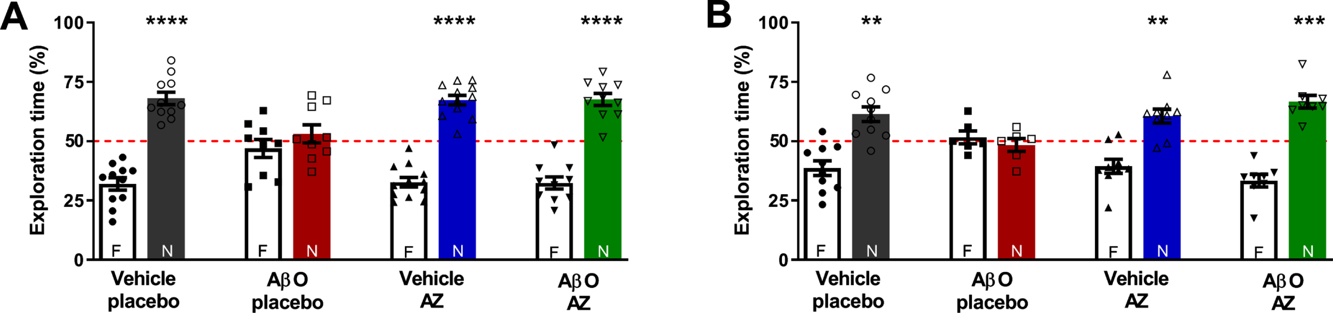
AZ formulation prevents AβO-induced memory impairment in mice. A,B) Mice were daily treated with placebo or AZ formulation by oral gavage for 30 days before i.c.v. infusion of AβOs (10 pmol) or vehicle. Animals were tested in the NOR task 24 h (A) and 7 days (B) after i.c.v. infusion of AβOs. Percentages of time spent exploring the novel object are represented by colored bars (labeled N for novel and F for familiar objects). Symbols correspond to individual mice (n = 6–10 mice per experimental group). Bars represent means±SEM. Asterisks denote statistically significant ** p < 0.01, *** p < 0.001, **** p < 0.0001 differences from 50%(chance level).
DISCUSSION
In the current study, we examined the protective effects of a nutritional formulation (denoted AZ) in a murine model of AD. Our results indicate that pretreatment of mice with AZ prevented hippocampal glial activation and memory impairment induced by i.c.v. infusion of AβOs.
AβOs are soluble Aβ aggregates that accumulate in AD brains and are thought to play a central role in AD pathogenesis [2, 3]. In both murine and macaque models, i.c.v. infusion of AβOs induces microglial and astrocyte activation [7, 8], consistent with observations in AD patients [53]. Microglia are ramified brain-resident immune cells known to modify their morphology under pathological conditions, decreasing ramification number and increasing cell body area, and are the major regulators of inflammatory mediators in the CNS [54, 55]. In AD brains, morphologically reactive microglia, with increased cell body area and retracted processes, are found surrounding amyloid deposits [56, 57]. Astrocytes also change their morphologies when activated, showing increased cell body area, number of ramifications and upregulation of GFAP [58]. Reactive astrocytes can be found in AD postmortem brain tissue and in transgenic mouse models of AD, both after and preceding the onset of amyloid deposition [59, 60]. We here verified that hippocampal microglial and astrocyte activation induced by AβOs persists for at least 10 days after a single i.c.v. infusion. Importantly, we found that pretreatment of mice with the AZ formulation prevented glial activation induced by AβOs.
The AZ formulation provides a combination of several neuroprotective nutrients that can also have anti-inflammatory actions. Fast-absorbing medium-chain fatty acids, for example, increase the concentration of circulating ketone bodies (KBs) [35, 36] that can cross the blood-brain barrier and become bioavailable in the CNS [61]. Previous studies have shown that KBs have anti-inflammatory actions and suppress glial activation [37]. In primed cultured microglia, the ketone body, β-hydroxybutyrate (BHB), induced microglial ramification and prevented the release of pro-inflammatory cytokines [62].
In addition, the AZ formulation contains docosahexaenoic acid (DHA) and vitamins, important nutrients with anti-inflammatory properties [63 –67]. DHA is an important fatty acid membrane component reported to prevent LPS-induced neuroinflammation by normalizing microglia activity in vitro and in vivo [68, 69]. Vitamin D, an essential steroid hormone, was shown to attenuate hippocampal neuroinflammation in AD [70]. Moreover, a study showed that vitamin C is important for microglial homeostasis by regulating nuclear translocation of the transcription factor NF-kB [71].
AβOs have been found to bind to several microglial receptors and to induce microglial activation through TLR-4/NFκB and inflammasome activation [7 , 73]. Interestingly, a previous report demonstrated that ketone bodies, such as BHB, suppress macrophage NLRP3 inflammasome in response to several structurally unrelated NLRP3 activators [37]. Furthermore, vitamin E was shown to inhibit LPS-induced microglial activation by suppressing activation of MAPK and NF-kB [74].
Glial activation and increased pro-inflammatory signaling have been found to be related to cognitive impairment in AD. For example, TNF-α released by microglia in AβO-infused mice has been shown to drive memory impairment [14]. A recent study showed that pretreatment with the AZ formulation prevented memory impairment in mice that received an i.c.v. injection of streptozotocin [75]. Here, we found that the AZ formulation prevented memory deficits induced by AβOs, and that this protective action persisted for at least 7 days after interruption of treatment.
The potential beneficial actions of the AZ formulation are not limited to anti-inflammatory effects, as previous studies have revealed cognitive protective effects of several AZ components analyzed in isolation [28–31 , 40–43]. In a randomized clinical trial with mild-moderate AD patients, ingestion of medium-chain fatty acids, mainly caprylic acid, resulted in elevated BHB and improved cognitive performance [76]. Treatment with B-complex vitamin reduced homocysteine levels and ameliorated cognitive decline in MCI patients [77]. Antioxidant nutrients such as vitamins C, E, selenium, magnesium, and zinc may also exert neuroprotection.
In addition, the AZ formulation provides phosphatidylserine, the major acidic phospholipid component in human membranes, which can cross the blood-brain barrier and is correlated with cognitive improvement [78, 79]. In a double-blinded placebo-controlled randomized clinical trial, oral phosphatidylserine supplementation improved cognitive impairment in elderly subjects exhibiting mild memory loss [80]. Thus, prevention of aberrant glial activation, combined with cognition-enhancing nutrients, provides a plausible mechanism to explain the protective action of AZ in our model.
In conclusion, oral treatment with the AZ formulation prevented aberrant glial activation, a central feature of AD pathogenesis, and, importantly, blocked memory impairment in an acute AD mouse model (Fig. 4). Since supplementation with the AZ formulation requires no special intervention in regular dietary intake, it may represent a novel and effective non-pharmacological approach to delay or attenuate the progression of AD.
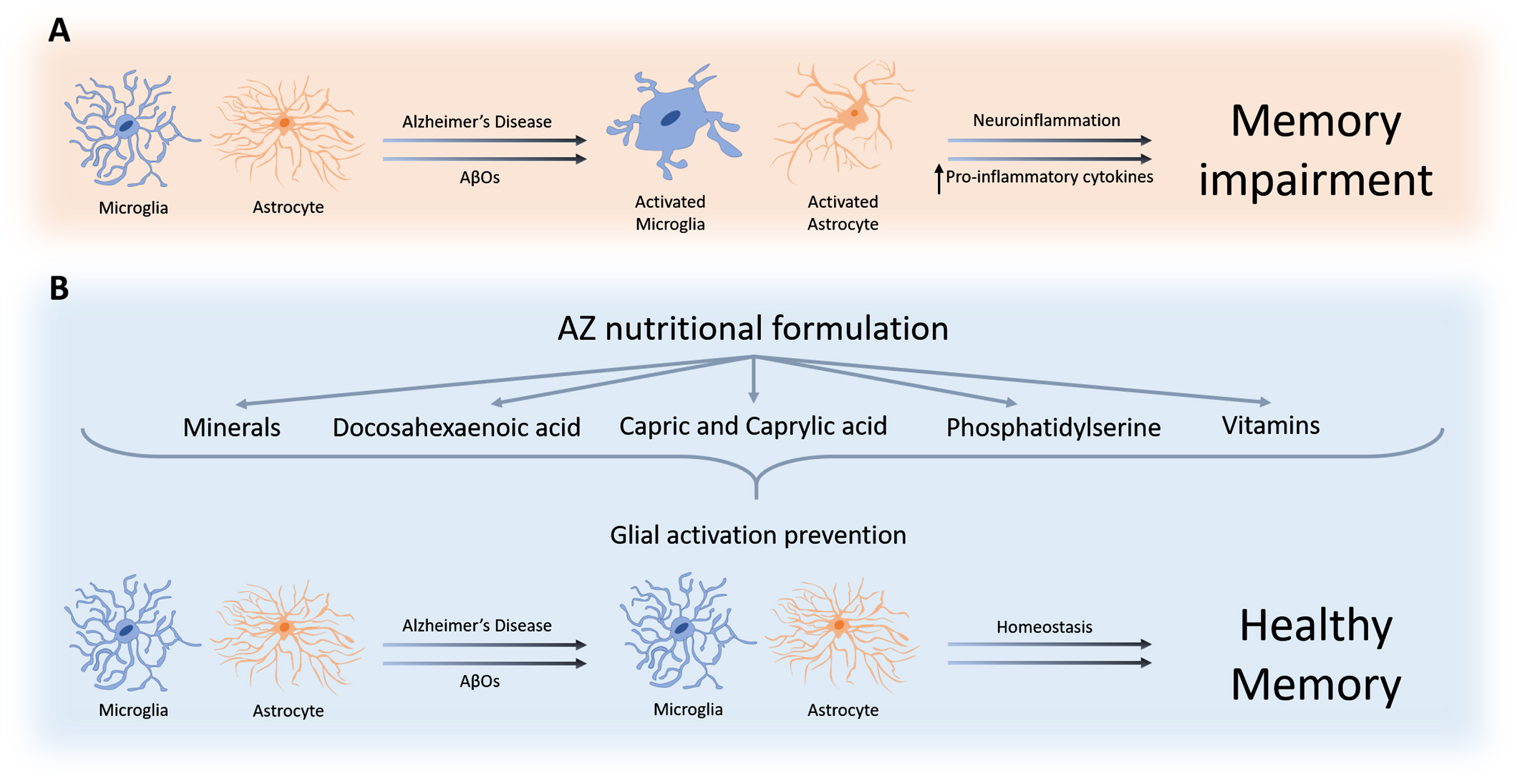
Graphical summary of the protective action of the AZ formulation. A) AβOs cause exacerbated glial activation, leading to increased production/release of proinflammatory cytokines [9 , 22] and resulting in memory loss. B) Treatment with the AZ formulation prevents glial activation, leading to maintenance of homeostasis and preservation of memory.
Footnotes
ACKNOWLEDGMENTS
This work was supported by the Brazilian funding agencies Conselho Nacional de Desenvolvimento Científico e Tecnológico (CNPq), Fundação de Amparo á Pesquisa do Estado do Rio de Janeiro (FAPERJ), National Institute for Translational Neuroscience, and by Prodiet Medical Nutrition.
