






Abstract
Background:
The strongest risk factor for the development of Alzheimer’s disease (AD) is age. The progression of Braak stage and Thal phase with age has been demonstrated. However, prior studies did not include cognitive status.
Objective:
We set out to define normative values for Alzheimer-type pathologic changes in individuals without cognitive decline, and then define levels that would qualify them to be resistant to or resilient against these changes.
Methods:
Utilizing neuropathology data obtained from the National Alzheimer’s Coordinating Center (NACC), we demonstrate the age-related progression of Alzheimer-type pathologic changes in cognitively normal individuals (CDR = 0, n = 542). With plots generated from these data, we establish standard lines that may be utilized to measure the extent to which an individual’s Alzheimer-type pathology varies from the estimated normal range of pathology.
Results:
Although Braak stage and Thal phase progressively increase with age in cognitively normal individuals, the Consortium to Establish a Registry for Alzheimer’s Disease neuritic plaque score and Alzheimer’s disease neuropathologic change remain at low levels.
Conclusion:
These findings suggest that an increasing burden of neuritic plaques is a strong predictor of cognitive decline, whereas, neurofibrillary degeneration and amyloid-β (diffuse) plaque deposition, both to some degree, are normal pathologic changes of aging that occur in almost all individuals regardless of cognitive status. Furthermore, we have defined the amount of neuropathologic change in cognitively normal individuals that would qualify them to be “resilient” against the pathology (significantly above the normative values for age, but still cognitively normal) or “resistant” to the development of pathology (significantly below the normative values for age).
Keywords
INTRODUCTION
First described in the modern literature by Alois Alzheimer in 1907 [1], Alzheimer’s disease (AD) is characterized clinically by symptoms such as executive dysfunction, declarative memory impairment, disorientation and language problems, as well as behavioral and emotional disturbances [2], and neuropathologically by neurofibrillary degeneration combined with diffuse and neuritic plaques [3, 4]. Neurofibrillary degeneration and plaque formation tend to progress through the brain in stereotypical patterns. Neurofibrillary tangles first appear in the brainstem (locus coeruleus), followed by the medial temporal lobe, limbic structures, and finally the neocortex [5, 6]. Plaque formation, on the other hand, begins in the neocortex and progresses to the entorhinal cortex, hippocampus, basal ganglia, brainstem, and cerebellum [7]. While AD is the most common cause of dementia worldwide [8, 9], it is often accompanied by other comorbid pathologies, including cerebrovascular disease, Lewy body pathology, and limbic-predominant age-related TDP-43 encephalopathy (LATE), amplifying symptom severity and often complicating the clinical picture [10–12].
Previous neuropathologic evaluation of 2,332 individuals aged 1–100 years old [6] demonstrated that p-tau deposition begins with threads and pre-tangles in some individuals 10 years old or less, and amyloid-β deposition begins around the 4th-5th decade of life. By age 60, 100% of individuals had some degree of p-tau pathology and 25% had amyloid-β pathology. Whereas, at age 90–100, 100% of individuals displayed p-tau pathology and 80% had amyloid-β pathology. This previous work evaluated neuropathologic findings in the general aging population without distinguishing between individuals with and without clinical symptoms, and so does not consider what degree of pathology would be expected in cognitively normal individuals.
The concepts of “resistance” and “resilience” have been described previously by multiple groups using several pathologic and cognitive correlates. These descriptions have been applied inconsistently across studies, but what can be agreed upon is that there are a subset of cognitively normal individuals with less Alzheimer-type pathology than would be expected for their age (i.e., “resistant” individuals) and a subset with more pathology than would be expected for their age, given the maintenance of their cognitive status (i.e., “resilient” individuals) [13–17]. The correct terminology for these successful aging cognitively normal older individuals has been debated [13]. There was a workshop (The Collaboratory on Research Definitions for Reserve and Resilience in Cognitive Aging and Dementia) to develop consensus definitions and nomenclature for the various terms used in the field. Within the context of the framework, our use of “resistance” would correspond to “brain maintenance”, whereas our use of “resilience” would correspond to “cognitive reserve” [18]. It is important to note that in this study, we are only referring to resistance and resilience against AD neuropathologic change, not resistance or resilience against other comorbid pathologies that could be present. It has therefore been proposed that “apparent resilience” be used when coexisting pathology is not considered [16]. In fact, some studies have demonstrated that many “resilient” individuals are simply resistant to comorbid pathologies [19]. However, other studies have demonstrated that there are additional contributing factors including lower levels of neuroinflammation and less evidence of oxidative stress in the resilient, as well as better maintenance of synapses and fewer hyperphosphorylated tau monomers and multimers in synapses [20–22].
Herein, we utilize the National Alzheimer’s Coordinating Center (NACC) dataset to investigate the level of Alzheimer-type pathology and other comorbid conditions in cognitively normal individuals (defined here as Clinical Dementia Rating (CDR) = 0; n = 542) from age 45 to > 100 years old. In doing so, we establish the approximate amount of Alzheimer-type pathology [Braak stage, Thal phase, Consortium to Establish a Registry for Alzheimer’s Disease (CERAD) neuritic plaque score, and overall Alzheimer’s disease neuropathologic change (ADNC)] that is compatible with normal cognition in each age group. We have also generated a linear model for Braak stage progression with age in cognitively normal individuals from which we were able to define a 90% prediction interval for each age. From this linear model and 90% prediction interval, we have defined “resistance” as cognitively normal individuals with Braak stages below the 90% prediction interval, i.e., bottom 5th percentile at each age (less pathology than expected for one’s age), and “resilience” as cognitively normal individuals with Braak stages above the 90% prediction interval, i.e., top 5th percentile at each age (more pathology than expected for one’s age, but still cognitively normal). We then evaluated the presence or absence of comorbid pathologies and other clinical factors that were statistically associated with resistance and resilience compared to the general population.
METHODS
Case selection
Using the Uniform Data Set (UDS) and neuropathology (NP) data set from the National Alzheimer’s Coordinating Center (NACC), established with funding from the National Institute on Aging (U01 AG016976), we identified 4,659 total patients with both NP data and CDR scores, as well as 542 cognitively normal patients (CDR = 0 at last visit) after excluding any subjects with an interval > 24 months between last visit and autopsy (NACCINT). The number of patients in each age group are listed in Table 1. UDS and NP data was requested and downloaded from NACC. Standardized UDS variable definitions [23] and NP variable definitions [24] from NACC were used, previously described in detail [25, 26]. All participants provided written informed consent at each Alzheimer’s Disease Research Center (ADRC) that contributed tissue and clinical data to NACC. Data on immunohistochemical stains used for tau (NPTAN) and amyloid-β (NPABAN), as well as modified Bielschowsky (NPHISMB), Gallyas (NPHISG), “other silver stain” (NPHISSS), and thioflavin (NPHIST) were recorded in the NP data set. No significant differences in Braak stage, Thal phase, or CERAD NP score were detected between these methods in the available cases. In addition, when comparing CDR = 0 and CDR > 0 individuals, there is no significant difference between these two groups in the distribution of stains (silver versus thioflavin) and IHC antibodies that were used for diagnosis.
Study subjects by age group
Data analysis
Statistical analyses were completed using custom R scripts (v4.2.0). Missing values (as indicated in the downloaded dictionaries) in each category were identified and removed before any analyses. Spearman correlation was used to calculate the correlation between age groups and Braak stage, Thal phase, CERAD NP score, and ADNC variables. A linear regression has been applied individually with age of death (NACCDAGE) as an independent variable and Braak, Thal, CERAD, and ADNC as dependent variables separately. Then we used this model to make a 90% prediction interval for different age groups using predict() function from package stats. Any individuals above the prediction interval will be termed “resilient” (upper 5% of Braak stage in each age group) and anyone below this interval will be termed “resistant” to the development of AD pathology (lower 5% of Braak stage in each age group). For selecting the variables associated with Braak in the resilient and resistant subgroups, we first removed any variable that had more than 5 missing values in the resistant or resilient and ended up with 168 and 223 variables, respectively. In these analyses, we only included cases with NACCDAGE above 45 years old (31 subjects were removed). In each class of resistant/resilient, we fitted a linear model individually for all the variables that remained in the dataset, with Braak stage (NACCBRAA) as a dependent variable. This model was adjusted by age at death (NACCDAGE). All the resulting p-values are adjusted for multiple comparisons using Bonferroni correction, and the cutoff was 0.05. All the visualizations were done using ggplot2 package.
RESULTS
Alzheimer-type neuropathologic findings in cognitively normal individuals
When plotting the progression of Alzheimer-type pathology in the cognitively normal individuals (CDR = 0 subgroup, n = 542), Braak stage progressively increases with age (Fig. 1A), with the median Braak stage rising only to Braak stage III. From the established standard line for age versus Braak stage in the CDR = 0 subgroup, a 65-year-old cognitively normal individual would be expected to have Braak stage I-II, and an 85-year-old cognitively normal individual would be expected to have Braak stage II-III (Fig. 1B). Thal phase (n = 253) increases variably with age, up to a maximum of Thal phase 3 (Fig. 1 C, D). However, CERAD NP score (n = 545) does not increase with age in cognitively normal individuals, and only reaches a CERAD neuritic plaque score of sparse (Fig. 1E, F). Therefore, although Braak stage and Thal phase both increase with age in cognitively normal individuals, CERAD neuritic plaque score does not rise above sparse. Median overall ADNC (n = 253) increases modestly from “not” at age 40–50 to “low” from age 50–90 and “intermediate” from 90–100 (where the average Braak stage is III and Thal phase is 3), after which it decreases back to “low” (Fig. 1G, H). Representative histologic images of pathologic findings in “normal aging” as compared to individuals with cognitive decline are shown in Fig. 2.
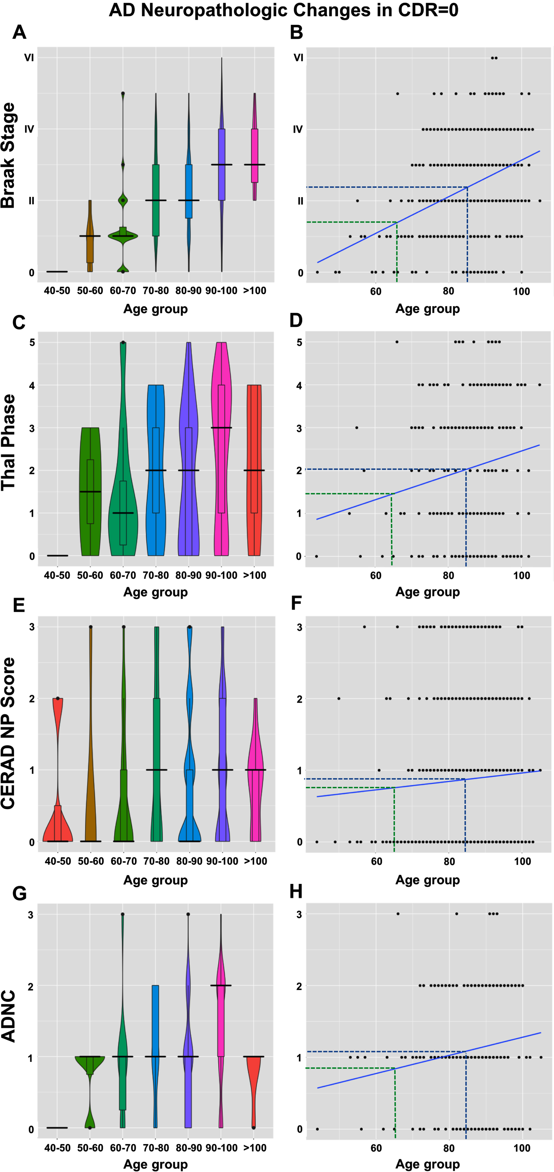
Violin and correlation plots illustrating age related changes in Braak stage, Thal phase, CERAD NP score, and ADNC in cognitively normal individuals (CDR = 0; n = 542). Braak stage and Thal phase progressively increase with age, but CERAD NP score and ADNC do not. Cognitively normal individuals aged 65 have an expected Braak stage of I-II (dashed green line), and individuals aged 85 have an expected Braak stage of II-III (dashed blue line).
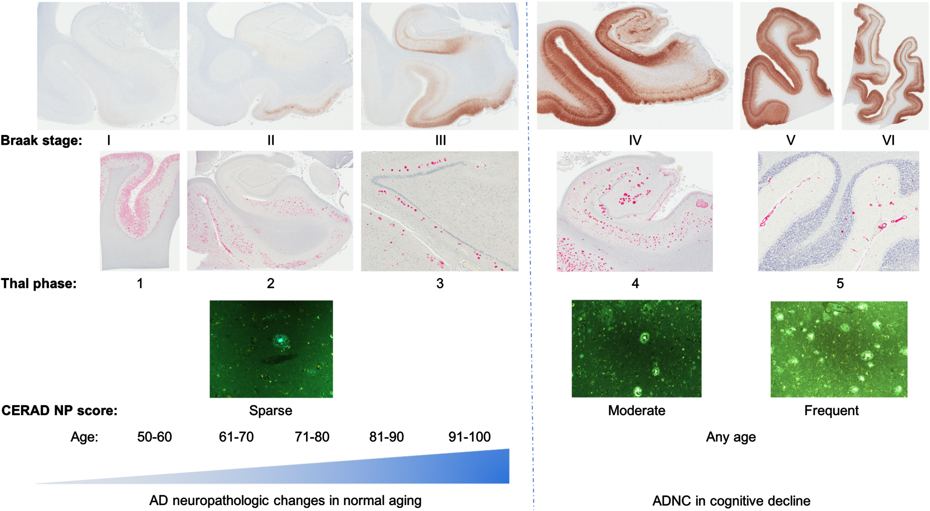
Summary figure demonstrating examples of increasing neuropathology with age (Braak stage (p-tau antibody, AT8), Thal phase (amyloid-β, 4G8), and CERAD NP score (Thioflavin-S) in cognitively normal individuals (“normal aging”; CDR = 0) and individuals with cognitive decline.
In comparison, when plotting the Alzheimer-type pathologic changes in all individuals (any CDR) from the NACC dataset who had neuropathology data available and had been evaluated within 2 years of death (n = 4,659), Braak stage progressively increased from a median stage of 0 at age 30–40 years old to I at age 40–50, II at 50–60, V at 60–90, and IV at 90–100+ (Supplementary Figure 1A). Based off of the established standard line for age versus Braak stage in all cases, a 65-year-old individual in this NACC cohort would be expected to have Braak stage III-IV, and an 85-year-old individual would be expected to have Braak stage > IV (Supplementary Figure 1B). This is at least 1 stage above the cognitively normal population at each age. Thal phase progressively rose from 0 at age 30–40 to a plateau of phase 4 from 60–100+ (Supplementary Figure 1C, D). CERAD NP score rose from 0 (“absent”) from age 30–50 to 1 (“sparse”) from age 50–60, 2 (“moderate”) from 60–70, 3 (“frequent”) from 70–80, and then back down to moderate from 80–100 and sparse above 100 (Supplementary Figure 1E, F). Subsequently, overall median ADNC increases from “none” at age 30–40 to a plateau of “high” from age 60–90, and then intermediate at 90+ (Supplementary Figure 1G, H). This suggests that these neuropathologic correlates of AD progressively increase with age; however, after 80–90 years of age, individuals are somewhat less likely to have high ADNC, as those with worse pathology are more likely to have passed away by this age.
Defining “resistance” and “resilience”
Using the CDR = 0 cognitively normal individuals, we established a working definition of “resistance” and “resilience” based off of the linear model made for Braak stage progression with age (Fig. 3). Braak stage was chosen as it was the neuropathologic variable recorded for the most individuals in the NACC NP dataset and is most closely associated with clinical symptoms [27]. “Resistant” individuals are defined here as cognitively normal individuals with less neuropathologic findings than would be expected for age, or bottom 5% of Braak stage in each age range (n = 23). Conversely, “resilient” individuals are defined as cognitively normal individuals with more neuropathologic findings than would be expected for age, or upper 5% of Braak stage in each age range (n = 31) (Fig. 3A). Similar definitions for “borderline resistant individuals” (bottom 10% of Braak stages in each age range; n = 48) and “borderline resilient individuals” (upper 10% of Braak stages in each age range; n = 52) (Fig. 3B) were defined. Thresholds for Braak stage in the resilient and resistant groups in each age range are shown in Supplementary Table 1.
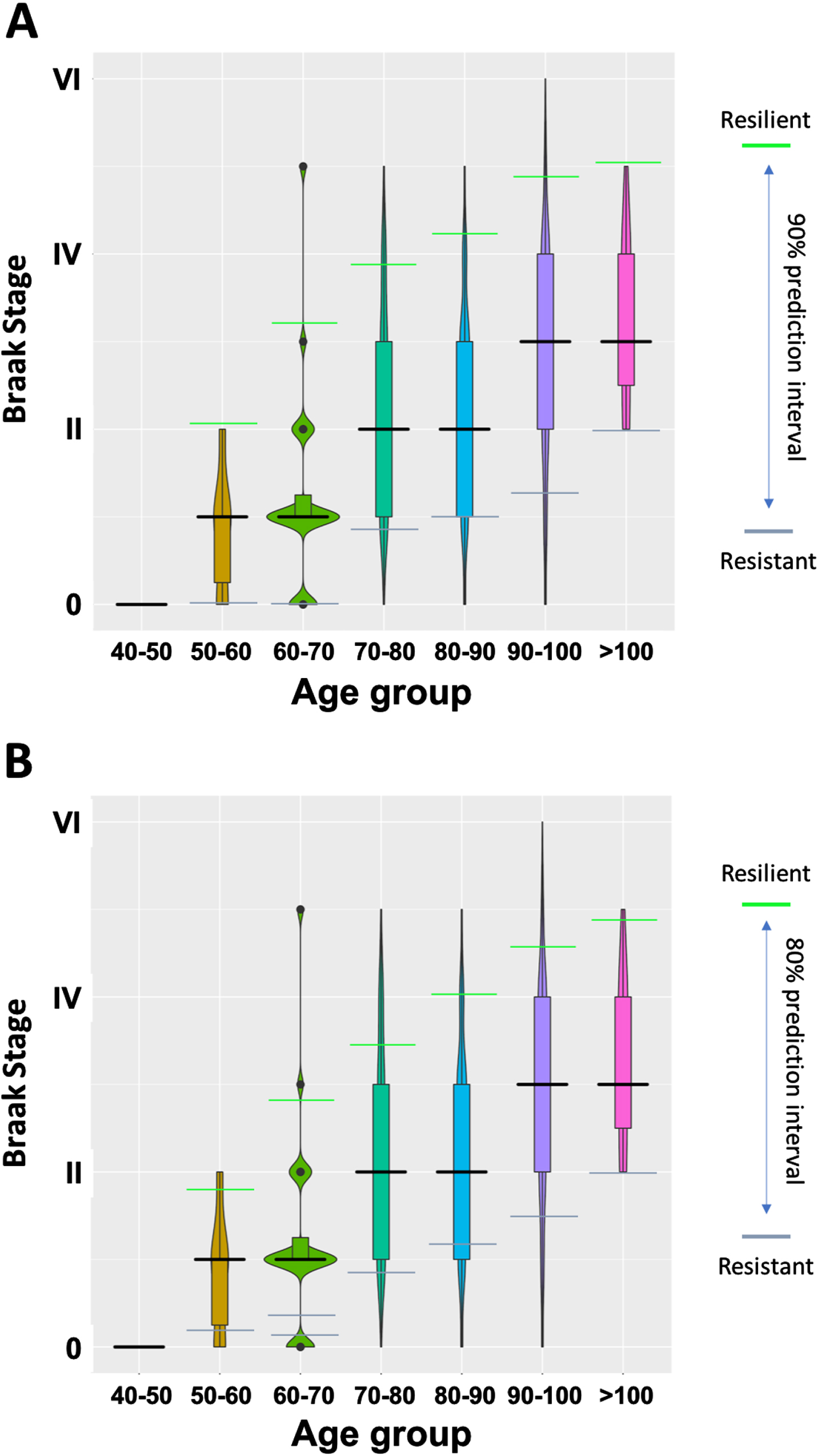
“Resilient” and “resistant” definitions based off of age and Braak stages in cognitively normal individuals (CDR = 0; n = 542). Using a 90% prediction interval (top panel), the 5% of cases at each age with the highest Braak stages are defined here as “resilient” (n = 31) and the 5% of cases at each age with the lowest Braak stages are defined here as “resistant” (n = 23). Using an 80% prediction interval (bottom panel), the 10% of cases at each age with the highest Braak stages are defined here as “resilient” (n = 52) and the 10% of cases at each age with the lowest Braak stages are defined here as “resistant” (n = 48).
Significant variable differences in resistant cases
When comparing the NACC UDS and NP variables [23, 24] in the resistant individuals to these variable measures in all other individuals, with and without cognitive decline (Fig. 4 and Supplementary Table 2), the resistant individuals displayed significantly lower Braak stages (as expected, given this is how we define them), as well as lower Thal phases, and CERAD NP scores (none of the resistant displayed frequent neuritic plaques). There was no Lewy body pathology in the resistant subgroup. In addition, the resistant group displays less cerebral amyloid angiopathy (CAA) with no cases of moderate or severe CAA. Intriguingly, the resistant have a higher frequency of reported transient ischemic attacks (TIAs), yet they have fewer infarcts and fewer microinfarcts, pathologically. They also display less arteriolosclerosis (no cases of severe, and very few moderate). In contrast, the resistant group has a slightly higher percentage of cases with severe atherosclerosis. Furthermore, compared to the other CDR = 0 individuals, the resistant group displays a trend toward less usage of lipid lowering drugs (Supplementary Figure 2). Interestingly, the resistant group also has a slightly higher incidence of CNS neoplasms (Fig. 4).
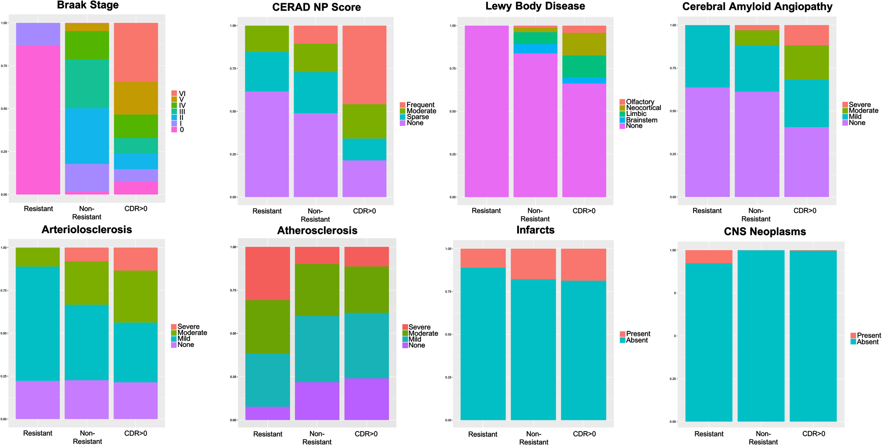
Bar plots of select variables demonstrating differences between the resistant, non-resistant but cognitively normal, and CDR > 0 subgroups, including Braak stage, CERAD NP score, amounts of Lewy body pathology, CAA, arteriolosclerosis, atherosclerosis, infarcts, and CNS neoplasms.
Significant variable differences in resilient cases
When comparing the NACC UDS and NP variables [23, 24] in the resilient individuals to all others (Fig. 5 and Supplementary Table 3), the resilient individuals have a significantly higher percentage of cases with Braak stage IV, V, and VI as compared to the non-resilient CDR = 0 group, and as compared to the CDR > 0 group, the resilient have higher levels of Braak stage IV and V, but less of Braak stage VI. Examination of Thal phase demonstrates that the resilient have a slightly higher percentage of Thal phases 2, 3, and 4, but a lower percentage of Thal 5 when comparing to the CDR > 0 group, but generally higher Thal phases than the non-resilient CDR = 0 group. The resilient display a lower percentage of “frequent” neuritic plaques and more “sparse” neuritic plaques as compared to the CDR > 0 group. Furthermore, the resilient primarily display intermediate ADNC and they harbor a lower percentage of high ADNC cases as compared to the CDR > 0 group (Fig. 5). These results demonstrate that the “resilient” individuals harbor higher levels of Alzheimer-type pathology than most cognitively normal individuals, however they display somewhat lower levels than cognitively impaired individuals.
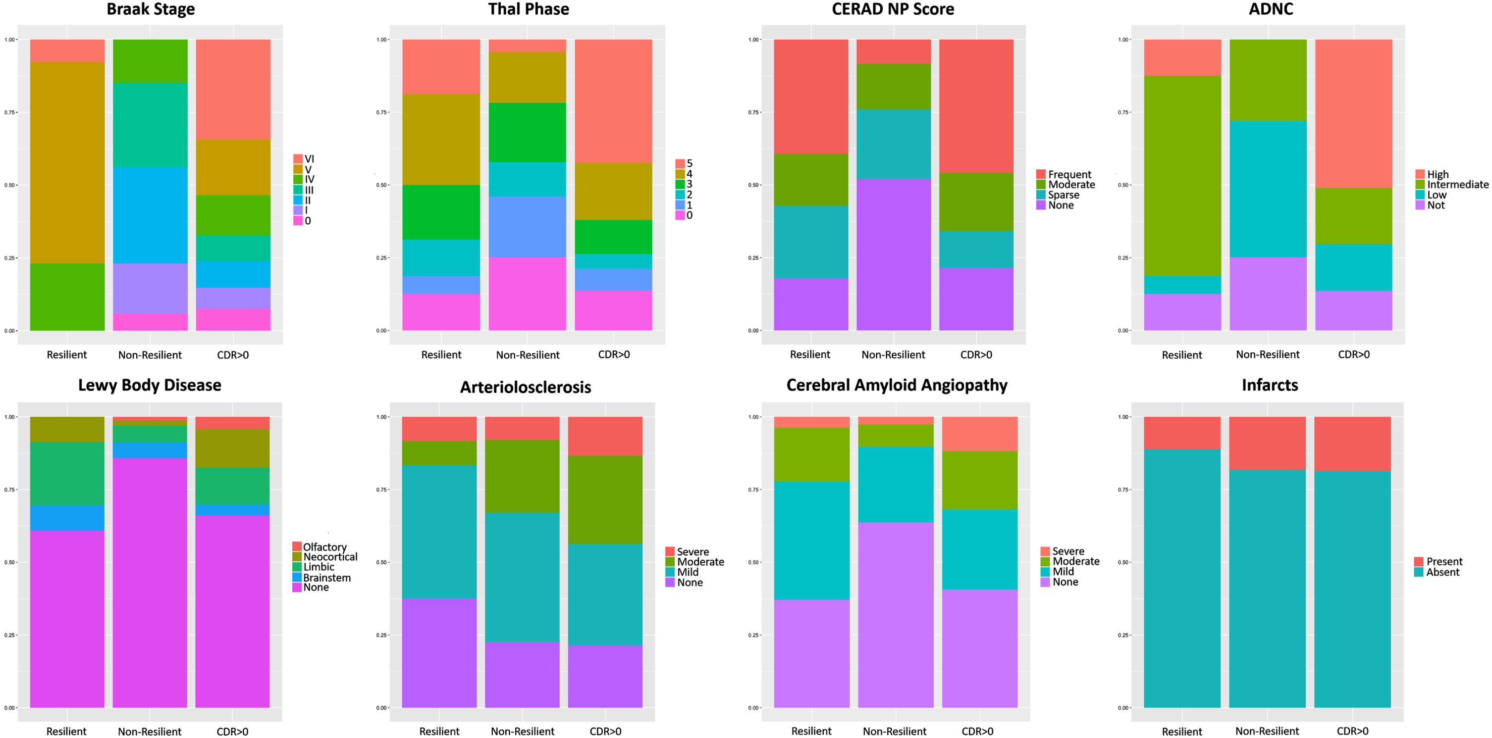
Bar plots of select variables demonstrating differences between the resilient, non-resilient but cognitively normal, and CDR > 0 cohorts, including Braak stage, Thal phase, CERAD NP score, ADNC level, amounts of Lewy body pathology, arteriolosclerosis, CAA, and infarcts.
Interestingly, there is more of brainstem and limbic Lewy body disease (LBD) in the resilient as compared to the other groups, but less of diffuse neocortical as compared to the CDR > 0 group. The resilient also have a lower frequency of severe CAA and more mild CAA as compared to the CDR > 0 group. In addition, the resilient have less of moderate and severe levels of arteriolosclerosis, and a lower percentage of cases with severe atherosclerosis. They also have fewer infarcts. As expected, both resistant and resilient individuals had less frequent incidence of clinical symptoms (based off of variables related to cognition and ability to perform activities of daily living) compared to the CDR > 0 group (Supplementary Tables 2 and 3). In addition, there was a higher percentage of females in the resilient subgroup as compared to the non-resilient and CDR > 0 group.
Compared to other cognitively normal (CDR = 0) individuals, the resilient individuals demonstrated a trend toward lower diastolic blood pressure, less atherosclerosis in the circle of Willis, less “additional pathology present”, more frequent use of anticoagulants and antiplatelet drugs, more frequent use of anti-depressants, as well as more frequent use of NSAIDs. In addition, the resilient demonstrated less usage of lipid lowering drugs (Supplementary Figure 3). Although TDP-43 pathology and hippocampal sclerosis were excluded from the larger analysis as there were > 5 cases with missing data in both the resilient and resistant groups, there are trends toward less hippocampal sclerosis and less LATE neuropathologic change in both the resistant and resilient (Supplementary Figures 4 and 5). However, the resilient do appear to have more TDP-43 pathology than the other cognitively normal individuals.
DISCUSSION
The cognitive symptoms of AD tend to progress in concert with the level of ADNC, in particular, with the Braak stage of neurofibrillary degeneration. Clinical severity increases as neurofibrillary degeneration progresses from the brainstem to entorhinal cortex and limbic system, and eventually to the neocortex [5, 28]. This progression occurs with normal aging as well, albeit at a slower pace and to a lesser extent. However, it is generally recognized that there are a small number of successful aging outliers, who either do not develop neuropathologic changes associated with age (“resistant” individuals) or who have these changes but do not develop the characteristic cognitive decline associated with the underlying pathology (“resilient” individuals) [22, 30]. These outliers include “SuperAgers”, individuals with remarkably good cognition despite advanced age (generally defined as patients of 80+ years of age with episodic memory similar to patients in their 50 s and 60 s) [31, 32]. However, the terms resistance and resilience have been used in describing successful aging populations without consensus criteria or strict definitions as to how much pathology would qualify one to be resistant or resilient. In addition, imaging studies have defined amounts of brain atrophy that are expected for one’s age; a so-called “brain age” [33]. Individuals who display less atrophy than expected for their age have been called “resilient” [34, 35]; however, the lower amount of atrophy may suggest less pathology than expected for their age, and thus, these individuals would be called “resistant” from a pathologist’s perspective. It is essential that this “resistance” and “resilience” terminology is standardized across clinical, imaging, and pathologic criteria and to establish the amount of AD-type neuropathology that can reasonably be expected at certain ages. The findings of PET imaging and biomarker investigators must be correlated with neuropathologic findings and detailed cognitive information to fully understand “normal amount” of pathology to better design clinical trials and more targeted therapy. The concept of resistant and resilient individuals across these specialties is crucial to understanding the disease process and what constitutes a diagnosis of “pre-clinical AD.”
While it is well-established that neuropathologic changes associated with AD are associated with age, and begin much earlier than clinical symptoms [6], the degree to which these changes occur in individuals who do not develop clinical symptoms has not been established. In this report, we define resistant and resilient individuals within the NACC dataset, where “resistant” individuals are those with normal cognition (CDR = 0) and Braak stages below the 90% prediction interval for each age group, and “resilient” individuals are those with normal cognition (CDR = 0) and Braak stages above the 90% prediction interval for each age group, despite a lack of clinical symptoms (Fig. 4A). We therefore determine Braak stage thresholds for classifying cases as resistant or resilient at any given age (Supplementary Table 1). Furthermore, we evaluated the expected amount of p-tau and amyloid-β pathology at each age in all cases (n = 4,659) (Supplementary Figure 1) and in cognitively normal-only cases (n = 542) (Fig. 1). With plots formulated from these data, we establish standard lines (similar to a growth chart) that may be utilized to measure the extent to which an individual’s Alzheimer-type pathology varies from the estimated normal range of pathology. While the literature suggests that Braak neurofibrillary tangle stage most closely correlates with cognition in the development of AD pathology, the finding that Braak stage and Thal phase progressively increase with age in cognitively normal individuals, but CERAD neuritic plaque score does not, suggests that an increasing burden of neuritic plaques is a strong predictor of cognitive decline, whereas some degree of increasing neurofibrillary degeneration and diffuse amyloid-β plaque deposition are common and expected pathologic changes of aging that occur (at least to some degree) in the vast majority of individuals [6], regardless of cognitive status.
Overall, we found that the resistant and resilient harbor fewer comorbid pathologies, including Lewy body pathology, infarcts, CAA, and arteriolosclerosis. This has previously been reported in multiple studies, and it has been suggested that resilience to ADNC may in part be due to resistance to the development of additional comorbid neuropathologic processes, and thus represent “apparent resilience” [12, 19]. Interestingly, brainstem and limbic Lewy body pathology are present in the resilient, but not in the resistant, which suggests that the more p-tau pathology one develops, the more Lewy body pathology they may develop. The concurrence of p-tau and α-synuclein pathology corroborates reports that these proteins may aggregate synergistically in some individuals [36–38]. It is also possible that some individuals who are resilient against one pathology (such as ADNC) have a tendency to be resilient against others (such as additional Lewy body pathology), and so are cognitively normal in the face of both disorders.
One interesting difference between the resistant and resilient is in levels of atherosclerosis. Whereas the resilient display both less arteriolosclerosis and less atherosclerosis in the circle of Willis, the resistant display less arteriolosclerosis, but a higher frequency of severe atherosclerosis. The resistant also have a higher frequency of TIAs, but they have fewer infarcts and microinfarcts. They also have lower usage of lipid lowering drugs. It is possible that the higher levels of atherosclerosis could be associated with the higher frequency of TIAs, and could be caused by the lack of lipid lowering drugs. However, these do not appear to be affecting their cognition in a negative way, and are not associated with increased AD pathology. The resilient also have lower usage of lipid lowering drugs, but higher use of anti-coagulants. In addition, the resilient display a trend toward lower diastolic blood pressure. Along the same lines, previous studies have demonstrated that higher diastolic blood pressure is associated with cognitive impairment [39].
Another intriguing finding in the resistant individuals is that they have a significantly higher incidence of CNS neoplasms compared to both non-resistant but cognitively normal individuals and cognitively impaired individuals (Fig. 4). This suggests that having (or having had) cancer may somehow impart a degree of protection against the development of AD pathology. The dichotomy between AD and cancer has been reported previously [40–47]. It appears that these disease processes are diametrically opposed, so if one develops AD, they are unlikely to have cancer, and if one has survived cancer, they are unlikely to develop AD. Several theories have been proposed to explain this phenomenon including a possible inverse relationship between senescence (in AD) and cell proliferation (in cancer) [48], as well as opposing transcriptomic profiles in AD and cancer for p53 and Wnt pathways, and the ubiquitin/proteasome system [49].
Differences in pharmaceutical usage in the resilient and resistant groups could be interpreted in multiple ways. For example, NSAID usage was lower in the resilient cohort as compared to both the non-resilient CDR = 0 and CDR > 0 group (Supplementary Figure 3), which could suggest that NSAIDs are protective against cognitive decline in the presence of pathology. However, since the resilient have more pathology than the non-resilient CDR = 0 group, it could also be argued that those subjects were cognitively normal to begin with, and increased use of NSAIDs caused them to develop more pathology. All of the medication usage differences could have contrasting arguments similar to this one, concluding that the drug is protective versus causative. However, with the example of the NSAIDs, since the CDR > 0 group uses less, it would favor the argument that the NSAIDs are protective. A clinical trial investigating NSAID usage in AD found that they are protective in asymptomatic individuals when taken for 2-3 years; however, they may have an adverse effect when taken in later stages of AD progression [50]. In addition, a study investigating the association between NSAID usage and AD neuropathologic changes found that NSAID use is associated with higher levels of Aβ42 in the frontal and temporal cortex, but not associated with higher p-tau levels in any of the cortical regions analyzed [51]. This could suggest that NSAIDs are protective or neutral with respect to neuritic plaque formation. As another example, for the lipid lowering drugs, both the resistant and the resilient appear to use less lipid lowering drugs, which could suggest that those people do not have lipid abnormalities and do not need to be on medication, or that these drugs are not protective against cognitive decline. Previous studies investigating the effects of statins on AD risk have varying outcomes, but primarily statins have been reported to reduce the risk of dementia [52, 53]. However, one study examined the association between statin use and AD neuropathologic change in clinically and pathologically diagnosed AD cases. This study found that statin use had no effect on the amounts of AD neuropathologic change, although no investigation into the effects of statins in cognitively normal individuals was performed [54].
There are several limitations to this study. One of the main limitations is the fact that the NACC cohort contains a disproportionately high number of dementia cases compared to community-based cohorts. This may skew the average pathology measured in the general population (Supplementary Figure 1) to a much higher level. That said, this would not affect the analyses that used only cognitively normal individuals. In future studies, we plan to repeat these analyses in larger community-based cohorts that would be more representative of the general population. In future studies, we also plan to use cohorts with lifestyle factors (social determinants of health), personality, physical performance and frailty data to reveal differences that the resistant and resilient may have in these areas (including physical activity, social engagement, attitude, mindset, etc.). Another limitation of this study is that we defined levels for resistance and resilience only off of the age versus Braak stage plot. ADNC may prove to be a better variable to use, however, not as many cases reported this measure in the NACC NP dataset. Additionally, it is important to note that there are variables in the NACC dataset that did not have sufficient data to be included in the main analyses. Examples of this include TDP-43 pathology consistent with LATE, hippocampal sclerosis, white matter rarefaction, and aging-related tau astrogliopathy. The lack of sufficient data in many of these pathologic variables in subsets of cases also precludes isolating cases with only ADNC in the absence of additional co-existing pathologies for additional analyses.
In future studies, we plan to assess the presence of LATE and its progression with age, as well as defining levels that would make one resistant to the development of LATE or resilient against it. LATE is a relatively new entity to be named [11], but studies have reported the presence of LATE in older individuals to be as high as 40% [55]. In general, the presence of LATE neuropathologic change is associated with amnestic symptoms similar to AD, but may be present in cognitively normal individuals as well. The trends we observe are suggestive of less hippocampal sclerosis and LATE in the resistant and resilient (Supplementary Figures 4 and 5). However, the resilient appear to have slightly more hippocampal sclerosis and LATE than the other cognitively normal individuals. The resilient, by definition, also have more AD pathology than the other cognitively normal individuals. Similar to the α-synuclein pathology, this could suggest that TDP-43 and AD pathology may develop synergistically in some people. It will be interesting to investigate the incidence of LATE in cognitively normal individuals in other cohorts. We also plan to assess the presence of LBD and cerebrovascular disease in cognitively normal individuals and their progression with age. With this, we could define the expected amount of LBD, LATE, and cerebrovascular disease pathology for one’s age if cognitively normal, and determine levels for resistance and resilience against those pathologic changes.
It would also be of interest to examine differences in the development of neurofibrillary degeneration in hippocampal subregions in normal aging as compared to those with cognitive decline. Previous studies have demonstrated that primary age-related tauopathy cases tend to have early vulnerability of the CA2 subregion of the hippocampus for neurofibrillary degeneration rather than CA1 and entorhinal cortex, as is observed in AD [56]. Having more in-depth quantitative assessments of neurofibrillary degeneration in each subregion would be informative because it is possible that developing neurofibrillary degeneration in CA2 first (rather than entorhinal and CA1) could be indicative of healthier aging.
In conclusion, we have developed standard lines and estimated normative values for the progression of Alzheimer-type pathology with age in cognitively normal individuals. In addition, we have defined levels that would qualify one to be called resistant to the development of AD neuropathologic change or resilient against it. While not a conclusive definition of resilience and resistance to neuropathologic changes, these data suggest a method for standardizing pathologic definitions of disease burden in “normal” individuals. We believe that this study will assist with harmonizing nomenclature and developing consensus definitions for “resistant” and “resilient” individuals in future studies.
Footnotes
ACKNOWLEDGMENTS
The NACC database is funded by NIA/NIH Grant U24 AG072122. NACC data are contributed by the NIA-funded ADRCs: P30 AG062429 (PI James Brewer, MD, PhD), P30 AG066468 (PI Oscar Lopez, MD), P30 AG062421 (PI Bradley Hyman, MD, PhD), P30 AG066509 (PI Thomas Grabowski, MD), P30 AG066514 (PI Mary Sano, PhD), P30 AG066530 (PI Helena Chui, MD), P30 AG066507 (PI Marilyn Albert, PhD), P30 AG066444 (PI John Morris, MD), P30 AG066518 (PI Jeffrey Kaye, MD), P30 AG066512 (PI Thomas Wisniewski, MD), P30 AG066462 (PI Scott Small, MD), P30 AG072979 (PI David Wolk, MD), P30 AG072972 (PI Charles DeCarli, MD), P30 AG072976 (PI Andrew Saykin, PsyD), P30 AG072975 (PI David Bennett, MD), P30 AG072978 (PI Neil Kowall, MD), P30 AG072977 (PI Robert Vassar, PhD), P30 AG066519 (PI Frank LaFerla, PhD), P30 AG062677 (PI Ronald Petersen, MD, PhD), P30 AG079280 (PI Eric Reiman, MD), P30 AG062422 (PI Gil Rabinovici, MD), P30 AG066511 (PI Allan Levey, MD, PhD), P30 AG072946 (PI Linda Van Eldik, PhD), P30 AG062715 (PI Sanjay Asthana, MD, FRCP), P30 AG072973 (PI Russell Swerdlow, MD), P30 AG066506 (PI Todd Golde, MD, PhD), P30 AG066508 (PI Stephen Strittmatter, MD, PhD), P30 AG066515 (PI Victor Henderson, MD, MS), P30 AG072947 (PI Suzanne Craft, PhD), P30 AG072931 (PI Henry Paulson, MD, PhD), P30 AG066546 (PI Sudha Seshadri, MD), P20 AG068024 (PI Erik Roberson, MD, PhD), P20 AG068053 (PI Justin Miller, PhD), P20 AG068077 (PI Gary Rosenberg, MD), P20 AG068082 (PI Angela Jefferson, PhD), P30 AG072958 (PI Heather Whitson, MD), P30 AG072959 (PI James Leverenz, MD).
The authors would also like to thank Drs. Heiko Braak and Kelly Del Tredici for their suggestions and thoughtful discussion.
J.M.W. and T.E.R. are supported in part by National Institute on Aging (NIA) R21 AG078505 and P30 AG066546. J.M.W. is also supported by the San Antonio Claude D. Pepper Older Americans Independence Center, P30 AG044271. The funders had no role in study design, data collection and analysis, decision to publish or preparation of the manuscript.
