






Abstract
Background:
Down syndrome (DS) is caused by an extra copy of all or part of chromosome 21. The patients with DS develop typical Alzheimer’s disease (AD) neuropathology, indicating the role of genes on human chromosome 21 (HSA21) in the pathogenesis of AD. Purkinje cell protein 4 (PCP4), also known as brain-specific protein 19, is a critical gene located on HSA21. However, the role of PCP4 in DS and AD pathogenesis is not clear.
Objective:
To explore the role of PCP4 in amyloid-β protein precursor (AβPP) processing in AD.
Methods:
In this study, we investigated the role of PCP4 in AD progression in vitro and in vivo. In vitro experiments, we overexpressed PCP4 in human Swedish mutant AβPP stable expression or neural cell lines. In vitro experiments, APP23/PS45 double transgenic mice were selected and treated with AAV-PCP4. Multiple topics were detected by western blot, RT-PCR, immunohistochemical and behavioral test.
Results:
We found that PCP4 expression was altered in AD. PCP4 was overexpressed in APP23/PS45 transgenic mice and PCP4 affected the processing of AβPP. The production of amyloid-β protein (Aβ) was also promoted by PCP4. The upregulation of endogenous AβPP expression and the downregulation of ADAM10 were due to the transcriptional regulation of PCP4. In addition, PCP4 increased Aβ deposition and neural plaque formation in the brain, and exuberated learning and memory impairment in transgenic AD model mice.
Conclusion:
Our finding reveals that PCP4 contributes to the pathogenesis of AD by affecting AβPP processing and suggests PCP4 as a novel therapeutic target for AD by targeting Aβ pathology.
Keywords
INTRODUCTION
Down syndrome (DS) occurs in people who carry an extra copy of all or part of human chromosome 21 (HSA21) and affects approximately 1 in 700–800 babies [1, 2]. Most patients with DS die in infancy or adulthood [3, 4]. However, some patients with DS can live to age of 60 or older. DS patients with trisomy-21 develop typical Alzheimer’s disease (AD) neuropathology [5]. AD is a chronic neurodegenerative disease with progressive cognitive impairment, personality changes, and language dysfunction [6, 7]. AD is the fifth-leading cause of death among people aged 65 years and older [8]. The main pathological features of AD are extracellular senile plaque formed by the deposition of amyloid-β protein (Aβ), intraneuronal neurofibrillary tangles formed by the hyperphosphorylation of tau protein, neuronal loss, and brain atrophy. Many key genes associated with AD pathogenesis including amyloid precursor protein (APP), the regulator of calcineurin 1 (RCAN1), BACE2, and Ubiquitin Specific Peptidase 25 (USP25) are located on HSA21, indicating dysregulation of HSA21 genes may play a critical role in the development of AD in DS [9–11].
To generate Aβ, AβPP is cleaved by β-secretase at amino acid number 1 (aspartic acid) in the Aβ domain of AβPP to produce the transmembrane-bound C99 fragment. Subsequently, C99 is cleaved by γ-secretase within the transmembrane to release Aβ40 or Aβ42 [12]. However, AβPP also can be cleaved by β-secretase at amino acid number 11 (glutamic acid) in the Aβ domain to produce the transmembrane-bound C89 fragment. γ-secretase cleaves C89 within the transmembrane to release only a smaller truncated Aβ fragment [13, 14]. Under normal conditions, AβPP is predominantly cleaved by α-secretase between amino acids 16 (lysine) and 17 (leucine) in the Aβ domain to release C83, and C83 is then cleaved by γ-secretase to produce P3 fragment [7, 15]. BACE2, the θ-secretase, cleaves AβPP at amino acid number 20 (Phenylalanine) in the Aβ domain to produce the transmembrane-bound C80 fragment in the non-amyloidogenic pathway[9, 16].
The gene encoding for Purkinje cell protein 4 (PCP4) is located on HSA21 [17, 18]. PCP4, also known as brain-specific protein 19 (PEP-19), is a 7.6 kDa calmodulin-binding protein with an IQ motif. PCP4 was initially isolated based on its developmental regulation in the rat cerebellum [19]. PCP4 is a key molecule in the development and functioning of the nervous system. It is a brain-specific polypeptide whose levels markedly increase during the later stages of rodent nervous system maturation [20]. PCP4 is abundant in cerebellar Purkinje cells and plays an important role in synaptic plasticity [21]. It is also expressed in other regions of the brain besides the cerebellum [22]. Overexpression of PCP4 induces precocious neuronal differentiation during mouse embryogenesis and causes motor and learning impairments [23, 24]. Expression of PCP4 in the brain occurs primarily after birth and has a neuronal expression pattern in adulthood. During embryogenesis, PCP4 is mainly expressed in the ectoderm and neuroectoderm, including cells of neural crest origin [17].
PCP4 plays an important role in the pathological process of many neurological diseases. PCP4 has calmodulin-related functions, which is one of the important factors leading to Huntington’s disease pathology [25]. In animal models of Parkinson’s disease commonly associated with MPTP injury, mRNA and protein expression levels of PCP4 in the striatum are decreased [26]. PCP4 is downregulated in alcoholics by neuronal microarray analysis and RNA level verification [27]. In addition, PCP4 regulates cardiac excitability through autonomic and central Purkinje cell mechanisms [28]. However, its role in the pathogenesis of AD is currently unclear.
In the current study, we demonstrated that PCP4 regulates AβPP processing and exacerbates AD-related amyloid pathology both in vivo and in vitro. Furthermore, the overexpression of PCP4 significantly increased amyloid deposition in APP23/PS45 mice. Our finding reveals that PCP4 contributes to the pathogenesis of AD and suggests PCP4 as a novel therapeutic target for AD.
MATERIALS AND METHODS
Cell culture
Cells were cultured in a 37°C incubator containing 5% CO2. The essence of 20E2 and 2EB2 cells were actually human embryonic kidney cells (HEK293 cell line). 20E2 cells were stably overexpressing human Swedish mutant AβPP (screening with 50μg/mL G418). 2EB2 cells were also stably overexpressing BACE1 on the basis of 20E2 (screening with 50μg/mL G418 and 100μg/mL zeocin). The essence of SAS cells were human neuroblastoma cell line SH-Sy5y cells. SAS cells were also stably overexpressing human Swedish mutant AβPP (screening with 100μg/mL zeocin). The essence of N2AAPP cells were mouse neuroblastoma cell line Neuro-2a (N2A) cells. N2AAPP cells were stably overexpressing human Swedish mutant AβPP (screening with 50μg/mL G418). 20E2 cells, 2EB2 cells, SH-Sy5y cells, SAS cells, N2A cells, and N2AAPP cells were cultured in Dulbecco’s Modified Eagle Medium (DMEM) (Gibco, A1443001) containing 10% fetal bovine serum (FBS) (ExCell Bio, FSB500). In addition, the human neuroblastoma cell line M17 (SK-N-BE) (2) (Shanghai Institute of Biochemistry and Cell Biology, China), was cultured in 45% MEM (Univ, abs9467) (included 1.5 g/L of NaHCO3 (Gibco, 25080094) and 0.11 g/L of Sodium Pyruvate (Gibco, 11360070)), 45% F-12 (Gibco, 21700075) (included 1.5 g/L of NaHCO3 and L-glutamine (Gibco, 25030081)) and 10% FBS.
Transgenic mice and intra-cerebroventricular (ICV) injection
The research plan has been approved by the relevant departments, namely the Animal Study Committee of the Children’s Hospital of Chongqing Medical University. In addition, the experiment was carried out based on the Guide for the Care and Use of Laboratory Animals of the Ethics Committee of Chongqing Medical University [29]. APP23 transgenic mice carry human APP751 cDNA with the Swedish double mutation at positions 670/671 (KM⟶NL). PS45 transgenic mice carry human presenilin-1 cDNA with the G384A mutation [30]. Genotypes were identified via polymerase chain reaction (PCR) using DNA extracted from tail tissues. The following primers were used for PCR. Thy1E2 (forward 5’ -CACCACAGAATCCAAGTCGG-3’), APP1082 reverse (5’-CTTGACGTTCTGCCTCTTCC-3’), and PS-1 reverse (5’-ATCACAGCCAAGATGAGC-3’).
Adeno-associated virus carrying PCP4 (AAV-PCP4) was constructed by OBiO Technology (Shanghai, China). AAV-PCP4 was injected into the ICV of mice at 7 weeks of age. After anesthesia with pentobarbital (1%, 50 mg/kg, i.p.) was completed, fixation and craniotomy were performed. The ICV was located 0.5 mm away from the anterior fontanelle (AF), 1.1 mm toward the AF, and 3.0 mm deep (coordinates: –0.5,±1.1, –3.0 mm). The X-axis (1.5 units) and Y-axis (1.1 units) knobs move the operating arm to the coordinate position. The injection dose was 2μL (injection on both sides, 1μL each), and the injection time was approximately 5 min per side. After injection, the needle was kept still for 3 min, and the syringe was slowly pulled out. Finally, the surgical wound was closed.
Construction of plasmids
The construction of the PCP4 plasmid involved the use of homologous recombination (Vazyme, C112-01). Specifically, first a pcDNA4.0-PCP4-myc-his-(A) plasmid was assembled, with the pcDNA4.0-myc-his-(A) vector serving as the base. The PCP4 cDNA sequence was derived from M17 cells and amplified using primers: forward 5’-TTGGTACCGAGCTCGGATCCGCCACCATGAGTGAGCGACAAGGTGC-3’ and reverse 5’ GAAGGGCCCTCTAGACTCGAGGGACTGAGACCCAGCCTTCT-3’. Subsequently, the T2A-EGFP sequence was inserted into this initial plasmid construction, amplified using primers forward 5’-CTGGGTCTCAGTCCCTCGAGGAGGGCAGAGGAAGTCTGCT-3’ and reverse 5’-TGTTCGAAGGGCCCTCTAGACTTGTACAGCTCGTCCATGCC-3’.
Western blotting
Cells or tissue were lysed using RIPA lysis buffer which added with protease inhibitors (Roche, 4693159001) and phosphatase inhibitors (Roche, 4906837001). After quantification, the dissolved products were diluted in 5×sample buffer (Yamei, LT101S) and boiled at 98°C. The lysates were analyzed in Tris-glycine or Tris-tricine gel. AβPP and C-terminal fragments (CTFs) were detected using the rabbit anti-AβPP C-terminal polyclonal antibody C20 (1:3000) [31]. BACE1 was detected using the anti-BACE1 monoclonal antibody (CST, 1:1000, D10E5). ADAM17 was detected using the anti-TACE antibody (CST, 1:10000, D22H4). ADAM10 was detected using the anti-ADAM10 antibody (Abcam, 1:1000, ab124695). PS1 was detected using the rabbit anti-Presinilin-1 polyclonal antibody (Abcam, 1:1000, ab15458). PCP4 was detected using the anti-PCP4 antibody (Proteintech, 1:1000, 14705-1-AP). Actin, GAPDH or α-tubulin was detected as the internal controls. β-actin was detected using the AC-15 (Sigma-Aldrich, 1:5000, A1978). GAPDH was detected using the anti-GAPDH antibody (Proteintech, 1:10000, 60004-1-Ig), and α-tubulin was detected using the anti-α-tubulin antibody (Proteintech, 1:10000, 66031-1-Ig). Target proteins were incubated with primary antibodies overnight at 4°C. Next day, they were followed by incubation with HRP-labeled secondary antibodies (PEL, 1:3000, NEF812001EA, and NEF822001EA) at room temperature for 1.5 h.
ELISA
After overexpression of control plasmid and PCP4 plasmid in 20E2 cells, the cell supernatant was collected and pre-treated. The collected supernatant was centrifuged at 5000 g for 10 min at 4°C and transferred to a new centrifuge tube. The pre-treated sample was then used as a test sample to measure the concentration of Aβ40 (Thermo Fisher, KHB3481) and Aβ42 (Thermo Fisher, KHB3544) in the cell supernatant using an ELISA kit, strictly following the manufacturer’s instructions.
Quantitative real-time PCR
BioTeke kit (BioTeke, RP1202) was used to extract the total RNA. After measuring the RNA concentration, cDNA was obtained using the reverse transcription kit (Takara, RR047A). Genes were amplified with TB Green Premix Ex Taq (Takara,RR820A) and specific primers. The following primers were used for AβPP (forward 5’-TTGTAAGTGATGCCCTTCTCGTT-3’ and reverse 5’-AGCAACATGCCGTAGTCATGCAA-3’); ADAM10 (forward 5’- ATGGGAGGTCAGTATGGGAATC-3’and reverse 5’-ACTGCTCTTTTGGCACGCT-3’);ADAM17 (forward 5’- GTGGATGGTAAAAACGAAAGCG-3’ and reverse 5’-GGCTAGAACCCTAGAGTCAGG-3’); BACE1 (forward 5’-CAGCTCCTTAAACTGACGCTA-3’ and reverse 5’-TCCTTTCTGCCTTTGATACTCT-3’); PS1 (forward 5’-CATATTTGCGGTTAGAATCCCA-3’ and reverse 5’-CAAAGTCCAATAGTGCAAGGT-3’). Bio-Rad CFX Manager software was used to process the data and obtain the 2-ΔΔCT.
Immunohistochemical staining
Immunohistochemical staining was performed according to previously published methods [32]. Brain samples were fixed in 4% paraformaldehyde and cut into 30-μm thick section. Eight to ten sections with the same reference position were retrieved from each brain for staining. Amyloid plaques were detected using 4G8, an ABC kit (vector, 26951), and a DAB kit (vector, SK-4105).
Behavioral tests
In previous articles, including the Morris water maze test (MWM-test) was carried out at the age of 4 months [33, 34]. On the first day, the platform was visible. The platform trial was conducted over the next 4 consecutive days. Another exploratory trial was performed on the sixth day. The platform search time of each mouse sample was set as 60 s, which was the same for both hidden and visible platform tests. In addition, the platform would be removed on the last day and the exploration duration would also be 60 s. Mouse behavior was recorded using the ANY-maze software (ANY-maze, Stoelting).
The Y-maze tests were performed according to previously published method. Each mouse was continuously recorded for 8 min while exploring patterns in the Y-maze. The frequency of orientation changes based on three consecutive visits to the mouse was also determined. The maze was washed and dried between tests to avoid the influence of previous mice on subsequent exploration. Mouse behavior was recorded using the ANY-maze software.
Statistical analysis
Statistical analysis was performed using SPSS Statistics, version 19. The results of the experimental analysis were expressed in the form of mean±SEM. These data were analyzed and processed, including independent sample t-test and analysis of variance (ANOVA). Differences were considered statistically significant at p < 0.05.
RESULTS
The levels of PCP4 in patients and mice with AD
To investigate the role of PCP4 in patients, we first examined data from online databases [35] (http://www.alzdata.org/Normalized_differential.php). It was expressed at low level in the brain of patients with AD (Fig. 1A). The RNA-seq (unpublished) data between C57 and APP23/PS45 mice showed that the RNA levels of PCP4 were significantly lower in APP23/PS45 mice (179.16±8.32) than in C57 mice (224.18±22.55) at 3 months of age (Fig. 1B). Brain cortex tissue samples from mice of different ages were collected for further exploration. The results showed that the protein level of PCP4 was significantly decreased in the brain of APP23/PS45 mice at 9 months (0.84±0.19) and 3 months of age (0.83±0.22) compared with C57 mice at the corresponding ages (1.59±0.43 and 1.16±0.09, respectively) (Fig. 1C, D, G, H), which was consistent with the results obtained in the database and RNA-seq. However, a different trend in PCP4 expression was observed in 8- and 6-week-old mice. Compared with C57 mice (0.89±0.10) at the same age, the expression of PCP4 protein was increased in APP23/PS45 mice (1.14±0.07) at 8 weeks of age, although the difference was not significant (Fig. 1E, I). When the brains of APP23/PS45 mice were collected at a younger age (6 weeks), the expression of PCP4 protein was significantly upregulated in these mice (0.68±0.17) compared with C57 mice (0.34±0.08) (Fig. 1F, J). The results advice that the expression of PCP4 may decrease with increasing age, therefore, we also suggest the expression of PCP4 in APP23/PS45 mice at different time points. As expected, it was highly expressed during the early stages of mice (Fig. 1K, L). Our previous study showed that in APP23/PS45 mice, Aβ deposition began at the sixth week, and behavior changed at 3 months of age. Therefore, we hypothesized that with the deposition of Aβ, the expression of PCP4 in the brain of APP23/PS45 mice gradually decreases. We performed a preliminary validation of this phenomenon in vitro. The expression of PCP4 decreased after stable human Swedish mutant AβPP overexpression in N2A cells (Fig. 1M). Based on this, we speculate that PCP4 is likely involved in AβPP expression, as well as Aβ production.
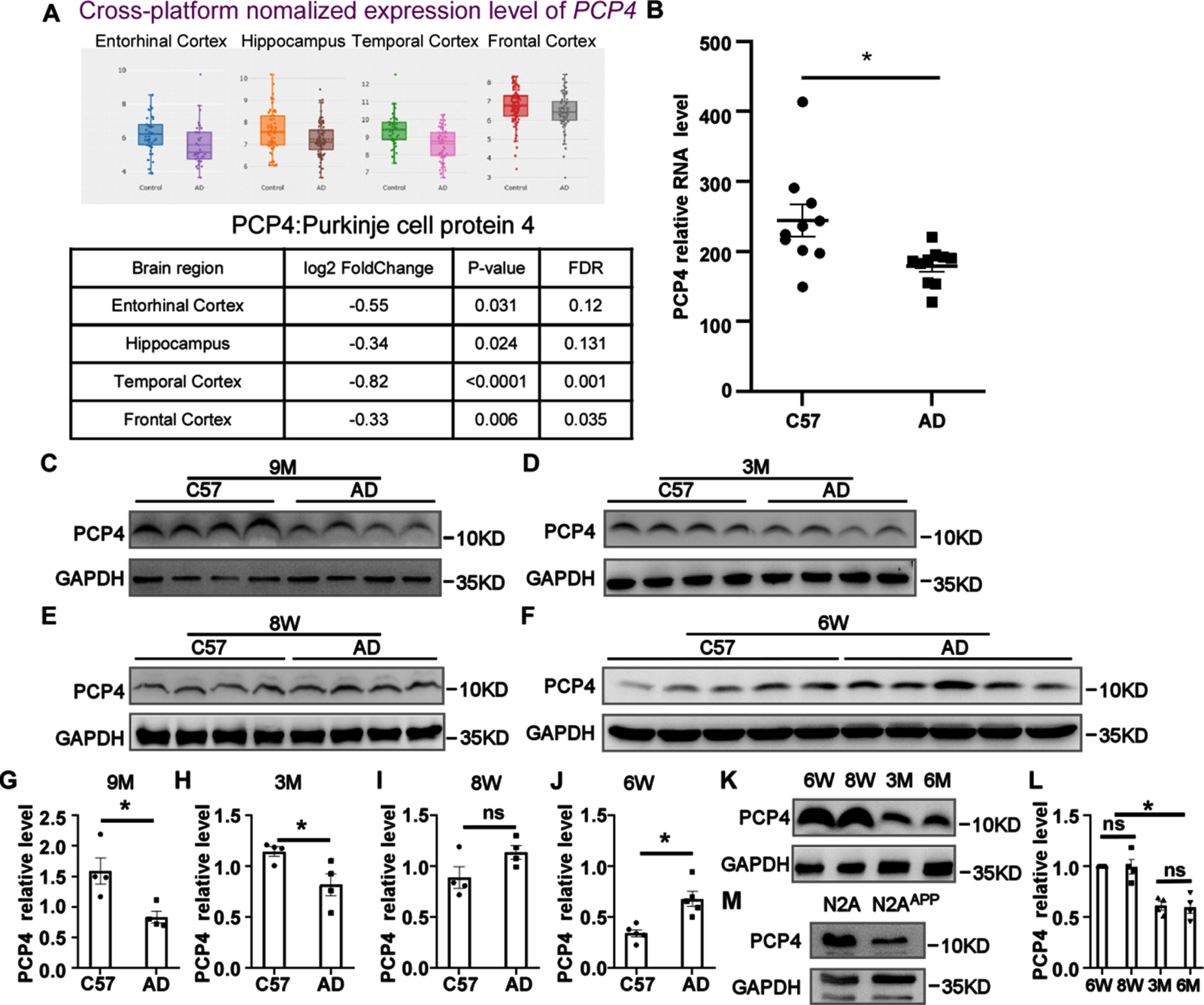
PCP4 levels were abnormal in patients and mice with AD. A) The level of PCP4 in the brains of humans from the database AlzData (https://www.AlzData.org). The data showed the difference in expression for the PCP4 gene in healthy- and AD- derived brains. Each dot represents one sample. B) The RNA relative level of PCP4 in the brains of C57 and APP23/PS45 mice at 3 months of age (n = 10). C) The protein level of PCP4 in the brains of C57 and APP23/PS45 mice at 9 months of age (n = 4). D) The protein level of PCP4 in the brains of C57 and APP23/PS45 mice at 3 months of age (n = 4). E) The protein level of PCP4 in the brains of C57 and APP23/PS45 mice at 8 weeks of age (n = 4). F) The protein level of PCP4 in the brains of C57 and APP23/PS45 mice at 6 weeks of age (n = 5). G-J) Quantitative analysis of C-F. K) The protein level of PCP4 in the brains of AD mice at different ages. L) Quantitative analysis of K (n = 4). M) The protein levels of PCP4 in the lysates of N2A and N2AAPP cells. Data were presented as mean±SEM. *p < 0.05, **p < 0.01, ***p < 0.001.
PCP4 altered AβPP processing
To explore the effect of PCP4 on AβPP processing, an overexpression plasmid of PCP4 was constructed. Because of its small size and difficulty in detection, the fluorescent tag EGFP was added, and the T2A peptide was also added to eliminate the effect of EGFP. After PCP4 was successfully overexpressed, AβPP processing was significantly affected. The expression of AβPP protein increased to 152.85% ±13.6% in SH-Sy5y cells (Fig. 2A), but the change of exogenous AβPP was not obvious in SAS cells (98.78% ±13.6%) (Fig. 2B) and 20E2 cells (96.58% ±4.77%) (Fig. 2C). The expression of exogenous AβPP in SAS and 20E2 cells was much higher than that of endogenous AβPP. In 20E2 cells, C83 expression was significantly decreased to 21.67% ±9.03% (Fig. 2D). At the same time, the levels of the β-secretase cleavage product C99 significantly increased to 127.00% ±6.89% (Fig. 2E) in 2EB2 cells. We found that the protein expression of C83 was significantly reduced to 80.54% ±9.61%, whereas the expression of C99 was significantly increased to 162.23% ±27.30% in SAS cells (Fig. 2F). The expression of α-secretase ADAM10 decreased to 71.26% ±8.51% and 76.70% ±7.54% in SAS (Fig. 2G) and 20E2 cells (Fig. 2H), respectively. The expression of ADAM17 decreased to 81.87% ±6.26% and 72.93% ±7.40% in SAS cells (Fig. 2I) and 20E2 cells (Fig. 2J). There was no significant difference in the expression of exogenous BACE1, endogenous neuronal BACE1, and non-neuronal endogenous BACE1 in 2EB2 cells, SAS cells, and 20E2 cells (Fig. 2K, L, M). In 20E2 cells, we observed a slight tendency of increased BACE1 to 114.60% ±11.57%, p = 0.275. γ-secretase PS1 was not affected by PCP4 overexpression in SAS (Fig. 2N) and 20E2 cells (Fig. 2O). Overexpression of PCP4 in 20E2 cells resulted in a significant increase of Aβ40 concentration to 158% ±19.74% (Fig. 2P) and a significant increase of Aβ42 concentration to 193.33% ±24.00% (Fig. 2Q) compared to the control group. These results suggest that with the overexpression of PCP4, the expression of α-secretase and its cleavage product C83 decreased, and the expression of the β-secretase cleavage product C99 increased. The most significant finding of this study was the increase in generation of Aβ40 and Aβ42 mediated by PCP4. Thus, AβPP processing could be affected by PCP4.
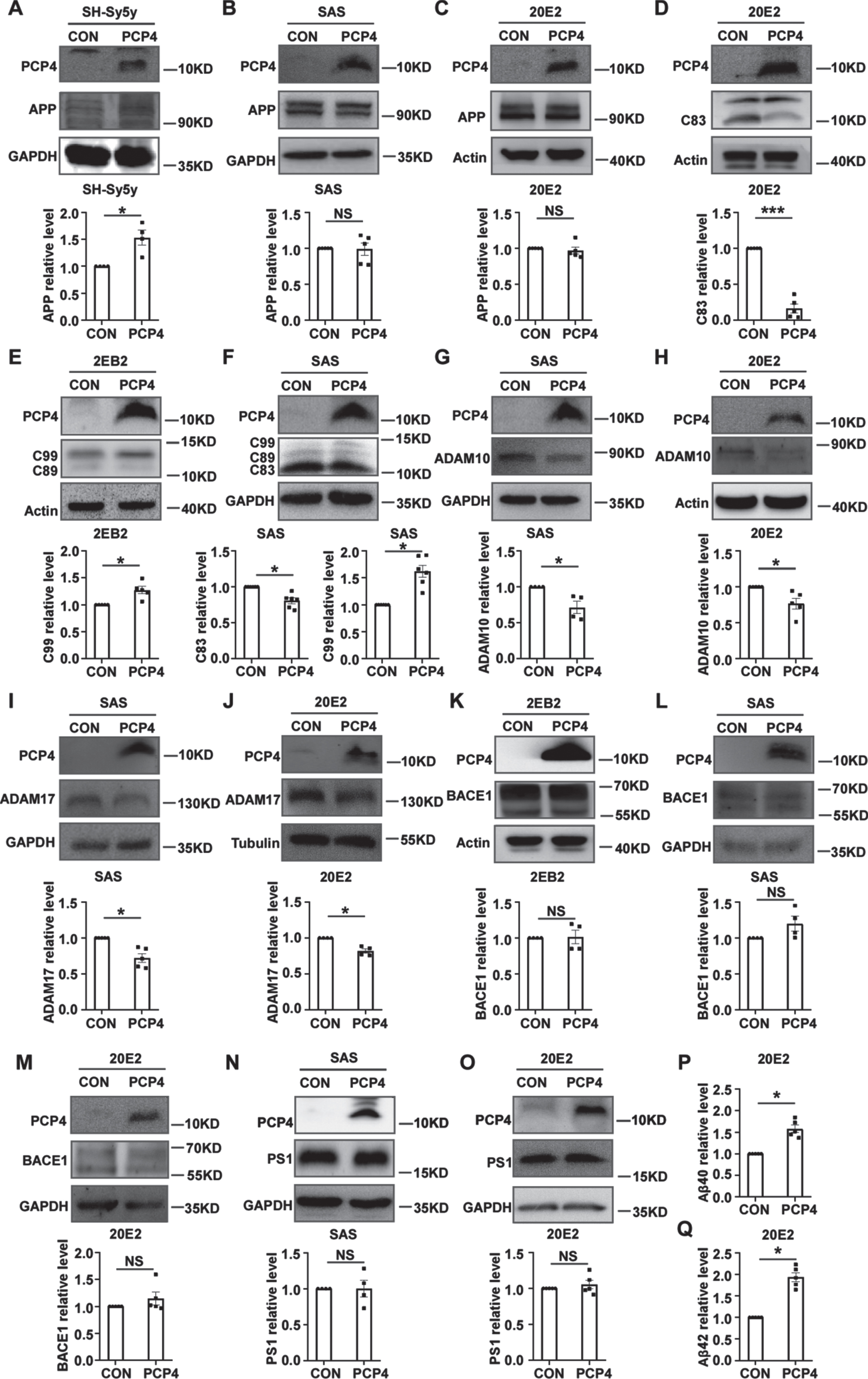
PCP4 altered AβPP processing. PCP4 regulated AβPP processing in 2EB2, 20E2, SH-Sy5y, and SAS cells. 2EB2 cells stably express exogenous human AβPP and BACE1. 20E2 and SAS cells stably express exogenous human AβPP. Control and PCP4 plasmids were transfected into 20E2, 2EB2, SH-Sy5y, and SAS cells to detect AβPP processing-related proteins. Cell lysates were analyzed by western blotting. Each batch of samples was divided into control group (CON) and PCP4 group (PCP4) according to the plasmid transfected. A) AβPP was detected by antibody C20, and GAPDH was detected as the internal control in SH-Sy5y cells (n = 4). B, C) Exogenous human AβPP was detected by antibody C20, and GAPDH or actin was detected as the internal control in 20E2 and SAS cells (n = 5). The results of gray value statistics showed that the endogenous AβPP of the two cells was basically the same between two groups. D) C83 was detected by antibody C20, and actin was detected as the internal control in 20E2 cells (n = 5). The results of gray value statistics showed that C83 was decreased in PCP4 group. E) C99 was detected by antibody C20, and Actin was detected as the internal control in 2EB2 cells (n = 5). The results of gray value statistics showed that C99 in 2EB2 cells was significantly increased with the overexpression of PCP4. F) The level of C83 and C99 in SAS cells. C83 and C99 were detected by antibody C20, and GAPDH was detected as the internal control. The results of gray value statistics showed that C83 in PCP4 group was significantly decreased, and C99 in PCP4 group was significantly increased (n = 6). G, H) ADAM10 was detected by an anti-ADAM10 antibody, actin or GAPDH was selected as the internal control in SAS (n = 4) and 20E2 (n = 5) cells. The results of gray value statistics showed that ADAM10 in SAS and 20E2 cells was significantly decreased with the overexpression of PCP4. I, J) ADAM17 was detected by an anti-ADAM17 antibody, and GAPDH or tubulin was selected as the internal control in SAS (n = 5) and 20E2 (n = 4) cells. The results of gray value statistics showed that ADAM17 in SAS and 20E2 cells were significantly decreased with the overexpression of PCP4. K-M) BACE1 was detected by an anti-BACE1 antibody and actin or GAPDH was detected as the internal control in 2EB2 cells (n = 4), SAS cells (n = 4), or 20E2 cells (n = 5). The results of gray value statistics showed that exogenous BACE1 in 2EB2 cells was not different between the PCP4 overexpression and control groups and endogenous BACE1 in SAS cells and 20E2 cells were also not different between the PCP4 overexpression and control groups. N, O) PS1 was detected by an anti-PS1 antibody, and GAPDH was detected as the internal control in SAS (n = 4) and 20E2 (n = 5) cells. The results of gray value statistics showed that PS1 in SAS and 20E2 cells were also not different between the PCP4 overexpression and control groups. P) After overexpression of the control plasmid or PCP4 plasmid in 20E2 cells, the concentration of Aβ40 (n = 5). Data quantitative analysis showed that the concentration of Aβ40 in the supernatant was increased in PCP4 group. Q) After overexpression of the control plasmid or PCP4 plasmid in 20E2 cells, the concentration of Aβ42 (n = 5). Data quantitative analysis showed that the concentration of Aβ42 in the supernatant was increased in PCP4 group. Data were presented as mean±SEM. *p < 0.05, **p < 0.01, ***p < 0.001.
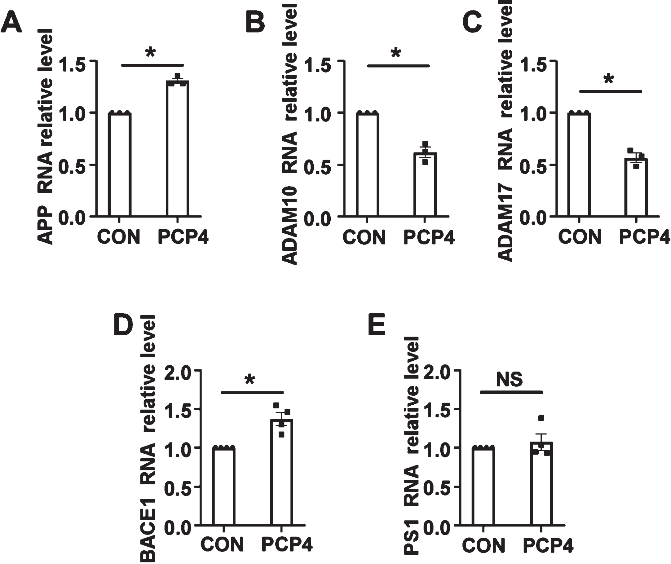
PCP4 affected AβPP processing gene transcription. Control (CON) and PCP4 plasmids were transfected into SH-Sy5y cells to detect AβPP processing-related gene transcription. Each batch of samples was divided into control group (CON) and PCP4 group (PCP4) according to the plasmid transfected. SH-SY5Y cells were transfected with PCP4 plasmids for 24 h, and AβPP, ADAM10, ADAM17, BACE1, and PS1 mRNA levels were measured by quantitative reverse transcription-PCR with specific primers. PCP4 significantly increased the mRNA levels of (A) AβPP (n = 3) and (D) BACE1 (n = 4), and significantly decreased the mRNA levels of (B) ADAM10 (n = 3) and (C) ADAM17 (n = 3). E) The mRNA levels of PS1 in SH-Sy5y cells were not different between the PCP4 overexpression and control groups (n = 4). Data were presented as mean±SEM. *p < 0.05.
PCP4 altered AβPP processing at the transcriptional level
After the overexpression of PCP4, the mRNA level of AβPP increased to 130.78% ±2.44% in SH-Sy5y cells (Fig. 3A). The mRNA levels of α-secretase ADAM10 and ADAM17 decreased to 61.71% ±5.13% and 56.62% ±4.50% in SH-Sy5y cells, respectively (Fig. 3B, C). The mRNA level of β-secretase BACE1 increased to 137.15% ±8.37% (Fig. 3D). γ-secretase PS1 was not affected at the transcriptional level by PCP4 overexpression in SH-Sy5y cells (Fig. 3E).
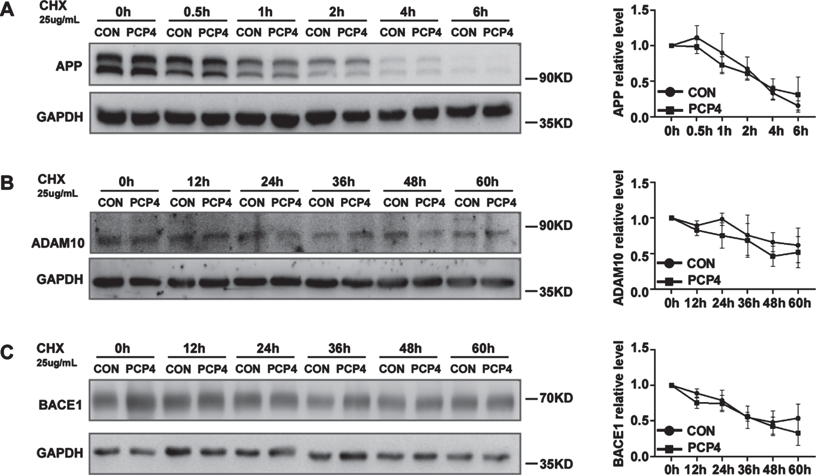
PCP4 did not affect AβPP, ADAM10 and BACE1 degradation. To determine AβPP degradations in the presence of PCP4, 20E2 cells were transfected with PCP4 plasmids for 24 h and harvested at 0 min, 30 min, 1 h, 2 h, 4 h, or 6 h after adding 25μg/mL CHX. To determine ADAM10 and BACE1 degradation in the presence of PCP4, 20E2 and 2EB2 cells were transfected with PCP4 plasmids for 24 h and harvested at 0, 12, 24, 36, 48, or 60 h after adding 25μg/mL CHX. Cell lysates were analyzed by western blotting. A) AβPP was detected using antibody C20. GAPDH was detected as an internal control. The results of gray value statistics showed the percentage of remaining AβPP in 20E2 cells was not different between the PCP4 overexpression and control groups at any time point (n = 3). B) ADAM10 was detected using an anti-ADAM10 antibody. GAPDH was detected to as an internal control. The results of gray value statistics showed that the percentage of remaining ADAM10 in 20E2 cells was not different between the PCP4 overexpression and control groups at any time point (n = 3). C) BACE1 was detected using an anti-BACE1 antibody. GAPDH was detected as an internal control. The results of gray value statistics showed that the percentage of remaining BACE1 in 2EB2 cells was not difference between the PCP4 overexpression and control groups at any time point (n = 3). Data were presented as mean±SEM. *p < 0.05.
The level of protein in cells is determined by the balance between its synthesis and degradation [29]. To further investigate whether the changes in AβPP, ADAM10, and BACE1 expression induced by PCP4 were due to impaired or enhanced degradation, cycloheximide (CHX), a compound that can inhibit the protein synthesis, was used to detect the degradation of target proteins (Fig. 4A-C). We found that PCP4 had little effect on the catabolism of AβPP, ADAM10, and BACE1 (p > 0.05). Hence, PCP4 does not contribute to AβPP, ADAM10, and BACE1 changes by affecting their degradation.
PCP4 affected AβPP processing and increased neuritic plaque formation in APP23/PS45 mice
APP23/PS45 mice were administered intraventricular injections of AAV-PCP4 for in vivo experiments. Mice were injected with the virus when they were 7 weeks old. Behavioral tests were performed at 4 months of age. At approximately 4.5 months of age, the mice were sacrificed, and brain tissues were removed for the validation of relevant parameters. In the mouse brain tissue, the indicators related to AβPP processing were consistent with those of the in vitro experiment. The change in exogenous AβPP was not obvious in mice (p > 0.05) (Fig. 5A). At the same time, the level of the β-secretase cleavage product, C99, was increased from 0.80±0.09 in the control group to 1.19±0.05 in the PCP4 overexpression group (Fig. 5A). In addition, the study found that the expression level of C89 increased from 0.89±0.35 in the control group to 1.14±0.46 in the PCP4 overexpression group. However, statistical analysis did not reveal any significant difference between the two groups (Fig. 5A). The effect in BACE1 expression was also not significant (Fig. 5A). Additionally, the expression of α-secretase ADAM10 was decreased from 1.18±0.14 in control group to 0.84±0.07 in the PCP4 overexpression group (Fig. 5A). It was observed that PCP4 can significantly promote the accumulation of neuritic plaques in APP23/PS45 mice by 4G8 immunostaining (Fig. 5B), and plaque numbers were increased by approximately 24.13% (121.78±7.78 to 151.17±6.16) (Fig. 5B). These results demonstrated that AβPP processing could be affected by PCP4 at the protein level, and that PCP4 could facilitate amyloid plaque accumulation in APP23/PS45 mice.
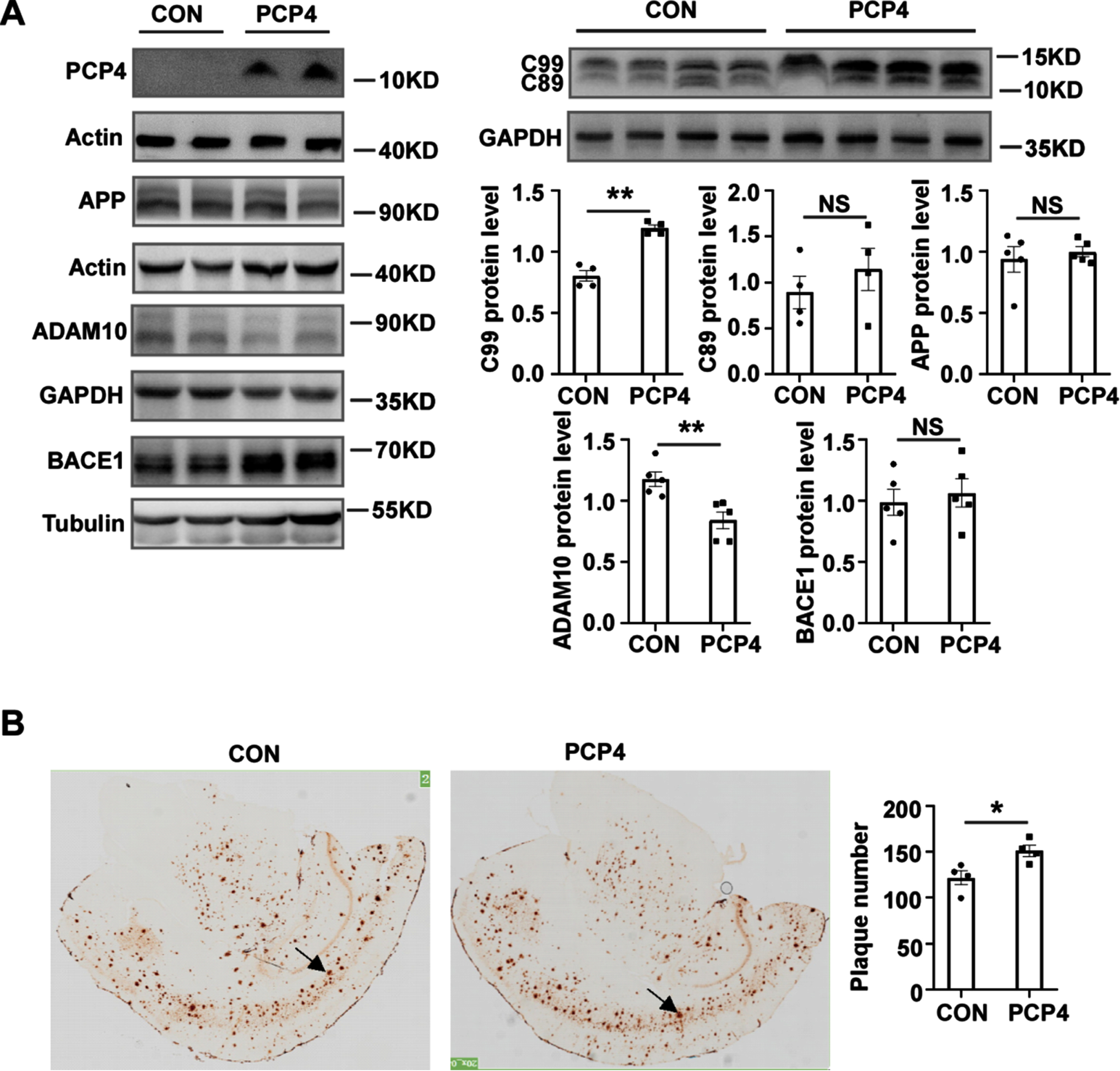
PCP4 affected AβPP processing and increased neuritic plaque formation in APP23/PS45 mice. PCP4 altered AβPP processing and increased Aβ production in viv o. Mice were injected with AAV-Control (CON) and AAV-PCP4 in the brain at 7 weeks of age. At approximately 4.5 months of age, the mice were sacrificed, and brain tissues were removed for western blotting and immunohistochemical staining. A) AβPP and CTFs were detected by antibody C20. BACE1 was detected by anti-BACE1 antibody D10E5. ADAM10 was detected by an anti-ADAM10 antibody. PS1 was detected by an anti-PS1 antibody. And GAPDH was detected as the internal control. The results of gray value statistics showed that C99 levels were significantly increased in PCP4 overexpression mice, and C89 levels were not different between the PCP4 overexpression and control groups (n = 4). ADAM10 expression significantly decreased with the overexpression of PCP4 (n = 5). AβPP and BACE1 levels were not different between the PCP4 overexpression and control groups (n = 5 for each group). B) Mice were evaluated using the Aβ specific monoclonal antibody, 4G8. Plaque was visualized by microscopy at 20× magnification and the arrows indicate plaque. Quantification of neuritic plaque in APP23/PS45 mice showed that the number of plaque significantly increased in PCP4 overexpression mice. Data were presented as mean±SEM. *p < 0.05, **p < 0.01.
PCP4 aggravated learning and memory impairment in APP23/PS45 mice
In order to explore whether the memory and spatial learning abilities of APP23/PS45 mice were related to PCP4, MWM-test was conducted on them, and the age was 4 months. Through experimental analysis, the control and PCP4 overexpression mice exhibited similar escape latency (34.07±2.58 and 40.07±3.78 s, respectively) (p > 0.05) (Fig. 6A) and swimming distance to the platform (6.06±0.61 and 6.19±0.58 m, respectively) (p > 0.05) (Fig. 6B) in the visible platform test. These tests found that the expression of PCP4 made the vision and motor ability of mice changed little. However, on the third and fourth day of the hidden platform tests, the escape latency was longer for the PCP4 overexpression mice (41.57±3.91 and 39.13±4.71 s on the third and fourth day, respectively) than that for the control group (29.96±3.29 and 25.83±2.03 s on the third and fourth day, respectively) (p < 0.05) (Fig. 6C). During the MWM-test on the last day of PCP4 overexpression mice, they showed fewer times crossing from the site of the hidden platform (control mice versus PCP4 overexpression mice, 2.33±0.26 versus 1.00±0.38 times) (p < 0.05) (Fig. 6D). We also investigated the effects of PCP4 on learning and memory in mice using the Y-maze test. We found that PCP4 significantly exacerbated the learning and memory deficits in the Y-maze task. The control mice showed a significantly higher alternation rate (control mice versus PCP4 overexpression mice, 0.63±0.21 versus 0.55±0.24) (p < 0.05) (Fig. 6E). In general, PCP4 exacerbates learning and memory deficits in APP23/PS45 mice.
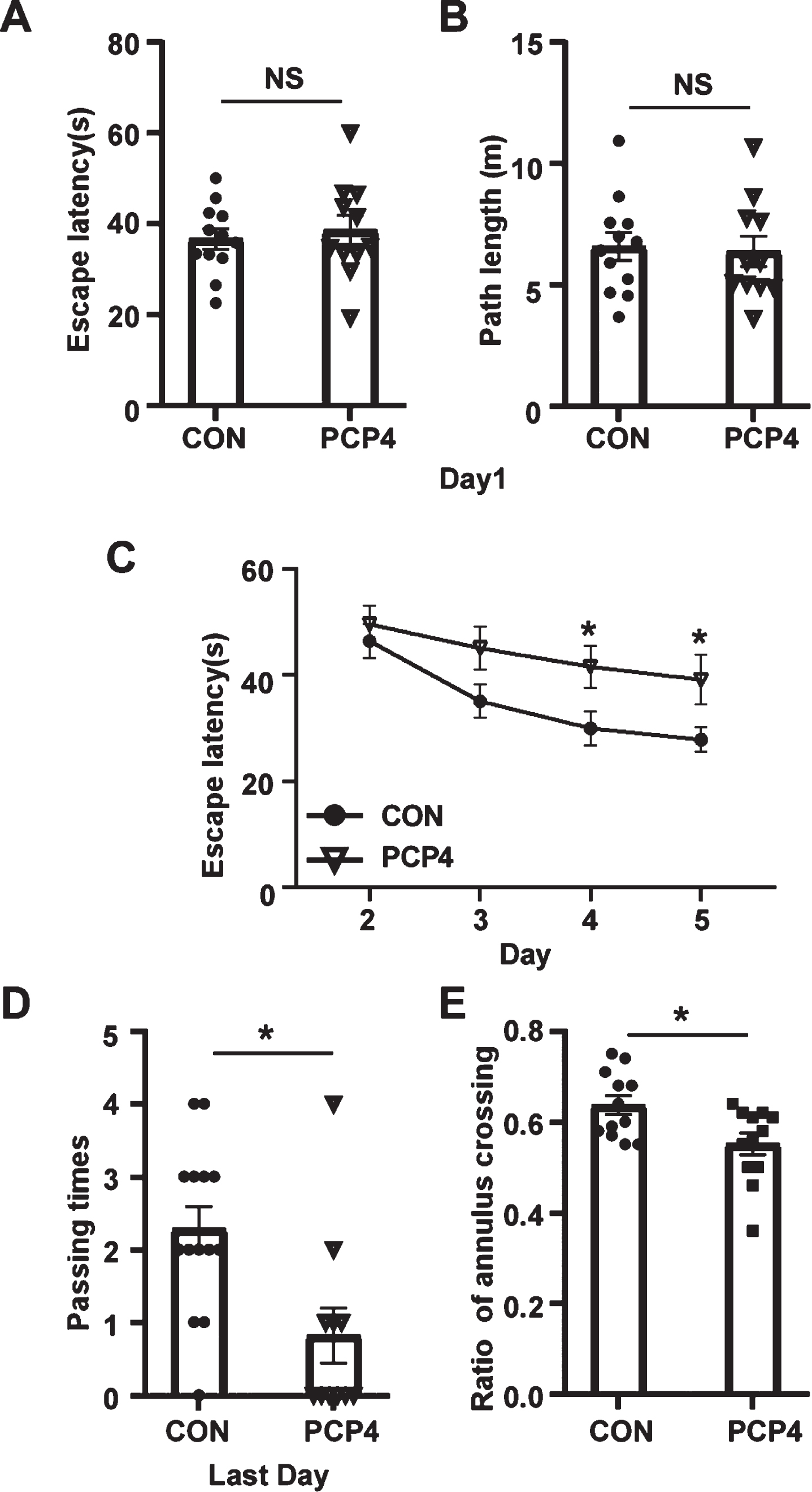
PCP4 aggravated learning and memory impairment in APP23/PS45 mice. APP23/PS45 mice at 4 months of age conducted MWM-test (control (CON), n = 12 and overexpression of PCP4, n = 11) and the Y-maze test (control, n = 12 and overexpression of PCP4, n = 12). ANY-maze tracking software was used to record mouse movement. The MWM-test consisted of 1 day of adaptive trials and 4 days of hidden platform trials, and 24 hours of probe test after the last hidden platform test. The study found that on the 1st day of the visible platform test, the overexpression of PCP4 and control mice showed basically the same (A) escape latency and (B) swimming distances to escape onto the visible platform. C) The hidden platform test lasted for 4 days and mice were trailed for five times per day. PCP4 overexpression mice need a longer time to escape to the hidden platform compared with control mice on the 4th and 5th days. D) In the probe trial, the frequencies of leaping over the platform among PCP4 overexpression mice were significantly lower than those among the control group. E) In the Y-maze test, PCP4 overexpression mice were found to have more exploration times that were repeated in one or two maze spaces than control mice. Data were presented as mean±SEM. *p < 0.05.
DISCUSSION
PCP4 is located on a critical region of HSA 21 and is present as three copies in individuals with DS [36–38]. The patients with DS develop typical AD neuropathology. In our study, we found a decrease in PCP4 expression in AD in late stages. Interestingly, we found that PCP4 expression increased in AD before Aβ generation in the early 6 weeks. In a previous study, using a transgenic mouse model to evaluate the consequences associated with the three copies of this gene, it was found that the overexpression of PCP4 induces precocious neuronal differentiation during mouse embryogenesis. In this mouse model, motor and learning impairments appeared gradually and became severe in adulthood [23, 39]. This might suggest that high expression of PCP4 may induce the development of AD pathology. To better understand the role of PCP4 in AD, especially in AβPP processing, we further explored the underlying mechanism through in vitro and in vivo experiments. In our study, the results indicated that overexpressing PCP4 in a mouse model did not improve AD, and the AβPP processing toward Aβ production has not been alleviated in related cell lines. In contrast, PCP4 aggravates AD progression and exacerbates memory impairment in mice with AD. PCP4 affects AβPP processing, especially ADAM10 and ADAM17, which promote AβPP cleavage toward the direction of no Aβ production.
At present, the role of PCP4 in AD has not been studied in depth except for the changes in its expression in AD. For further study, we overexpressed PCP4 in APP23/PS45 double-transgenic mice and stable cells expressing human Swedish AβPP [40]. We used 20E2 and 2EB2 cells to investigate the effect of PCP4 on AβPP cleavage products. Meanwhile, except for 20E2 and 2EB2, we also selected the neural cells SH-Sy5y and SAS to observe the expression of cleaving enzymes in AβPP processing. ADAM10 and ADAM17 were significantly decreased by PCP4, whereas PCP4 had no effect on PS1. We further confirmed that PCP4 regulated AβPP processing in vitro. Interestingly, the expression of exogenous AβPP did not change significantly in 20E2 and SAS and including APP23/PS45 mice. However, there was an increase in endogenous AβPP levels in neural cell lines. Moreover, the effect of PCP4 on BACE1 expression was greater at the transcriptional level. Lastly, PCP4 did not affect the degradation of related proteins; therefore, we speculate that PCP4 is important for its effect on AβPP processing at the transcriptional level. According to our experimental results, PCP4 plays a significant role in the alteration of transcription level of AβPP processing. Taking the protein level results as a guide, it is likely that PCP4 mainly affects the expression of α-secretase, leading to increased AβPP and feedback towards the β-cleavage pathway. PCP4 itself can exist as a transcription factor [41]. In this process, whether it plays a role as a transcription factor will be further explored.
PCP4 expression is decreased in AD patients [42]. However, in our study, the overexpression of PCP4 did not improve the symptoms of AD but resulted in more severe consequences. First, this may be due to a different stage of AD development than the exploration. In early stage of AD, we found that PCP4 promoted memory decline and cognitive dysfunction. Combined with the changes in PCP4 expression in the brain tissues of different aged mice, we speculated that the decrease in PCP4 expression in AD is limited by the course of the disease. PCP4 levels gradually decreased when AD symptoms were more obvious. However, in the primary stage, PCP4 was highly expressed. This promotes the development of AD and indicates a more severe direction, and PCP4 is consumed or inhibited by feedback in this process. But the process of nerve damage was irreversible, the aggravated pathological phenomenon could not be reversed. Our study provides insights into the diagnosis and treatment of AD; however further research is required to clarify the specific mechanism by which PCP4 regulates AβPP processing to aggravate the development of AD.
In conclusion, we found that PCP4 altered AβPP processing and aggravated AD related phenotypes. These data suggest that PCP4 increases the risk of AD development, and can help to determine detection indicators and therapeutic targets for AD.
Footnotes
ACKNOWLEDGMENTS
The authors have no acknowledgments to report.
FUNDING
The authors have no funding to report.
CONFLICT OF INTEREST
Weihong Song is an Editorial Board Member of this journal but was not involved in the peer-review process nor had access to any information regarding its peer-review.
DATA AVAILABILITY
All data generated or analyzed during this study are included in this published article. All data available upon request.