






Abstract
Background:
Some human studies have identified infection with cytomegalovirus (CMV), a member of the alpha herpesvirus family, as a risk factor for Alzheimer’s disease and related dementias (ADRD). To our knowledge, no studies have evaluated associations of CMV seropositivity with plasma biomarkers of ADRD risk in middle-aged adults.
Objective:
In participants recruited for an exercise study, we evaluated cross-sectional associations of CMV seropositivity with: Aβ42/Aβ40 ratio, a low ratio suggestive of central nervous system Aβ accumulation; glial fibrillary acidic protein (GFAP), a measure of neuroinflammation; and neurofilament light (NfL), a measure of neurodegeneration.
Methods:
Anti-CMV IgG was quantified by ELISA. Plasma ADRD biomarkers were quantified using the ultrasensitive SIMOA assay. We used linear regression to evaluate associations of CMV seropositivity with the ADRD biomarkers, adjusting for age, sex, and race (n = 303; Age = 55.7±9.2 years). For ADRD biomarkers significantly associated with CMV seropositivity, we evaluated continuous associations of anti-CMV IgG levels with the ADRD biomarkers, excluding CMV seronegative participants.
Results:
53% of participants were CMV seropositive. CMV seropositivity was associated with a lesser Aβ42/Aβ40 ratio (β=–3.02e–03 95% CI [–5.97e–03, –7.18e–05]; p = 0.045). In CMV seropositive participants, greater anti-CMV IgG levels were associated with a lesser Aβ42/Aβ40 ratio (β=–4.85e–05 95% CI[–8.45e–05, –1.25e–05]; p = 0.009). CMV seropositivity was not associated with plasma GFAP or NfL in adjusted analyses.
Conclusions:
CMV seropositivity was associated with a lesser plasma Aβ42/Aβ40 ratio. This association may be direct and causally related to CMV neuro-cytotoxicity or may be indirect and mediated by inflammatory factors resulting from CMV infection burden and/or the immune response.
INTRODUCTION
A growing body of literature suggests that common viral infections may increase the risk of late-life dementias [1]. In particular, some studies suggest that infection with herpes family viruses may be a risk factor for Alzheimer’s disease and related dementias (ADRD) [2, 3]. The herpes virus family comprises eight viruses that are endemic to humans, each of which can establish lifelong reservoirs of latently infected cells. Additionally, emerging evidence also suggests that amyloid-β (Aβ), whose aggregation initiates the pathologic cascade leading to Alzheimer’s disease (AD), may function as an antimicrobial peptide in the innate immune response against viral infection, including herpes viruses [4, 5]. Chronic infection with herpesviruses may therefore contribute to ADRD risk through its effects on Aβ metabolism.
The strongest evidence for an association of herpesvirus infections with incident dementia and its prodrome, mild cognitive impairment, is with the alpha herpesviruses: herpes simplex virus (HSV)-1, HSV-2, and varicella zoster virus (VZV) [2, 6]. There is less evidence for cytomegalovirus (CMV), a member of the beta herpesvirus family [7, 8]. CMV is endemic, with adult seroprevalence estimated at 83% globally and at 48% in the United States [9, 10]. CMV primarily infects myeloid cells, including monocytes; however, all resident cells of the central nervous system (CNS), including microglia and astrocytes, are susceptible to CMV infection [11, 12].
A 2019 systematic review and meta-analysis failed to identify an association of prior CMV infection with incident dementia or mild cognitive impairment [2]. Relatively few studies have evaluated associations of measures of CMV seropositivity with biomarkers of ADRD pathology [7, 13]. One study evaluated associations of plasma Aβ with anti-CMV Immunoglobulin G (IgG) in individuals with AD and healthy controls. This study found no significant association between measures of CMV seropositivity and Aβ pathology, however the study’s methodology for plasma Aβ quantification may not have been sufficiently sensitive for comprehensive detection of Aβ levels [8].
To our knowledge, no studies have evaluated the association of anti-CMV IgG with plasma biomarkers of ADRD risk in middle-aged adults using ultrasensitive assay methodologies. The objective of this analysis was to determine, in a sample of young and middle-aged adults recruited for an exercise study, the cross-sectional associations among measures of CMV seropositivity with plasma biomarkers of ADRD risk, including Aβ (the ratio of Aβ42/Aβ40 with a lesser ratio reflecting greater risk of CNS Aβ accumulation), neurofilament light (NfL, a marker of axonal neurodegeneration) and glial fibrillary acidic protein (GFAP, a marker of neuroinflammation).
METHODS
Study design
STRRIDE (Studies Targeting Risk Reduction Interventions through Defined Exercise, conducted 1998–2014) was a series of three randomized trials of supervised exercise training. The objective of STRRIDE was to determine the impact of varying amounts and intensities of aerobic training, resistance training, and diet, alone or in combination, on measures of metabolic and cardiovascular health in sedentary adults with cardiometabolic risk factors [14–16]. Exclusion criteria included current smoking, medications altering carbohydrate or lipid metabolism, the presence of diabetes or uncontrolled hypertension, inability to exercise, or concurrent heart disease. Sex balance was achieved in all three studies. STRRIDE was approved by the Duke and East Carolina University Institutional Review Boards; subjects provided written informed consent. Data and samples used in this analysis were from the STRRIDE pre-exercise training baseline assessment.
Biomarker quantification
Plasma Aβ40, Aβ42, GFAP, and NfL were quantified in singlicate from non-fasted EDTA plasma samples using the ultrasensitive Simoa Neuro 4-Plex E kit on the Simoa HD-X instrument (Quanterix, Billerica, MA, USA). A high and low concentration quality control sample and a pooled human plasma control sample were included in duplicate on each plate to assess intra- and inter-assay coefficients of variation (CV). Intra- and inter-assay CVs for NfL were 4.7% and 11.9%; for GFAP were 5.0% and 10.2%; for Aβ40 were 5.8% and 10.35%; and for Aβ42 were 4.5% and 11.8%, respectively.
Semi-quantitative assessment of anti-CMV IgG levels in serum was performed in singlicate by indirect ELISA using an assay certified for clinical use (Diamedix, Cat#720-320). Samples with values exceeding the upper limit of quantification (>160 EU/mL) were re-run at a 10-fold dilution to obtain a value within the linear range of the assay and corrected by the dilution factor. C-reactive protein (CRP) was quantified in singlicate by sandwich immunoassay (Cat#K151STD; Meso Scale Discovery, Rockville, MD). Intra- and inter-assay CVs for CRP were 3.0% and 5.1%, respectively. Normality of plasma biomarker distributions was evaluated by visual inspection; non-normally distributed biomarkers were log-transformed.
Statistical analysis
All analyses were conducted in R Version 4.1. We used the ratio of Aβ42/Aβ40 for all analyses. Given the small number of STRRIDE participants who self-identified as a race other than African American or White (n = 9), we recoded race as African American and Non-African American. Of the nine who did not self-identify as African American or White, 3 identified as Asian, 4 identified as Hispanic, and 2 identified as Native American. Anti-CMV IgG values more than 5 standard deviations (SD) from the mean, which could reflect either acute infection or acute reactivation, were designated as outliers and removed. Plasma GFAP, NfL, and CRP were not normally distributed and were log-transformed; normality was confirmed by visual inspection. To evaluate associations among demographic factors, anti-CMV IgG levels, plasma ADRD biomarkers, and CRP, we calculated Pearson’s r correlation coefficient for continuous variables and student’s t-test for categorical variables.
To determine the association of measures of CMV seropositivity and plasma ADRD biomarkers, we used simple linear regression, adjusting for age, sex, and race. First, to determine whether CMV seropositivity was associated with the plasma ADRD biomarkers, we fitted models coding anti-CMV IgG as a binary variable, using the manufacturer’s cutoff of ≥8 EU/mL to denote CMV seropositivity. Second, for plasma ADRD biomarkers that were significantly associated with CMV seropositivity, we evaluated models with anti-CMV IgG as a continuous variable, excluding participants who were CMV seronegative. We conducted additional sensitivity analyses adjusting for CRP to further account for confounding due to inflammatory status. For ADRD biomarkers that were significantly associated with anti-CMV IgG levels in CMV seropositive participants, we fitted models with an interaction term to evaluate, separately, whether associations were moderated by age, sex, or race.
RESULTS
Sample characteristics
Descriptive statistics for STRRIDE participants included in this analysis are shown in Table 1. Based on the manufacturer’s cutoff of ≥8 EU/mL, 52% of STRRIDE participants were CMV seropositive. Three STRRIDE participants had anti-CMV IgG levels more than 5 SD above the mean, consistent with acute infection or reactivation, and were excluded from the analysis. Anti-CMV IgG was weakly positively correlated with age (r = 0.16; p = 0.004), but not log-CRP (r=–0.02 p = 0.787). Anti-CMV IgG was greater in women compared to men (92.94 EU/mL versus 65.97 EU/mL; p = 0.007) and African Americans compared to non-African Americans (116.23 EU/mL vs. 75.76 EU/mL p = 0.010).
STRRIDE Sample Descriptive Statistics. STRRIDE participants with an anti-CMV IgG≥0.08 EU/mL were designated as CMV seropositive. Untransformed values are shown for NfL, GFAP, and CRP. To evaluate differences between CMV seronegative and seropositive participants, student’s t test was used for continuous variables and the Chi squared test was used for categorical variables. Mean (SD) provided for continuous variables. Count (%) is shown for categorical values
NfL, neurofilament light; GFAP, glial fibrillary acidic protein.
Greater age was correlated with a lesser Aβ42/Aβ40 ratio (r=–0.19; p < 0.001) and greater concentrations of log-GFAP (r = 0.52; p < 0.001) and log-NfL (r = 0.65; p < 0.001). Plasma GFAP was greater in women compared to men (83.07 pg/mL versus 66.06 pg/mL [original units]; p < 0.001), but there were no significant sex differences for the Aβ42/Aβ40 ratio or NfL (all p≥0.151). There were no significant differences in any of the plasma ADRD biomarkers comparing African Americans to non-African Americans (all p≥0.065). Log-CRP concentrations were not correlated with the plasma ADRD biomarkers (all p≥0.058).
Association of anti-CMV IgG with plasma ADRD biomarkers
We first evaluated whether CMV seropositivity was associated with the plasma ADRD biomarkers. CMV seropositivity was associated with a lesser plasma Aβ42/Aβ40 ratio, adjusting for age, sex, and race (Table 2). There were no significant associations of CMV seropositivity with plasma log-NfL or log-GFAP (all p≥0.622). We next evaluated associations of anti-CMV IgG levels with the plasma Aβ42/Aβ40 ratio, excluding participants who were CMV seronegative. In CMV seropositive participants, greater anti-CMV IgG levels were associated with a lesser Aβ42/Aβ40 ratio, adjusting for age, sex, and race (Table 3). Adjusting for log-CRP did not affect our findings (data not shown). To determine whether associations of anti-CMV IgG levels with the Aβ42/Aβ40 ratio differed by age, sex, or race in CMV seropositive participants, we refitted models with an interaction term. There was no evidence that associations were significantly moderated by age (p = 0.086), sex (p = 0.501), or race (p = 0.931). Scatter plots showing associations of anti-CMV IgG levels with the plasma ADRD biomarkers in CMV seropositive participants are shown in Fig. 1.
Association of CMV Seropositivity with the Plasma ADRD Biomarkers. Univariate linear
CI, confidence interval; NfL, neurofilament light; GFAP, glial fibrillary acidic protein.
Association of Anti-CMV IgG Levels with the Plasma
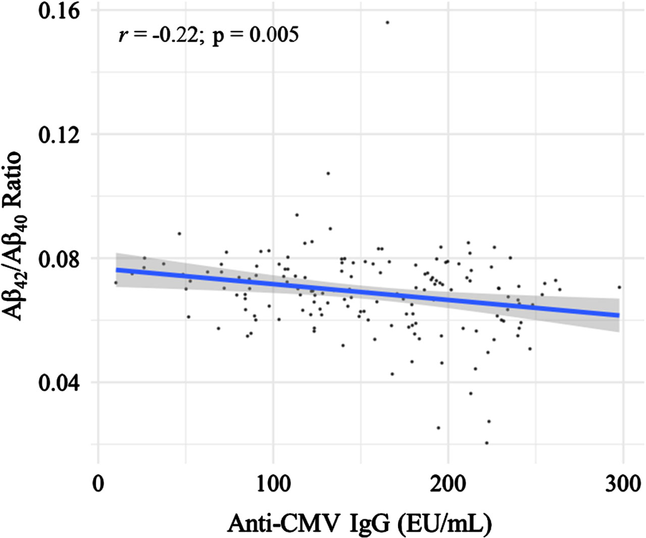
Association of anti-CMV IgG levels with the plasma ADRD biomarkers in CMV seropositive participants. Scatterplot showing the association of anti-CMV IgG levels (EU/mL) with the plasma Aβ42/Aβ40 ratio in CMV seropositive participants. Pearson correlation coefficient and corresponding p-value are shown.
DISCUSSION
To our knowledge, this is the first study to evaluate associations between anti-CMV IgG levels and ultrasensitive plasma measures of ADRD pathology. Here, in a sample of mostly middle-aged adults recruited for an exercise intervention, anti-CMV IgG levels were not associated with plasma GFAP or NfL. However, in CMV seropositive participants, anti-CMV IgG levels were associated with a lesser plasma Aβ42/Aβ40 ratio. This association may be direct and causally related to CMV neuro-cytotoxicity, indirect and mediated by inflammatory factors resulting from CMV infection burden and/or the immune response, or a combination of direct and indirect factors.
The inverse association between anti-CMV IgG levels and plasma Aβ42/Aβ40 may result from an endogenous protective response to CMV infection. This association is consistent with work in animal and in vitro AD models indicating human Aβ-amyloidosis serves as a protective response against herpesvirus infection [4]. Aβ is upregulated by pattern- and damage-associated molecular patterns (PAMPs/DAMPs), likely functioning as a cytokine in the innate immune response [17]. CMV may induce CNS Aβ production, CNS Aβ accumulation, and plasma Aβ42/Aβ40 reduction. Also, the immune response to CMV viral activity could induce peripheral Aβ production [17] from the adrenal gland, kidney, heart, liver, and various peripheral cell types including thrombocytes and endothelial cells [18]. Increases in peripheral Aβ could lead to increased CNS Aβ and CNS Aβ aggregation while reducing Aβ42/Aβ40 [18]. To our knowledge, as compared to HSV-1, HHV-6, and HHV-7, there is no evidence of CNS CMV infection in post-mortem human AD brain tissue [19–21]. Thus, it is possible that in the CNS, periphery, or both, CMV induces an immune response that produces Aβ and resolves CNS CMV infection; however, residual accumulation leads to ADRD development without post-mortem evidence of prior CMV infection.
Our findings are consistent with human population studies showing ADRD associations with clinical diagnoses of herpesvirus infections generally, and CMV specifically. Based on data from the FinnGen and UK biobanks [1], risk of dementia is increased after diagnosis of herpes and other viral infection, with greatest risk in the initial year following infection. However, in this study, over time, the increased risk of dementia was attenuated, suggesting these findings were impacted by an observation bias whereby recently infected individuals have greater healthcare exposure and a greater likelihood of dementia diagnosis. Also, a greater 10-year risk of developing AD is associated with the combination of CMV and HSV-1 infection [22], implying a potential synergistic effect of multiple herpesvirus infections. Consistent with CMV-specific contributions to ADRD pathology, an increased risk of dementia is associated with a prior history of CMV-associated end-organ disease [23].
Our findings are also consistent with connections between herpesvirus infections and ADRD pathology measures including brain imaging, cognitive performance, and ADRD biomarkers. In data from the Baltimore Longitudinal Study of Aging, a clinical diagnosis of varicella-zoster, HSV-1, or HSV-2 is associated with longitudinal CNS white matter volume decline, accelerated declines in attention, and elevated plasma GFAP; furthermore, treatment with antivirals attenuate declines in occipital white matter [24]. However, herpesvirus-related diagnoses were not associated with plasma Aβ42/Aβ40 ratio or NfL.
In contrast to clinical diagnoses of herpes or CMV infection, seropositivity and antibody concentrations are less consistently related to dementia [2, 25], cognitive performance [26–28], or plasma Aβ [8], consistent with our work. Further, CMV antibody concentrations may reflect successful endogenous Aβ responses to CMV infection, reducing the likelihood of clinical CMV infection, dementia, or cognitive impairment diagnoses.
CMV infection and the response to infection is very likely to be one of many factors including genetics and cardiometabolic risk factors that contributes to Aβ production, CNS Aβ aggregation, and eventual development of CNS Aβ amyloidosis, tau pathology, and dementia. These multiple etiologies for ADRD explain, in part, that in the general population, the plasma Aβ42/Aβ40 ratio is poorly predictive of future dementia risk [29], and significant CNS Aβ accumulation without tau pathology or cognitive impairment occurs in up to 25% of cognitively unimpaired older adults [30].
Future studies are required to determine whether CMV seropositivity is associated with gold standard assessments of CNS Aβ-amyloidosis using CSF or Aβ PET imaging. Additionally, longitudinal studies are required to determine whether anti-CMV IgG levels predict future CNS Aβ-amyloidosis. If CMV infection is associated with future CNS Aβ-amyloidosis, the next step would be to determine whether anti-CMV antivirals can delay or prevent CNS Aβ-amyloidosis in high-risk individuals. There is currently a clinical trial underway evaluating high dose valacyclovir—an anti-viral medication with efficacy against HSV-1, HSV-2, VZV, and CMV—in mild AD dementia [31].
Strengths and limitations
Our work has several strengths, including the relatively large sample size and the use of the ultrasensitive Quanterix SIMOA platform to quantify biomarkers of ADRD risk. In addition, to our knowledge, this is the first study to evaluate associations of anti-CMV IgG with plasma ADRD biomarkers in a sample of mostly middle-aged adults. Limitations of our work include the cross-sectional design, singlicate quantification of ADRD biomarkers, lack of assessment of other herpesviruses, and absence of gold standard assessment of AD pathology using positron emission tomography imaging or cerebrospinal fluid. STRRIDE also did not collect data on educational attainment or socioeconomic status, which may be important confounders of CMV infection and adverse neurocognitive outcomes [32].
Conclusions
In conclusion, we report, in a study of middle-aged adults recruited for an exercise study, that in CMV seropositive participants, greater anti-CMV IgG levels are associated with a lesser plasma Aβ42/Aβ40 ratio. Our findings provide support for the hypothesis that Aβ plays a role in the anti-microbial response to CMV infection and that CMV-mediated alterations in Aβ metabolism may contribute to CNS Aβ-amyloidosis. Further work is needed to determine whether anti-CMV IgG levels predict longitudinal CNS Aβ accumulation and whether treatment with anti-CMV antivirals can attenuate CNS Aβ accumulation. Interventions targeting CMV and other chronic infections may favorably affect Aβ homeostasis, thereby reducing CNS Aβ-amyloidosis and future AD dementia risk.
AUTHOR CONTRIBUTIONS
Daniel Parker (Conceptualization; Data curation; Formal analysis; Funding acquisition; Investigation; Methodology; Project administration; Visualization; Writing – original draft; Writing – review & editing); Heather E. Whitson (Writing – review & editing); Patrick J. Smith (Writing – review & editing); Virginia B. Kraus, MD, PhD (Writing – review & editing); Janet L. Huebner (Investigation; Methodology; Writing – review & editing); Rebecca North (Methodology; Writing – review & editing); William E. Kraus (Funding acquisition; Investigation; Resources; Writing – review & editing); Harvey Jay Cohen (Writing – review & editing); Kim M. Huffman (Writing – review & editing).
Footnotes
ACKNOWLEDGMENTS
The authors would like to thank Melissa Hurdle, Grace Link, Stephanie Arvai, Karen Abramson, and the study participants for their contribution to this research.
FUNDING
This study was supported by a Lina Mae Edwards Young Investigator Research Grant from the Dementia Alliance of North Carolina and an NIA GEMSSTAR 5R03-AG067897-02 to D.C.P. STRRIDE I (NCT00200993) and STRRIDE AT/RT (NCT00275145) were funded by NHLBI grant HL-057354 to W.E.K. STRRIDE-PD (NCT00962962) was funded by NIDDK grant DK-081559 to W.E.K. This work was additionally supported by an NIH R01-AG054840 to V.B.K.; an NIH P30-AG028716 to R.N., V.B.K., W.E.K., and H.E.W.; an NIH P30-AG072958 to H.E.W; and NIH R01-HL153497 to W.E.K. and K.M.H.
CONFLICT OF INTEREST
D.C.P. is an Editorial Board Member of this journal but was not involved in the peer-review process of this article nor had access to any information regarding its peer-review.
All other authors have no conflict of interest to report.
DATA AVAILABILITY
Data supporting the conclusions of this article will be made available by the authors. Requests to access this dataset should be directed to D.C.P.