






Abstract
Background:
The link between allergic diseases and dementia remains controversial, and the genetic causality of this link is unclear.
Objective:
This study investigated the causal relationship between allergic diseases and dementia using univariate and multivariate Mendelian randomization (MR) methods.
Methods:
We selected genome-wide association studies including 66,645 patients with allergic diseases and 12,281 patients with dementia, with statistical datasets derived from the FinnGen Consortium of European origin. After a rigorous screening process for single nucleotide polymorphisms to eliminate confounding effects, MR estimation was performed mainly using the inverse variance weighting method and the MR-Egger method. Sensitivity analyses were performed using Cochran’s Q test, MR-PRESSO test, MR Pleiotropy residuals and leave-one-out analysis.
Results:
Univariate and multivariate MR together demonstrated a causal relationship between atopic dermatitis and reduced vascular dementia (VaD) risk (OR = 0.89, 95% CI: 0.81–0.99, p = 0.031; OR = 0.85, 95% CI: 0.76–0.95, p = 0.003). MVMR confirmed asthma was associated with a reduction in the risk of Alzheimer’s disease (AD) (OR = 0.82, 95% CI: 0.71–0.94, p = 0.005) and may be associated with a reduction in the risk of VaD (OR = 0.80, 95% CI: 0.65–0.99, p = 0.042); allergic rhinitis may be causally associated with an increased risk of AD (OR = 1.16, 95% CI: 1.00–1.35, p = 0.046) and VaD (OR = 1.29, 95% CI: 1.03–1.62, p = 0.027). In sensitivity analyses, these findings were reliable.
Conclusions:
MR methods have only demonstrated that allergic rhinitis dementia is associated with an increased risk of developing dementia. Previously observed associations between other allergic diseases and dementia may be influenced by comorbidities and confounding factors rather than causality.
INTRODUCTION
Dementia, often defined as a syndrome rather than a specific disease, is associated with impaired ability to perform activities of daily living and cognitive dysfunction, and is one of the greatest challenges to global public health [1, 2]. According to estimates, 50 million people globally have dementia as of 2018, and as life expectancy rises, that number is projected to triple to 152 million by 2050 [1]. Alzheimer’s disease (AD) is the most common dementia in the aging population, accounting for approximately 60–70% of all dementia cases [3, 4]. Other subtypes of dementia include vascular dementia (VaD), frontotemporal dementia (FTD), and dementia with Lewy bodies (DLB). The etiology of dementia currently remains unclear, and the classic amyloid hypothesis suggests that the accumulation of amyloid-β (Aβ) and tau in the brain triggers downstream neuronal damage, synaptic dysfunction, and ultimately neurodegeneration [5, 6]. A growing body of new evidence now suggests that the pathophysiological mechanisms of dementia is significantly influenced by immunological dysregulation and chronic neuroinflammation in the brain [3, 5–7].
The incidence of allergic diseases (asthma, allergic rhinitis, and atopic dermatitis) has increased dramatically over the last 30 years and currently around 20% of the global population suffers from allergic diseases, which are now considered to be one of the major chronic diseases, creating a considerable burden on society [8–10]. Studies have shown that neuroinflammatory mechanisms mediate the negative effects of allergy on the brain, possibly through activation of the immune system thereby driving pro-inflammatory cytokines (e.g., interleukin-17, tumor necrosis factor-α, C-reactive protein, interferon gamma) [11, 12] that are not only present at the site of the allergic reaction, but also result in the pronounced neuroinflammation observed around the Aβ plaques, which may have a detrimental effect on the brain [13, 14]. For example, allergen exposure in the asthma mouse model will activate microglia and cause the expression of proinflammatory cells in areas related to dementia, resulting in harmful changes in brain structure [15, 16]. The results of several epidemiological studies indicate that allergic diseases increases the risk of dementia [17–20], while there are still several studies that suggest no significant correlation between allergic diseases and dementia [21, 22]. Therefore, the causal relationship of allergic diseases and dementia is largely inconclusive.
Unlike cohort study or randomized controlled trials, genetic association studies are not affected by confounding factors or reverse causality, because genes are randomly assigned at conception [23]. Mendelian randomization (MR) provides a novel method for causal inference based on observational studies that integrate pooled data from genome-wide association studies (GWAS) using single nucleotide polymorphisms (SNPs) as instrumental variables (IVs) to infer causal relationships between exposures and outcomes, with the advantage of minimizing bias from confounding and reverse causation [24–27]. To further explore the possible causal relationship between allergic diseases and dementia, we used univariate MR analysis in this study. In addition, we conducted a multivariate Mendelian randomization study (MVMR) to provide even stronger evidence of an independent causal relationship between allergic diseases and dementia.
METHODS
Study design
Using pooled data from GWAS, we used MR analyses to identify causal relationships between allergic diseases and dementia. Figure 1 briefly summarizes the univariate and multivariate MR designs between allergic diseases (asthma, allergic rhinitis, and atopic dermatitis) and dementia (including AD, VaD, FTD, and DLB). In addition, to determine whether there is a genetic association between allergic diseases and the staging of AD, we performed subgroup analysis to explore the causal relationship between allergic diseases and early-onset AD (EOAD) and late-onset AD (LOAD) using MVMR. This study did not require informed consent or ethical approval because it was an analysis of publicly abstracted available summary information.
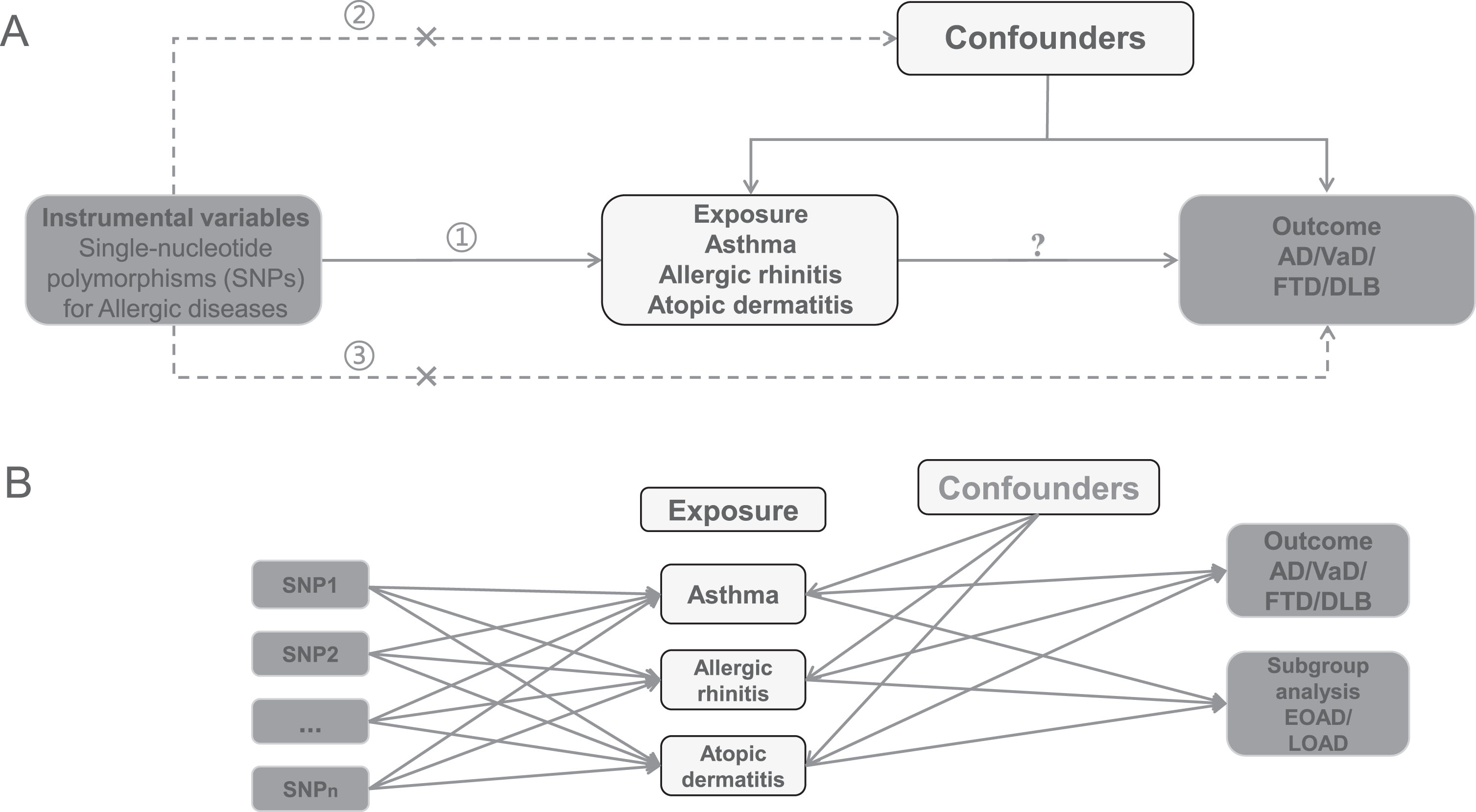
A brief explanation of the design of the Mendelian randomization (MR) model. Study design diagram for the present univariate and multivariate MR. Univariate MR was analyzed using allergic diseases as exposures and the 4 dementia subtypes as outcomes (A). Respectively, and given the interactions between allergic diseases, we also performed Multivariate MR (MVMR) to independently assess the impact of these allergic diseases on dementia risk (B). ➀, ➁, ➂ represent the three core hypotheses of MR, namely the association hypothesis, the independence hypothesis, and the exclusivity hypothesis, respectively. SNPs, single-nucleotide polymorphisms; AD, Alzheimer’s disease; VaD, vascular dementia; FTD, frontotemporal dementia; DLB, dementia with Lewy bodies; MR, Mendelian randomization.
Selection of instrumental variables
Based on the 3 core assumptions of MR analysis, as well as to ensure the accuracy and completeness of the research results, we have chosen the high-quality optimal IVs: 1) Genetic variation should be significantly correlated with exposure [28]. SNPs known to be significantly associated with allergic disease (asthma, allergic rhinitis, and atopic dermatitis) and dementia (AD, VaD, FTD, DLB) and with genome-wide significance (p < 5 ×10⁻⁸) were extracted as IVs [29]. We also used Steiger filtering to avoid interference from reverse causal associations [30]. The correlation strength of the IVs was assessed by F-statistic, calculated using the equation: F = BETA2 (exposure)/SE2 (exposure), and a strong correlation between exposures and outcomes were defined as F-statistic >10 [31]. 2) Genetic variations were not associated with any other confounding variables related to the result, simply the exposure [24]. Our study used the phenoscanner database (http://www.phenoscanner.medschl.cam.ac.uk/) to examine and exclude confounding variables associated with exposure and outcome as well as pleiotropic SNPs [32, 33]. When SNPs were initially screened, we used the phenoscanner database to find that some SNPs were associated with multiple confounders at the same time, so these SNPs associated with confounders were excluded from this study. 3) The only channel by which genetic variations can affect outcome is through exposure, not through any other factor [34]. To retain suitable SNPs, IVs were clustered within a 10 Mb genetic window using a stringent linkage disequilibrium (LD) threshold of r2 = 0.001, as the presence of strong LD may lead to bias of the results. We reconciled effect estimates for variation in exposure and outcome, screening of palindromic structures was implemented based on the harmonize_data function. We set action = 3 (dealing with more stringent levels of SNPs) to correct the strand of non-palindromic SNPs and removed all palindromic SNPs from the analysis, which made the results more conservative and accurate.
Data sources and IVs selection for allergic disease and dementia
GWAS summary statistics for allergic diseases and dementia were from the R9 version published by the FinnGen Consortium (r9.finngen.fi) [35]. The FinnGen Consortium was a study based on the collection of electronic health records and genetic data from the Finnish National Health Registry, with little overlap between exposure and outcome GWAS, thus minimizing type I error rates [36]. Exposure and outcome cases were from Finnish descent. All cases of allergic diseases were diagnosed according to the International Classification of Diseases (ICD) codes ICD8 (493), ICD9 (493), and ICD10 (J45, J46) for 42,163 patients with asthma; ICD9 (477), and ICD10 (J30.10, J30.19, J30.2, J30.3, J30.4) for 11,009 allergic rhinitis patients; ICD8 (691), ICD9 (6918), and ICD10 (L20) for 13,473 atopic dermatitis patients. In addition, all cases of dementia GWAS were defined according to ICD10 (G30) for 9301 patients with Alzheimer’s disease; ICD9 (4378), ICD10 (F01) for 2335 patients with vascular dementia; ICD10 (F020 & G310) for 111 patients with frontotemporal dementia; ICD10 (F02.3, F02.39) defined 534 patients with dementia with Lewy bodies. The detailed information of the relevant SNPs ultimately included can be found in Supplementary Tables 1 and 2. Summary-level GWAS statistics for both EOAD and LOAD in the subgroup analyses were also derived from the R9 version of the Finngen Consortium, with EOAD and LOAD comprising 1,314 patients and 6,489 patients, respectively, as well as both 170,429 controls.
Univariable MR and MVMR analyses
For univariable MR, Wald ratios for each of the auxiliary variables were estimated using SNP outcome regression divided by SNP exposure regression [37]. If more than one SNP was included, an inverse variance weighted (IVW) model with an assumed zero intercept was used and a fixed-effects meta-analysis was used to estimate potential two-sample causality between allergic disease and dementia [38, 39]. IVW can assume that IVs can only influence outcomes through specific exposures, and in the absence of horizontal pleiotropy, this approach enables unbiased causal effects and provides the most accurate assessment of outcomes [40, 41]. However, IVs can affect outcomes through other routes, suggesting a potential level pleiotropic effect, and causal estimates of IVW may be biased [42]. In order to reliably quantify causality, we devised the MR-egger regression [42] intercept and its 95% confidence interval (CI) to study the level of bias in the estimates by chance caused to directional pleiotropy, by which we can accurately estimate causality.
We also utilized weighted median, weighted mode, simple mode, MR-PRESSO, and leave-one-out analyses as sensitivity analyses to validate the overall estimates. The MR-PRESSO test was used to exclude outliers and correct for the IVW method [43], subsequently a leave-one-out analysis to examine the impact of eliminating a single, chosen SNP on the total results [44]. If an abnormal SNP was found, we chose to rerun the MR analysis after exclusion until there were no outliers and the presence of an outlier. Sensitivity analyses were essential to analyze potential heterogeneity and horizontal pleiotropy. In order to check for heterogeneity among the SNPs included in each analysis, Cochran’s Q test and funnel plot were utilized [45]. The intercept of the MR-egger method tested for pleiotropy and its slope coefficient estimated the causal effect, indicating that horizontal pleiotropy was observed when pintercept < 0.05 [37]. A flowchart of the entire analysis process can be seen in Fig. 2.
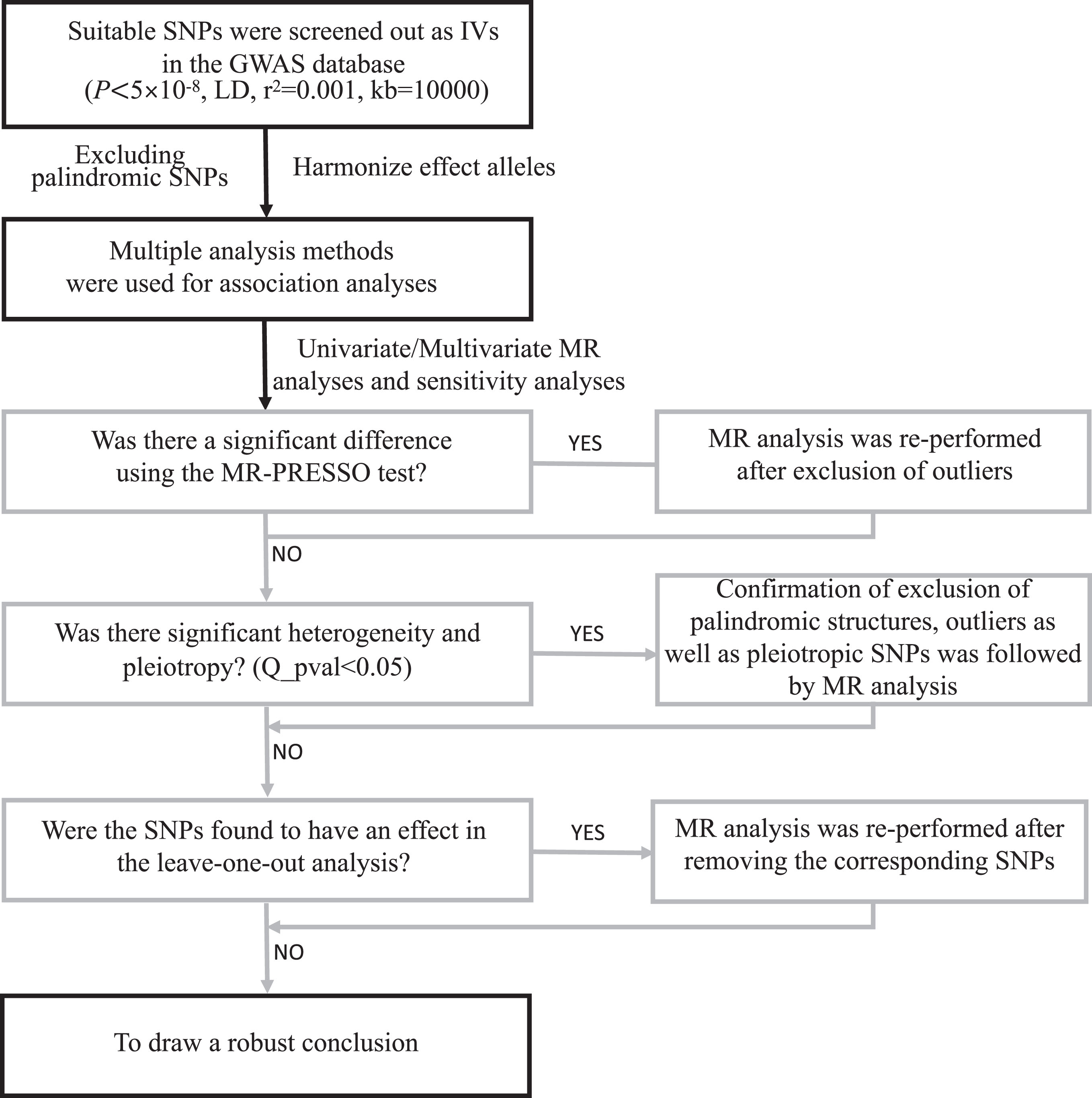
Flow diagram of the MR analysis process. MR, Mendelian randomization; SNPs, single-nucleotide polymorphisms; IVW, inverse-variance weighted; MR-PRESSO, MR-Pleiotropy Residual Sum and Outlier; LD, linkage disequilibrium.
When performing univariate MR analyses, we found shared SNPs between the three allergic diseases that could not be excluded because they were highly correlated with asthma, allergic rhinitis, and atopic dermatitis, respectively. To eliminate this effect, we applied MVMR to estimate the direct effect of each allergic disease on each dementia. In general, univariate MR estimate the “total” effect of an exposure on an outcome, whereas MVMR are typically used to estimate the causal effect of multiple exposures to genetic instruments on the same outcome variable, and their estimates represent the “direct” effect of individual exposures on the outcome [46, 47]. Thus, MVMR is particularly useful when there are two or more potentially relevant exposures of interest, and the researcher wants to know whether the two exposures have separate causal effects on the outcome. Meanwhile, we used MVMR to conduct subgroup analyses to explore the causal association between allergic diseases and AD staging (EOAD/LOAD).
To calculate the degree of binary causation, odds ratios (OR) and 95% confidence intervals were used. We adjusted for multiple testing with Bonferroni correction (p < 0.05/3), and p < 0.017 were considered statistically significant. A p-value between 0.017 and 0.05 was considered as a possible potential association. All analyses adhere to the STROBE-MR guidelines [48] and by applying the TwoSampleMR (version 0.5.6) and MRPRESSO packages (version 1.0), all of which are implemented by R (version 4.2.1, www.rproject.org/) for analysis reporting. For both univariable MR and MVMR results, scatter plots, leave-one-out analysis plots, and forest plots were available in the Supplementary Figures.
RESULTS
Table 1 showed the specific characteristics of participants from the FinnGen database, including GWAS IDs for exposure and outcome, population characteristics, sample size, and consortium.
Details of the GWAS included in the Mendelian randomization
EOAD, early-onset Alzheimer’s disease; LOAD, late-onset Alzheimer’s disease; GWAS, Genome Wide Association Study; SNPs, single-nucleotide polymorphisms. All included data can be found for specific information on the following website: https://r9.risteys.finngen.fi/.
The association between allergic diseases and dementia: univariate MR results
Based on the screening process in Fig. 2, the number and specific information of the relevant SNPs finally included in the univariate MR were shown in Supplementary Table 1, with F-statistics >29. Indicated that the specific information from the F-statistic and weak instrumental variables have little effect on the results. All univariate MR results were detailed in Fig. 3.
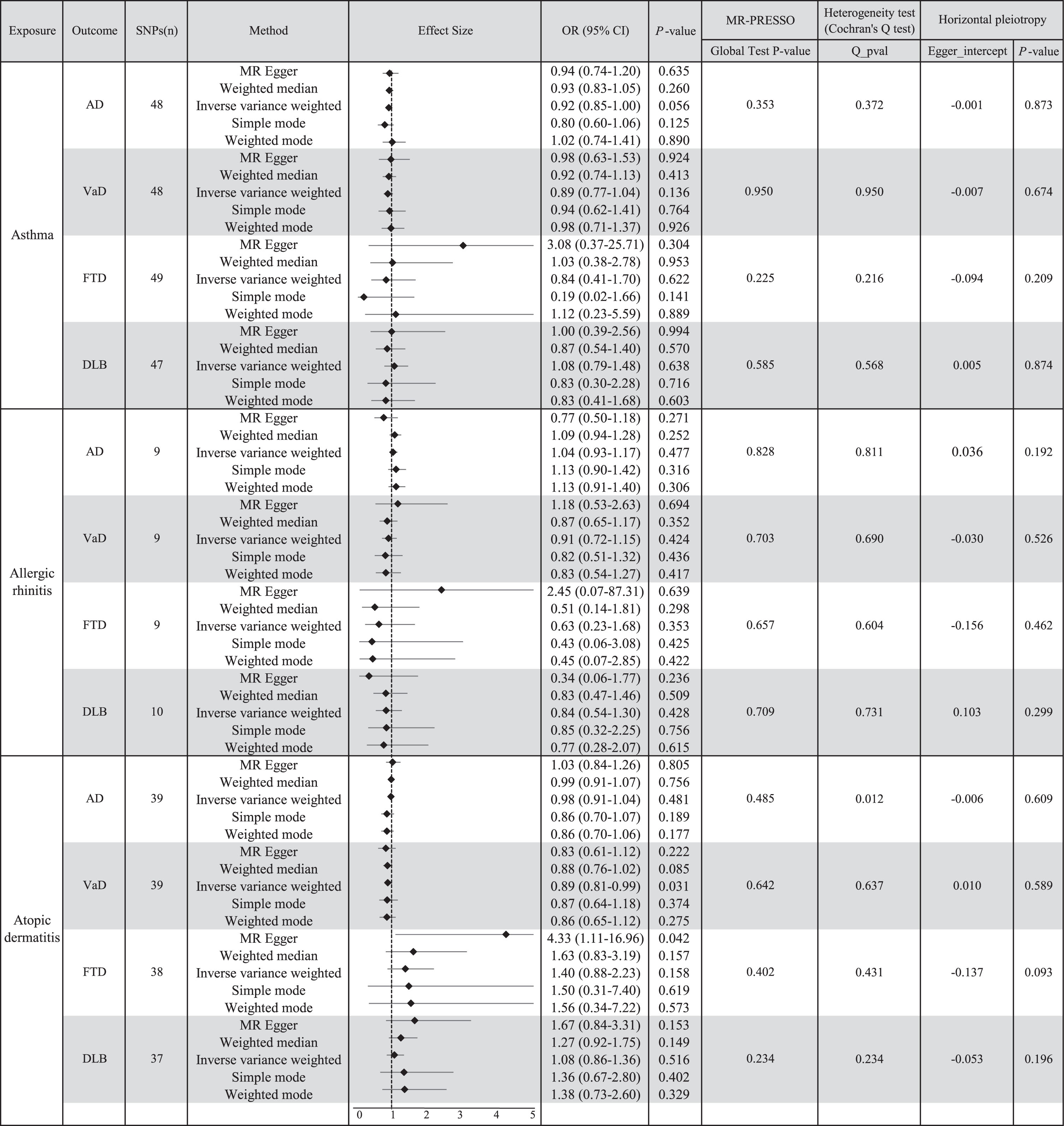
Causal effect information from univariate MR analysis. CI, confidence interval; OR, odds ratio; SNPs, single-nucleotide polymorphisms; AD, Alzheimer’s disease; VaD, vascular dementia; FTD, frontotemporal dementia; DLB, dementia with Lewy bodies; MR, Mendelian randomization.
According to the standard IVW method, a total of 49 relevant SNPs were included for asthma, no significant correlations were found between asthma and AD (OR = 0.92, 95% CI: 0.85–1.00, p = 0.056; Supplementary Figure 1), VaD (OR = 0.89, 95% CI: 0.77–1.04, p = 0.136; Supplementary Figure 2), FTD (OR = 0.84, 95% CI: 0.41–1.70, p = 0.622; Supplementary Figure 3) or DLB (OR = 1.08, 95% CI: 0.79–1.48, p = 0.638; Supplementary Figure 4). For allergic rhinitis, 10 SNPs were included in the correlation analysis but there was no proof of a possible relationship of causation to dementia risk (AD: OR = 1.04, 95% CI: 0.93–1.17, p = 0.477, Supplementary Figure 5; VaD: OR = 0.91, 95% CI: 0.72–1.15, p = 0.424, Supplementary Figure 6; FTD: OR = 0.63, 95% CI: 0.23–1.68, p = 0.353, Supplementary Figure 7; DLB: OR = 0.84, 95% CI: 0.54–1.30, p = 0.428, Supplementary Figure 8). For atopic dermatitis, 39 rigorously screened SNPs were included in the MR study. The IVW results, corrected for multiple tests, implied that there may be a correlation between atopic dermatitis and VaD (OR = 0.89, 95% CI: 0.81–0.99, p = 0.031, Supplementary Figure 10), which can be seen as a negative correlation in Supplementary Figure 10B. For other dementias, there was no evidence of an association between atopic dermatitis (AD: OR = 0.98, 95% CI: 0.91–1.04, p = 0.481, Supplementary Figure 9; FTD: OR = 1.40, 95% CI: 0.88–2.23, p = 0.158, Supplementary Figure 11; DLB: OR = 1.08, 95% CI: 0.86–1.36, p = 0.516, Supplementary Figure 12). MR-Egger, as a complementary method, was largely consistent with the IVW results, but still implied that atopic dermatitis may be associated with FTD (OR = 4.33, 95% CI: 1.11–16.96, p = 0.042), but between the IVW method being more reliable for this result, we concluded that atopic dermatitis was not significantly associated with FTD. In contrast, no evidence of an association was found when weighted median, weighted mode, and simple mode methods were used for outcome assessment.
According to sensitivity analysis, the analysis of atopic dermatitis on AD suggested potential heterogeneity, so we chose a multiplicative random-effects model to adjust for this, and the results were still heterogeneous (Cochran’s Q p = 0.012, Fig. 3). For the other outcomes, Cochran’s Q test failed to detect any appreciable heterogeneity. Both MR-PRESSO and MR-Egger regression failed to detect any horizontal pleiotropy. The leave-one-out analysis also revealed that our results were robust. Detailed results can be found in the Supplementary Figures.
The association between allergic diseases and dementia: MVMR analyses
In univariate MR analysis, we found that some IVs were shared between the three allergic diseases, and the results of univariate MR analysis may suffer from questioning. We therefore performed MVMR analysis for validating the association between allergic diseases and dementia. As shown in Table 2, there was a significant negative correlation between genetically predicted risk of asthma and AD in the standard IVW approach (OR = 0.82, 95% CI: 0.71–0.94, p = 0.005), and a possible negative correlation with risk of VaD (OR = 0.80, 95% CI: 0.65–0.99, p = 0.042), while no causal association with FTD and DLB; Allergic rhinitis seems likely to be positively associated with risk of AD (OR = 1.16, 95% CI: 1.00–1.35, p = 0.046) and VaD (OR = 1.29, 95% CI: 1.03–1.62, p = 0.027), while there was no causal relationship with FTD and DLB; There was a significant negative correlation between atopic dermatitis and the development of VaD (OR = 0.85, 95% CI: 0.76–0.95, p = 0.003), whereas there was no causal relationship with AD, FTD, and DLB. We validated this using both MR-PRESSO and MR-lasso methods, the results of MR-lasso showed no correlation between asthma and the onset of VaD, and all the rest of the results were consistent with the IVW method.
Multivariable MR Results of allergic disease on dementia
OR, odds ratio; SNPs, single nucleotide polymorphism; AD, Alzheimer’s disease; VaD, Vascular dementia; FTD, Frontotemporal dementia; DLB, Dementia with Lewy bodies. The bolded font in the values represents the p < 0.05.
As shown in Supplementary Table 3, IVW results of subgroup analyses showed no genetic association between EOAD and allergy; whereas asthma may be negatively associated with the onset of LOAD (OR = 0.83, 95% CI: 0.69–0.99, p = 0.042), and allergic rhinitis may be positively associated with the onset of LOAD (OR = 1.24, 95% CI: 1.02–1.52, p = 0.035).
DISCUSSION
To the best of current knowledge, this is the first MR study of the association between certain allergic diseases (asthma, allergic rhinitis, and atopic dermatitis) and types of dementia (AD, VaD, FTD, DLB). In the present study, we performed univariate MR and MVMR, and together the results suggest that atopic dermatitis reduces the risk of VaD. In addition, MVMR verified that asthma significantly reduced the risk of AD, while asthma may also reduce the risk of VaD; allergic rhinitis may lead to an increased risk of AD and VaD. We validated subgroup analyses of AD staging to explore whether EOAD and LOAD differ in the association of allergy and dementia. The results showed that asthma may reduce the risk of LOAD, whereas allergic rhinitis may increase the risk of LOAD.
Among the three allergic diseases, the more severe the condition at the time of consultation, the higher the risk of dementia, an effect relationship that suggests that the underlying pathophysiological mechanisms of allergic diseases may be closely related to the pathogenesis of dementia. However, few studies have determined whether allergic diseases contribute to dementia susceptibility, and to date, the findings remain controversial. Several nested case-control studies in Taiwan have reported an elevated risk of any dementia in middle and late life with asthma (HR = 2.17, 95% CI: 1.87–2.52) and atopic dermatitis (HR = 2.02, 95% CI: 1.24–3.29) [19, 49]. An elevated incidence of dementia (HR = 1.13, 95% CI: 1.12–1.14) was found to be significantly related with allergy diseases in a cohort study that focused on a national Korean population [20], and the incidence of allergic diseases is linearly correlated with the risk of dementia. However, another nested case-control study in Korea found no association between asthma and dementia (OR = 0.97, 95% CI: 0.92–1.02, p = 0.207) and showed consistent results across all age and gender subgroups [22]. A non-significant correlation between self-reported asthma and dementia (HR = 1.88, 95% CI: 0.77–4.63) was seen in another small European cohort study [21], and asthma that was discovered later in life appeared to be negatively related to cognitive impairment (HR = 1.88, 95% CI: 0.77–4.63). These above studies may be limited by small sample sizes, self-reported allergies, and the inability to explain causality; therefore, caution is needed in interpreting the results of observational studies. Most of the previous studies were from Asia, but our study population was mainly from Europe and the results were similar to the European small cohort studies [21]. In general, environment and lifestyle mediate the association between allergic diseases and dementia. Allergic diseases are frequently associated with an inflammatory response, which in turn is linked to the pathogenesis of dementia. Living environment and lifestyle, especially diet and exercise, can modulate the activity of the immune system and influence the level of inflammation between allergic diseases and dementia [50–52]. Secondly, certain environmental factors, such as exposure levels of PM2.5 in air pollution, may also produce high levels of pro-inflammatory mediators leading to allergic diseases, which can be risk factors for dementia [53–55]. Finally, interactions between an individual’s genes and the environment may play a key role in the link between allergic disease and dementia. For example, IL-13 may make individuals more sensitive to allergens, leading to abnormal activation of the immune system and thus affecting the association between the two [56–58]. In our study, minimizing the influence of these confounding factors on the results, it was demonstrated that only allergic rhinitis increased susceptibility to LOAD and VaD; asthma, on the contrary, may reduce the occurrence of LOAD and VaD; and atopic dermatitis also reduced only the occurrence of VaD, with no association with other dementias.
There are a number of seemingly plausible pathophysiologic mechanisms that might explain the link between allergic rhinitis and LOAD/VaD (Fig. 4A). Chronic systemic inflammation and immune alterations in long-term allergic rhinitis may lead to neuroinflammation and immune dysregulation in the brain [6]. Elevated amounts of immune mediators in allergic rhinitis leads to the activation of microglia, which ultimately leads to chronic neuroinflammation and neurodegeneration [7]. In addition, pro-inflammatory mediators of allergic rhinitis impair Aβ clearance by microglia in the brain, thereby accelerating Aβ accumulation [5, 6]. There is another theory: The sensation of stimulation of the trigeminal nerve endings within the nose as well as the orthonasal and postnasal olfactory pathways is referred to as olfactory function [59]. Chemosensory activation of the trigeminal nerve is closely associated with neuroinflammation. Patients with allergic rhinitis enhance the activation of trigeminal chemosensory function, which increases the risk of neuroinflammation and subsequently increases the risk of developing dementia [60, 61].
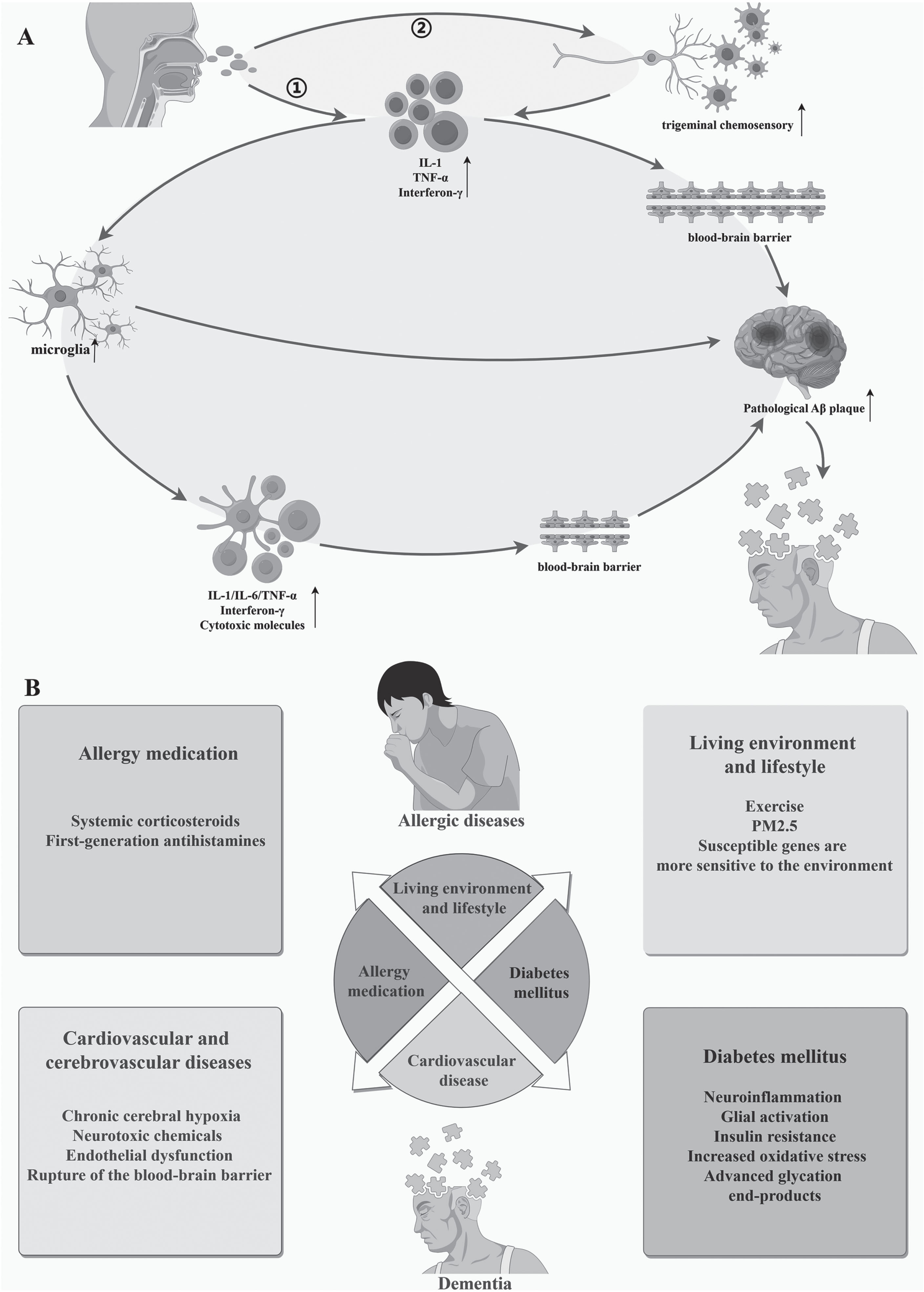
Pathway diagram of the association between allergic diseases and dementia. Panel A illustrates the pathophysiological mechanisms by which allergic rhinitis may lead to dementia; Panel B shows that the relationship between allergic diseases and dementia may be mediated by multiple confounding factors. Image production by Figdraw.
Apart from allergic rhinitis, we have no evidence that other allergic diseases increase the likelihood of dementia. We hypothesize that disease comorbidities or confounding factors, rather than allergic disease-specific factors, may largely increase the risk of developing dementia (Fig. 4B). To our knowledge, dementia has been demonstrated to be strongly correlated with both diabetes. In 2019, Kim et al. [22] grouped patients by income, age, gender, and residence, excluded patients with a history of asthma comorbid with diabetes mellitus and dyslipidemia, and investigated dementia patients with a prior history of asthma. The results of the study showed a negligible correlation between asthma and dementia. In addition to asthma, a study by Woo et al. [62] demonstrated that people with both atopic dermatitis and diabetes have a higher chance of developing dementia. In addition, many studies have found a correlation between allergic diseases and hypertension, atherosclerosis, and cardiovascular disease, all of which are also risk factors for dementia [63, 64]. Cerebrovascular injury causes chronic cerebral hypoxia and accumulation of neurotoxic chemicals while promoting endothelial dysfunction and rupture of the blood-brain barrier, events that ultimately lead to neurovascular dysfunction [3, 65]. Just as when Joh et al. [20] excluded vascular factors as potential confounders, the association between allergy and dementia was greatly attenuated, especially for VaD, which further supports the above mechanism. Finally, the association between allergy medication and dementia is also an unavoidable factor, and systemic corticosteroids or first-generation antihistamines are frequently used for more severe allergy symptoms, which confuses the link between pharmaceutical use and the risk of dementia [66, 67]. That is, treatment of allergies can also lead to cognitive changes in mild and moderate patients, and it may be the medication, rather than the allergy itself, that is associated with dementia.
Indeed, there is little research on the protective effect of asthma against dementia, and Rusanen et al. [21] found that a late-life diagnosis of asthma appeared to be negatively associated with cognitive impairment (HR = 0.42, 95% CI: 0.19–0.93), which the researchers attributed to survival bias, i.e., the number of people who suffer from asthma who go on to develop dementia is lower than the number of those who suffer from dementia in the healthy population. Another population-based study using a twin cohort suggests that people with asthma are not at higher risk of developing dementia [8]. We speculate that the heterogeneous biology and phenotypic characteristics of asthma could mitigate the effects of asthma on dementia [22]. Although early-onset asthma is predominantly associated with atopic responses, late-onset asthma includes a significant proportion of non-atopic asthma and has a different pathophysiology to early-onset asthma [68]. Therefore, the effect of asthma on dementia through atopy may be attenuated in older adults. It is hypothesized that atopy may increase the risk of dementia by increasing the inflammatory burden [8]. However, asthma alone does not lead to a higher risk of dementia.
Although our study demonstrated a causal association between allergic rhinitis and dementia only, given the high prevalence of allergic diseases, there is still a need for society to improve the management of allergic diseases and the early detection of cognitive decline in allergic patients. Since allergic diseases are preventable at both the individual and population levels, controlling allergic diseases may be a potentially effective strategy to prevent dementia. Clinicians should rationalize the use of medications and pay close attention to comorbidities associated with allergic diseases and dementia to avoid other confounding factors that may ultimately lead to cognitive impairment in patients.
The key strength of this study is primarily the MR design, which avoids confounding by various types of confounding factors. Moreover, we rigorously screened the included SNPs according to the three core assumptions of MR, and all of the included SNPs’ F-statistics were above 10, arguing that there were no weak instrumental variables in the IVs. However, there are still undeniable limitations. First, because all the populations from the GWAS database that were chosen were of European descent and because we were unable to gather complete patient demographic and clinical data, no analyses by sex or age were conducted, our study may have been biased in this regard. Second, epigenetic aspects such as transposon inactivity, RNA editing, or DNA methylation are non-negligible drawbacks of MR analysis. Thirdly, the presence of a Beavis effect (or Winner’s Curse) still cannot be ruled out in this study[69], but the bias was minimized as much as possible. Finally, patients from different ancestral backgrounds differ significantly in terms of pathogenesis, clinical presentation, and genetic susceptibility. However, some alleles in different ancestral backgrounds have not been sequenced and therefore important pathogenic genes may still be buried.
Conclusions
In conclusion, univariate MR and MVMR together demonstrated a causal relationship between atopic dermatitis and reduced VaD risk. In addition, MVMR confirmed that asthma was associated with a significant reduction in the risk of AD and may be associated with a reduction in the risk of VaD; allergic rhinitis may be causally associated with an increased risk of AD and VaD. In subgroup analyses, asthma may reduce the risk of LOAD, whereas allergic rhinitis may increase the risk of LOAD. The correlation between allergic diseases and dementia explored in previous studies may be influenced by comorbidities and confounding variables. In the future, more large-sample, multicenter prospective studies and follow-ups should be further conducted, and more updated MR studies will be more effective in validating our findings as long as less biased MR estimation techniques or larger GWAS pooled datasets are available.
AUTHOR CONTRIBUTIONS
YuanYing Wang (Conceptualization; Data curation; Formal analysis; Methodology; Resources; Validation; Writing – original draft; Writing – review & editing); ShiHao Wang (Conceptualization; Data curation; Formal analysis; Methodology; Resources; Software; Validation; Writing – review & editing); JiaXin Wu (Conceptualization; Data curation; Formal analysis; Resources; Writing – original draft; Writing – review & editing); XinLian Liu (Funding acquisition; Investigation; Methodology; Project administration; Supervision); LuShun Zhang (Conceptualization; Formal analysis; Funding acquisition; Investigation; Methodology; Project administration; Supervision; Writing – review & editing).
Footnotes
ACKNOWLEDGMENTS
The authors have no acknowledgments to report.
FUNDING
This work was supported by the National Natural Science Foundation of China (No. 81401161), the Development and Regeneration Key Laboratory of Sichuan Province (SYS13-006); Chengdu Medical College-Chengdu Eighth People’s Hospital Clinical Science Research Fund Project (YLLNZD2301); the National College Student Innovation and Entrepreneurship Training Program Project (202213705036); Sichuan Provincial College Student Innovation and Entrepreneurship Training Program Project (S202313705089; S202313705066).
CONFLICT OF INTEREST
The authors have no conflict of interest to report.
DATA AVAILABILITY
The original article includes the URLs of the public domain sites from which the study’s supporting data were acquired.
