






Abstract
BACKGROUND:
Lower risk of digestive tract cancer development has been associated with polyphenol intake. Bactris guineensis is an edible endemic palm that grows in Central and South America.
OBJECTIVE:
This study performs a phenolic characterization of Bactris guineensis and evaluates the bioactivity of this fruit.
METHODS:
The phenolic compounds of B. guineensis were characterized by HPLC-UV-HRMS analyses and the antioxidant activity was measured by chemical and cellular methods. Additionally, cytotoxicity of B. guineensis polyphenols was performed on 4 cancer cell lines and the pro-apoptotic effect was evaluated by flow cytometry using annexin staining.
RESULTS:
The major phenolic compounds of B. guineensis were proanthocyanidins. The extract IC50 for DPPH was 3.3±0.2 μg/mL and for induced intracellular ROS was 153±13 μg/mL. MTT cytotoxic assays demonstrate IC50 values between 16.6 and 24.9 μg/mL for the colon and hepatic adenocarcinomas, with high selectivity effects towards cancer cells compared to non-tumor cells. A 20 to 50% early apoptotic effect was observed in cancer cells lines by Annexin/PI staining.
CONCLUSIONS:
B. guineensis evidenced an important radical scavenging activity and a strong cytotoxic activity against hepatic and colorectal carcinoma cells, showing better values than procyanidin extracts from other fruits previously described.
Introduction
Epidemiological studies from the last years had associated the nutritional and functional properties of fruit and vegetable consumption with lower risk of cardiovascular diseases [1], type 2 diabetes [2] and different types of cancer [3]. Many fruits and vegetables contain bioactive compounds with positive effects on health, additionally to basic nutrition. Some of the most recognized and documented functional foods include berries, nuts, teas, fiber-enrich grains, probiotics products and omega-3 polyunsaturated fatty acid [4].
The current knowledge about cancer confirms that multiple diverse factors are involved in the initiation and progression of the disease. However, particularly in the digestive tract cancers, vegetable and fruit consumption has been associated with a lower risk of cancer development [3]. A study performed in Italy suggests that 20–40% of oral, laryngeal, pharyngeal and esophageal cancers are attributed to low consumption of vegetable and fruits, and these values are higher for stomach cancer (60%) and colorectal cancer (43%) [5].
The anticancer potential of many fruits and vegetables has been attributed to phytochemicals known as polyphenols. There are several polyphenol mechanisms of action proposed, some involve anti-inflammatory properties, carcinogen detoxification and modulation of cellular signaling pathways, particularly the MAP kinases, such as p38 and ERK, that are involved in the cell proliferation and survival. Another suggested mechanism of polyphenols is the relief of oxidative stress mainly by activation of glutathione peroxidase, catalase, glutathione-S- transferase and NADPH-quinone oxidoreductase [6].
Most of the well-characterized functional fruits are grown in template climate zones. However, it is known that the tropical zones are rich in plant biodiversity and in edible species that have been rarely studied. One of this unknown species is Bactris guineensis, which is an endemic palm that grows in Central America and the north part of South America. This palm grows in dry marine zones usually below 1000 meters above sea level. The fruit of this species is known as “uvita”, or “güiscoyol” in Central America, and as “corozo“ in Colombia. Usually the fruits of this species are consumed fresh and in some countries the pulp is used to prepare wine, jams and deserts [7, 8].
A few previous studies had reported bioactivity and chemical composition of pulp extracts from B. guineensis. Some publications reported a scavenging activity against synthetic radicals [8, 9], and more recently, antioxidant and cytotoxic activity in fibroblastomas [10] and cytoprotective action in astrocytes and neurons [11]. About the composition of the bioactive metabolites present in B. guineensis, it has been reported that cyanidin-3-rutinoside and cyanidin-3-glucoside represent together 87.9% of the identified pigments, and other metabolites such as: cyanidin-3-sambubioside, cyanidin-3-(6-O-malonyl) glucoside, peonidin-3-glucoside and peonidin-3-rutinoside were present in smaller amounts [7].
The objective of this study is to continue the research on bioactive compounds of B. guineensis. To accomplish this, the research performed a characterization of the phenolic compounds, and because this species is an edible fruit, we focused in the assessment of cytotoxic activities of polyphenols on common digestive tract cancers. The study also evaluates the antioxidant activity of B. guineensis polyphenols because direct relationship between oxidative stress and cancer development is widely known.
Materials and methods
Chemicals and reagents
Solvents used for polyphenol extraction were purchased from JT Baker (Griesheim, Germany). For the HPLC-MS analysis, water ultrapure and acetonitrile ultrapure (MS grade) were acquired from Romil (Cambridge, UK), and formic acid was provided by Sigma-Aldrich (Milan, Italy). Amberlite XAD-7, tert-butyl hydroperoxide (TBHP), 6-hydroxy-2,5,7,8-tetramethylchroman-2-carboxylic acid (Trolox), thiobarbituric acid (TBA), 2,2 diphenyl-1-picrylhydrazyl (DPPH), 2,2-azobis-2-methyl-propion-amidine-dihydrochloride (AAPH), quercetin, 2,7-dichlorodihydrofluorescein diacetate (DCFDA), catechin and fluorescein were obtained from Sigma Aldrich (St. Louis, MO, USA). The reagents used for cell culture: EMEM medium, fetal bovine serum, L-glutamine, streptomycin, penicillin and trypsin were purchased from Thermo Fisher Scientific (Waltham, MA, USA). The chemicals for the Griess reagents: sodium nitroprusside (SNP), sulfanilamide, naphthylethylenediamine dihydrochloride and sodium nitrite were provided by Merck (Darmstadt, Germany).
Plant material and polyphenols extract preparation
Ripe fruits of Bactris guineensis were collected from Guanacaste, Costa Rica (10°17’03.2"N 85°34’19.0"W). The fruit without seeds was frozen at –80°C and lyophilized. For the phenolic compounds purification, the dry B. guineensis powder (190.1 g) was extracted with 400 mL of acetone/water (70:30, v/v) three times. Each extraction was stirred for 30 min and then filtered through a cellulose membrane with a particle retention of 12–15 μm. Later, the acetone was removed using a rotary evaporator under vacuum at 37°C, and the extract, namely crude extract, was purified using a column (150 mm×20 mm i.d.) packed with the non-ionic hydrophobic polystyrene polymer, Amberlite XAD-7HP. The crude extract was fully loaded into the column and washed three times with distilled water, to remove carbohydrates, minerals and other fruit compounds. Finally, phenolic compounds were eluted with methanol/water (80:20, v/v) and the colored eluents were collected. The methanol of this extract rich in polyphenols was removed using a rotary evaporator under vacuum at 37°C, freeze-dryed and stored in glass vials protected from light at –80°C until further analysis. For all assays, the lyophilized extract was diluted in aqueous solutions (water, PBS or cell culture medium). To calculate the yield of lyophilized extract the following formula was used: lyophilized extract weight (g)/fresh fruit weight without seeds (g) * 100.
HPLC-UV-HRMS analysis
In order to characterize the presence of proanthocyanidins (PAs) and anthocyanin glycosides (AGs) in extracts of Bactris guineensis, accurate mass measurements were carried out using LTQ OrbiTrap XL mass spectrometer (ThermoFisher Scientific, Milan, Italy) equipped with an ESI source operated in negative ionization mode. Full mass spectra were acquired in profile mode with a setting resolution of 60000 at m/z while the dependant scan resolution was set at 7500 m/z. Instrumental mass spectrometry parameters were: source voltage 4.0 kV, capillary voltage –50 V, tube lens voltage –136.5 V, capillary temperature 300°C, sheath and auxiliary gas flow (N2) 35 and 10 (arbitrary units), respectively. Full scan mass range was from m/z 150 to 1500. For fragmentation study, a data dependant scan was applied by deploying the collision-induced dissociation (CID). The normalized collision energy of the collision-induced dissociation (CID) cell was set at 30 eV and the isolation width of precursor ions at m/z 2.0.
Chromatographic analyses were carried out by using a Platin Blue UHPLC system (Knauer, Labservice Analytica, Bologna, Italy), consisting of two Ultra High-Pressure Pumps, an autosampler and a diode array detector. The extract was chromatographed on a Kinetex column (50×2.1, 2.6 micron, Phenomenex, Bologna, Italy) at flow rate of 500 μL/min and 10 μL was injected by an autosampler. The column temperature was set at 30°C and mobile phase consisted of water (A) and acetonitrile (B), both with 0.1% formic acid. The linear gradient elution was as follows: 2–20% B 0–10 min, 20–80% B 10–15 min, 80–98% B 15–16 min, 98% B 16–20 min. The UV spectra were recorded in the range from 200 to 600 nm and the wavelengths 254, 280 and 520 nm were selected for the detection. Some spectroscopic information such as retention time, UV wavelength maxima (λmax), mass spectra information, accurate mass error, characteristic fragmentation pattern was used to identify fourteen compounds. MS data acquisition and processing were carried out by Xcalibur software (version 2.2), whereas UV data acquisition as well as HPLC system control were performed by EZChrom Elite software (ChromGate®).
Antioxidant activity assays
DPPH radical scavenging activity (RSA)
Scavenging activity on DPPH free radicals by the B. guineensis polyphenol extract was measured as described previously [12]. The reaction mixture containing 1 mL of a diluted sample in methanol (1–6 μg/mL) and 0.5 mL of 0.25 mM DPPH dissolved in methanol, was shaken and incubated in the darkness at room temperature for 30 min. A control tube (without extract) and a sample blank (without DPPH) were prepared for each sample dilution to eliminate color interference. After incubation, the absorbance of the reaction mixture was measured at 517 nm. The percentage of radical scavenging activity (% RSA) was calculated by using the following equation: % RSA = [1-(Abssample- Abs blank /Abscontrol)]* 100.
The % RSA was plotted against the extract concentration to calculate the IC50, defined as the concentration (expressed as μg/mL) of extract that produces 50% of radical scavenging activity. A low IC50 indicates higher radical scavenging activity. The assessment was carried out for three independent experiments. For each experiment the different doses of B. guineensis were analyzed in triplicate.
Oxygen radical absorbance capacity (ORAC)
The ORAC assay is based on a procedure described previously [13], using a spectrofluorometric analyzer (Biotek Instruments, USA). Fluorescence is measured at 565 nm with the excitation wavelength at 540 nm. AAPH (1.34 mM) is used as peroxyl radical generator and fluorescein (61 nM) was used as target; fluorescence loss is an indication of the extent of damage from its reaction with the peroxyl radical. The ORAC results were calculated based on the calibration curves obtained in each run and reported as μmol of Trolox equivalents (TE) per gram of extract. Sample was analyzed in triplicate.
Inhibition of intracellular radical oxygen species (ROS)
The assessment of the inhibition of intracellular ROS by B. guineensis polyphenol extract was performed using the fluorescent probe 2,7 dichlorodihydrofluorescein diacetate (DCFDA) [12]. The DCFDA probe is permeable for the cells and in the cytosol is cleaved by intracellular esterases producing a non-fluorescent molecule. Subsequently the probe is oxidized by ROS, induced by tert-butyl hydroperoxide (TBHP), and becomes a highly fluorescent product (DCF) that can be measured by flow cytometry.
Monkey normal epithelial kidney cells (Vero) were grown in minimum essential Eagle’s medium (MEM) containing 10% fetal bovine serum (FBS) in the presence of 100 IU/mL penicillin, 0.25 μg/mL amphotericin B, 100 μg/L-streptomycin and 2 mmol/L glutamine.
Two hundred and fifty microliters of 4×10⁵ cells/mL Vero cells suspension was seeded in a 48-well plate for 4 h and then was treated with different concentrations of the polyphenol extract (16–250 μg/mL) for 20 h. After incubation, the cells were washed twice with 1 mL PBS and oxidative stress was induced for 2 h with 4.2 μL TBHP (0.7 mM). Thirty minutes previous to the end of the TBHP oxidative induction, 300 μL of DCFDA probe (5 μM) was added to the cells. After washing with PBS twice, the % DCF positive cells was measured using a flow cytometer (FACSCalibur, Becton-Dickinson, Franklin Lakes, New Jersey, USA). For each analysis, 10 000 events were counted and analyzed with the Cell Quest program.
The % DCF positive cells were plotted against the extract concentration to calculate the IC50, expressed as μg/mL polyphenol extract that decreased 50% of DCF positive cells. A low IC50 indicates higher inhibition of intracellular ROS. A control was made without extract (100% DCF positive cells). The assessment was carried out in three independent experiments, for each experiment the different doses of B. guineensis were analyzed in duplicate.
Inhibition of lipid peroxidation assays
Inhibition of lipid peroxidation in liver homogenates
This procedure was approved by the Institutional Committee for Care and Handling of Experimental Animals at University of Costa Rica (CICUA N° 036-15). Five male Sprague-Dawley rats (220±20 g) were anesthetized with CO2 and decapitated. The liver was extracted and homogenized in PBS (20%) with an ultraturrax T-25 (Ika-Labortechnik, Staufen, Germany). Then, it was centrifuged at 9000×g for 15 min at 4°C. Seven hundred fifty microliters of liver homogenate were mixed with 75 μL of different concentrations of polyphenol extract (11–171 μg/mL) and incubated at 37°C for 30 min. The oxidative stress was induced with 0.5 mL TBHP in a final concentration of 1.7 mM and incubated at 37°C for 1 h. The TBARS final products of the lipid peroxidation were measured mixing 0.25 mL liver homogenate with 0.25 mL 35% TCA (v/v) and 0.25 mL Tris-HCl buffer (50 mM, pH 7.4) and incubated at room temperature for 10 min. Then, 0.5 mL of 0.75% TBA was added and heated in a water ebullition bath for 45 min. Once the mixture was at room temperature, 0.5 mL of 70% TCA was added, vortexed and centrifuged at 2500×g for 15 min. The supernatant absorbance was measured at 532 nm and the TBARS concentration was calculated using the MDA molar absorption coefficient: 1.5×105 cm–1* M–1. A sample blank was prepared for each extract dilution to subtract the color interference.
Results were expressed in nmol MDA/g of liver tissue. The MDA concentration was plotted against extract concentration to calculate the IC50, expressed as polyphenol extract (μg/mL) that reduces 50% of the liver lipid peroxidation. A low IC50 indicates a higher inhibition of peroxidation. The assessment was carried out in three independent experiments.
Erythrocyte cellular antioxidant activity (ERYCA)
ERYCA assay was described previously [14]. It relies on differences in turbidity, between intact and lysed human erythrocytes solution, to deflect the light. AAPH, a peroxyl radical generator, enhances the lipid peroxidation of the erythrocytic membranes and provokes hemolysis, as a result, the absorbance at 700 nm decays. When antioxidant compounds are present, they scavenge peroxyl radicals and inhibit the hemolysis. The area under the absorbance decay curve (AUC) is linearly proportional to the concentration of antioxidant compound.
Fifty microliters of a human erythrocytes suspension (2% in PBS), 100 μL of different concentrations of polyphenol extract (3.1–12.5 μg/mL) and 100 μg/mL AAPH (100 μM) were added to a flat-bottom, transparent, 96-well microplate. The absorbance at 700 nm was measured in a Cytation 3 (BioTek Instruments, Winooski, VT, USA) at 37°C every 5 min for 6 h, with a gentle shake for 15 s before each measure. A standard calibration curved was made with quercetin. The AUC was calculated using the program GraphPad Prim 5. ERYCA results were expressed in micromole of quercetin equivalents per gram of sample. The assessment was carried out in three independent experiments. For each experiment the different doses of B. guineensis were analyzed in triplicate.
Cytotoxic activity assays
Cell culture
For the cytotoxicity assays, different cancer cell lines were cultured and a non-tumor cell line was used as control. The cell lines evaluated include: human gastric adenocarcinoma (AGS), human metastatic colorectal adenocarcinoma (SW-620), human colorectal adenocarcinoma (HT-29), human hepatocellular carcinoma (HepG2) and monkey normal epithelial kidney cells (Vero). All the cell lines were obtained from American Type Culture Collection (ATCC, Rockville, MD, USA) and were grown in minimum essential Eagle’s medium (MEM) containing 10% fetal bovine serum (FBS), 100 IU/mL penicillin, 0.25 μg/mL amphotericin B, 100 μg/L-streptomycin and 2 mmol/L glutamine. The cells were kept in an atmosphere containing 5% CO2 at 37 °C and sub-cultured with Trypsin 0.05% -EDTA 0.54 mM.
Assessment of cytotoxicity by MTT assay
Each cell line was cultured in a 96-flat bottom well culture plate, with 2×10⁴ cells in a volume of 100 μL MEM medium per well. After 24 h of incubation, the cells were treated with final concentrations between 1.5 and 250 μg/mL of B. guineensis polyphenol extract. After 48 h, the medium was eliminated, cells were washed twice with 100 μL of PBS and then 100 μL of MTT solution (0.5 mg/mL) was added to each well. After 2 h with MTT, the supernatant was removed, and the formazan crystals were dissolved in 100 μL ethanol 95%. The absorbance was measured at 570 nm in a microplate reader (BioTek Instruments, Winooski, VT, USA).
The percentage of viable cells was calculated using the absorbance of the control (cells incubated without extract) as 100% and the IC50 was calculated from dose-response curves. The extracts were tested in three independent experiments for each cell line. For each experiment the different doses of B. guineensis were analyzed in triplicate.
Apoptosis assay
The apoptosis assay was performed only for the cell lines that showed better cytotoxic effect in the MTT assay. Cells were cultured in 6 well plates, adding 3×10⁵ cells of SW-620 and HepG2 cell lines and 1.5×10⁵ cells for HT-29 on each well. In all wells, 1 mL of MEM medium was the final volume used. After 24 h of incubation, cells were treated with different concentrations of B. guineensis polyphenol extract and incubated for 48 h. Then, the cell medium of each well was collected in tubes and the cells detached with Trypsin 0.05% -EDTA 0.54 mM and transferred to the same tube. The apoptosis induced in each tube was quantified using Annexin V-Alexa Fluor®488 dead cell apoptosis kit (Invitrogen). For this, all tubes were centrifuged at 300×g, washed twice with PBS and resuspended in 100 μL annexin binding buffer. Then to stain the cells, 5 μL of annexin and 1 μL of propidium iodide (1 mg/ml) were added and incubated for 15 min, in darkness at room temperature. Finally, 400 μL of binding buffer was added to each tube and the apoptotic cells were analyzed with a flow cytometer (FACSCalibur, Becton-Dickinson, Franklin Lakes, NJ, USA). It was defined as: lower left quadrant, living cells (Annexin V–/PI–); lower right quadrant, early apoptotic cells (Annexin V+/PI–); upper right quadrant, late apoptotic cells (Annexin V+/PI+); upper left quadrant, primary necrotic cells (Annexin V–/PI+).
For each extract concentration, the assay was performed in three independent experiments. Camptothecin (10 μM) was used as a positive apoptotic control. The data was analyzed with Cell Quest software.
Statistical Analysis
To evaluate the effect of B. guinnensis extract concentrations with the different methodologies, data was analyzed using one-way ANOVA, followed by a Tukey post hoc analysis. Values of p < 0.05 were considered significantly different.
Results and discussion
Characterization of phenolics and derivatives
In this work, polyphenol extract was prepared from the fruit of B. guineensis obtaining a lyophilized powder with a yield corresponding to a 0.9% of the fresh fruit without seed. Later, the polyphenol content of B. guineensis extract was determined by spectrophotometric methods and the major compounds were identified by HPLC-UV-HRMS analyses.
Table 1 shows the list of 14 identified compounds, detected by HRMS Orbitrap MS analyzer. Qualitative analysis indicated that the main compounds in B. guineensis extract belong to the class of proanthocyanidins (PAs) and anthocyanin glycosides (AGs). PAs are oligomers or polymers, also named as condensed tannins. PAs are classified depending on their flavan-3-ol unit, so that (epi)-catechin (eC), (epi)-afzelechin (eA), and (epi)-gallocatechin (eG) are associated to procyanidins (PCs), propelargonidins (PPs), and prodelfinidins (PDs), respectively. PAs from B. guineensis are mainly PAs B-type, linked at C4⟶6 or C4⟶8 positions, or doubly linked through an extra bond between C2⟶O7 (A-type). The PAs average degree of polymerization (DP) is 2–7, but also heteropolymers of procyanidins and prodelphinidins are present. PAs B-type are more common in foods than A-type [15]. PAs contained in B. guineensis extract are flavan-3-ols B-type, which are commonly found in palms of the family Arecaceae [16].
Retention times, [M – H]– ions, molecular formulas and MS/MS product ions of the identified compounds in Bactris guineensis extract
Retention times, [M – H]– ions, molecular formulas and MS/MS product ions of the identified compounds in Bactris guineensis extract
Cat = catechin; eCat = epicatechin; eCat-eCat = procyanidin B-type dimer; eCat-eCat-eCat procyanidin B-type trimer; eCat-eCat-eCat-eCat procyanidin B-type tetramer.
A previous paper indicated that mass spectrometry has been effectively employed to analyze complex PAs combinations from various plant sources [17]. Full-scan high resolution accurate mass spectrometry analyses are able to define the nature and the proportion of flavanol units and also the polymerization degree (DP). In addition, in order to obtain more information about the chemical formula of detected compounds, HPLC-HRMSn analyses were performed in negative ion mode acquisition due to PAs acidic nature.
Diagnostic fragment ions used to characterize target analysis derived from retro Diels-Alder reaction (RDA) (loss of 152), heterocyclic ring fission (HRF) of the extension unit (loss of 126) and quinone methide (QM) cleavage [15, 18]. Monomers, dimers, trimers and tetramers were identified. RDA fragmentation pathway of PAs B-type dimers produces a diagnostic ion of m/z 425 with subsequent water elimination (ion of m/z 407). The fragment ion of the dimer at m/z 451 via HRF indicates loss of a phloroglucinol molecule. QM cleavage produces fragmentation between two catechin or epicatechin and forms monomeric ions of m/z 287 or m/z 289. In the case of trimers there are two possibilities: QM cleavage of the upper interflavanoid bond produces ions of m/z 287 and m/z 577, while cleavage of the lower interflavanoid bond forms ions of m/z 289 and m/z 575.
Even when anthocyanins have been identified as a characteristic compound on Arecaceae family [16], few information has been found prior 2010 in regards to B. guineensis extract. The only information has been reported by Osorio and collaborators [7, 9] who identified anthocyanins and highlighted the sensory properties, health benefits and its possible use as hydrosoluble natural dye. The anthocyanidins are the basic structures of the anthocyanins. The anthocyanidins consist of an aromatic ring [A] bonded to a heterocyclic ring [C] that contains oxygen, which is also bonded by a carbon–carbon bond to a third aromatic ring [B]. When the anthocyanidins are found in their glycoside form (bonded to a sugar moiety) they are known as anthocyanins [19].
In our study, two anthocyanin glycosides were identified: cyanidin-3-O-glucoside and cyanidin-3-O-rutinoside, which coincide with Osorio et al. publication [7]. These compounds with a maximum absorbance at 520 nm are mainly responsible of red-brown color and are easily determined due to sugar moiety loss. It should be pointed out that also quercetin-3-O-glucoside was found and confirmed by HRMS in B. guineensis extract.
The HPLC–UV (280 nm) profiles of phenolic derivatives obtained from the extracts and a representative MS/MS spectrum of PAs dimer are presented in Supplementary Material (Fig. S1.)
To determine the antioxidant capacity, the radical scavenging activity of B. guineensis extract was evaluated using different synthetic radicals, and the results are presented in Table 2. Commercial catechin and quercetin were used as control. The values for both assays showed a high antioxidant capacity. However, the radical scavenging activity of B. guineensis was significantly lower in the ORAC method compared to commercial controls. For the scavenging activity against DPPH, B. guineensis extract showed significant better activity than catechin, but a lower activity than quercetin.
Free radical scavenging activity of Bactris guineensis polyphenol extract
Free radical scavenging activity of Bactris guineensis polyphenol extract
Each value is the mean±SE of three experiments. Dissimilar letters (a, b, c) in the columns mean significant differences (p < 0.05).
Previous publications reported that pulp extract of B. guineensis has a scavenging activity against synthetic free radicals [9, 11]. According to López et al. (2017) a crude hydro-alcoholic pulp extract of B. guineensis exhibits high radical scavenging activity against DPPH and ABTS comparable to Trolox [11]. Also for purified polyphenols of B. guineensis extracted with a similar method to our study, the DPPH radicals were scavenged to zero in less than 15 min, with a concentration of 514 μg/mL [9]. The radical scavenging activity of B. guineensis extract could be attributed to the presence of catechins and its polymers, which are the predominant compounds in the extract. These flavonoids are remarkably stronger scavengers when compared to other natural and synthetic antioxidants [20].
The synthetic free radical assays are important to establish a preliminary antioxidant capacity of an extract. However, the deleterious effects of ROS occur in the cellular context, and because of this it is important to measure the antioxidant capacity in a cellular model that includes factors as membrane permeability and the compartmentalization occurring within the cell. To achieve this goal, our study used a cellular model to evaluate the protection of B. guineensis polyphenol extract against intracellular ROS. Vero cells where pre-treated with the extract and afterwards exposed to an oxidative stress challenge with TBHP. Later, the intracellular ROS was measured using the probe DCFDA. The Fig. 1 shows how the pre-incubations with B. guineensis extract reduce the intracellular ROS in a dose-dependent manner. The extract concentration necessary to scavenge 50% of the induced intracellular ROS was 153±13 μg/mL. Considering the yield of the extract, it could be calculated that 17 mg of fresh fruit are required to achieve this IC50. However, to know the real consumption requirement is necessary to consider the polyphenols bioavailability.
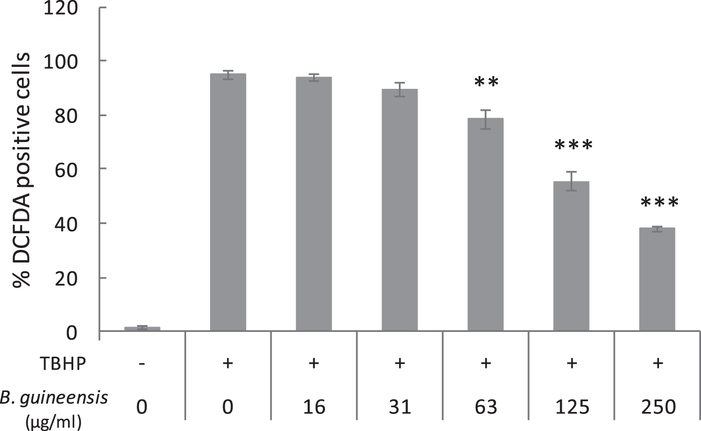
Effect of the B. guineensis extract against intracellular ROS. Each value is the mean±S.E. (three independent experiments). **p < 0.01, *** p < 0.001 compared to control with TBHP and without extract.
The capacity to inhibit intracellular ROS has been reported previously for seed extract of B. guineensis. Concentrations of 10 to 50 μg/mL inhibited significantly O2– radicals induced in neuroblastoma cells with rotenone. However, the same publication described that this inhibition was not observed for the pulp extract of the same fruit [11]. The capacity to inhibit intracellular ROS has been also assessed for compounds similar to the ones identified in B. guineensis extract in Table 1. For example, pre-treatments with 20 μg/mL of catechins and procyanidins from grape pulp reduced more than 50% UV-induced ROS production in keratinocytes [21]. Also, 1.42 μg/mL and 3.48 μg/mL of pure catechin and epicatechins respectively, provide a 50% protection against intracellular ROS induced with AAPH in erythrocytes [20].
To evaluate the protective capacity of extracts against intracellular ROS, some authors used models that measure cell viability in oxidative stress conditions instead of measuring the intracellular ROS. This kind of models has been used to evaluate B. guineensis extracts. Pre-incubations of 100 μg/mL of pulp extract increase in 26% the cell viability of fibroblastomas (HT1080) treated with rotenone to induce free radicals [10]. Also, pre-incubations of 10 μg/mL of B. guineensis pulp extract increase in 33% the cell viability of astrocytes treated with rotenone [11].
The B. guineensis inhibitory capacity against intracellular ROS reported in this study showed less activity against oxidative stress than other extracts cited in the previous paragraphs. However, there is a large variability in the conditions used in each model, such as the particularities of each cell line and the kind of pro-oxidant molecule used. All these factors determine the success of the extract against the oxidative challenge.
The oxidation of lipids by free radicals provokes membrane and tissue damage that is associated with the development of many diseases such as: cardiovascular illnesses, cognitive dysfunctions and cancer [22]. As a result, it is important to assess the role of antioxidants in the reduction of lipid peroxidation. This study evaluated the inhibition of lipid peroxidation using two models: one that measures MDA concentrations in rat liver homogenates and another that quantifies erythrocytes hemolysis caused by lipid peroxidation (Fig. 2).
Figure 2A shows the inhibition of the B. guineensis extract on MDA concentration measured in liver homogenates treated with TBHP to induce the oxidative stress. The extract concentration that was effective to decrease 50% of the MDA concentration (IC50), compared to the non-treated homogenate, was 52.8±3.7 μg/mL. To our knowledge, the inhibitory effect of lipid peroxidation had not been evaluated previously for B. guineensis. However, this capacity has been reported for procyanidins similar to the ones identified in Table 1. A meta-analysis of in vivo studies demonstrated that MDA levels were 3.06-fold lower in the experimental groups treated with procyanidins that in the control groups without treatments. Another conclusion of the meta-analysis is that procyanidins protection was more effective in tissues than in serum [23].
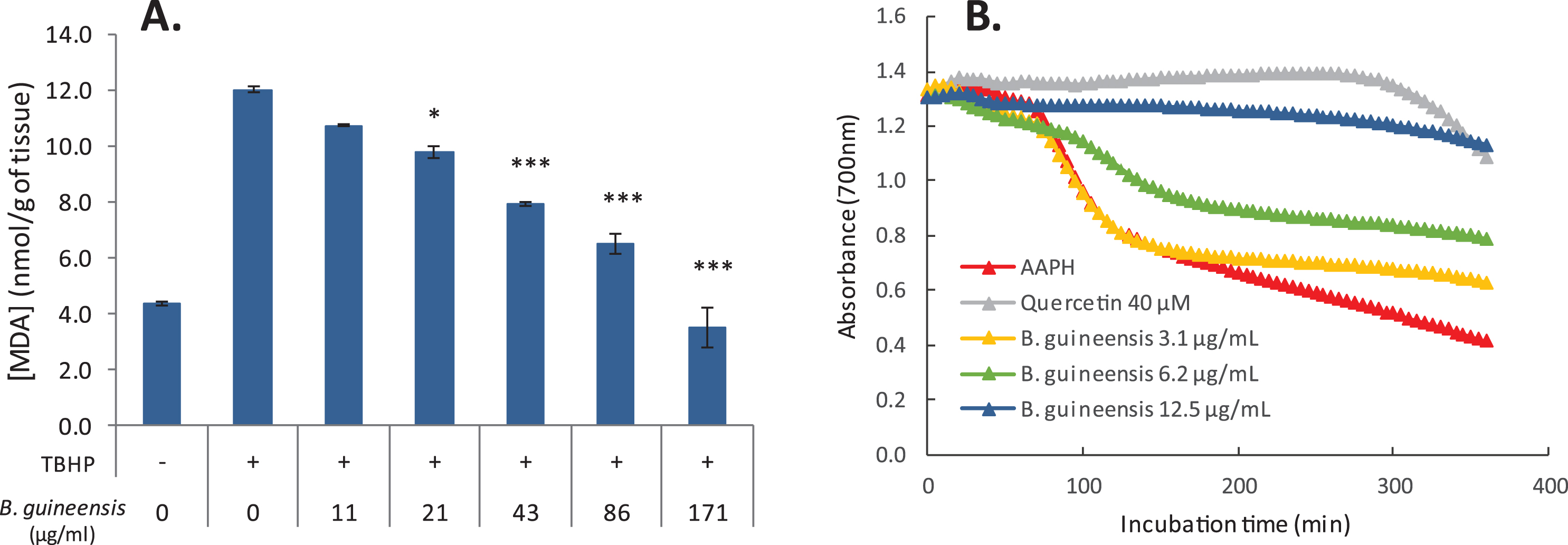
Effect of B. guineensis extract against lipid peroxidation in liver homogenates (A) and erythrocytes (B). Values represent the mean±S.E of three experiments. *p < 0.05, ***p < 0.001 compared to control without extract.
Figure 2B shows how the absorbance of erythrocytes decrease due to the hemolysis caused by lipid peroxidation induced by AAPH. This decrease in absorbance was counteracted when the erythrocytes were co-incubated with different doses of B. guineensis extract. A concentration of 12.5 μg/mL of B. guineensis extract was able to inhibit the erythrocytes hemolysis during 5 hours to similar levels than the inhibition showed by the commercial antioxidant quercetin (40 μM). The inhibitory capacity of B. guineensis extract was assessed in 3.65±0.15 mmol of QE/g (quercetin equivalents per gram of extract). This capacity of B. guineensis extract to inhibit the erythrocytes hemolysis showed similar values to the ones reported previously for pure catechins. Grzesik et al. (2018) pre-incubated erythrocytes with pure catechins prior submitting them to an oxidative challenge with AAPH and demonstrated that concentrations of 14.5 μg/mL significantly prolonged the time of hemolysis by more than 100%.
The protection against lipid peroxidation observed for catechins and their conjugates has been explained because of the interaction of these antioxidants with the membrane bilayer. These membrane-active flavonoids accumulate in the internal and external part of the bilayer increasing the membrane rigidity and hinder the access of free radicals. In this way, the flavonoids reduce the oxidation reaction kinetics and inhibits lipid peroxidation [24,25, 24,25]. Also, the direct scavenging activity of flavonoids against free radicals could contribute to the reduction of lipid peroxidation levels.
The B. guineensis extract was tested for its cytotoxic effect against human gastric adenocarcinoma cell lines (AGS), human hepatocellular carcinoma (HepG2) and two types of human colon adenocarcinoma cell lines (SW-620 and HT-29). Also, monkey normal epithelial kidney cells (Vero) were evaluated as non-tumor control. Table 3 demonstrates that the cytotoxic effect varies according to the cancer cell line. The variable response of each cell line to treatment evidences its differences in physiological, biochemical, histological, and immunological characteristics depending of the tissue of origin and also the degree of genetic modification and mutant profile of the cells during tumor development [26]. These differences in cells lines may be responsible for variances in uptake and retention amounts of the extract compounds. Treatments of black raspberry extracts in rat esophageal epithelial cells have demonstrated that variances in the apoptotic effect between highly and weakly tumorigenic cell lines are due to distinct uptake of anthocyanins [27].
Effect of Bactris guineensis polyphenol extract on cancer cell lines viability
Data are means±S.E. of three independent experiments, each performed in triplicates. Different superscript letters indicate significant differences (p < 0.01). SI*: specific index. AGS: gastric adenocarcinoma, SW-620: metastatic colon adenocarcinoma, HT-29: colon adenocarcinoma, HepG2: hepatocellular carcinoma, Vero: epithelial monkey non-tumor cell line (control).
The most sensible cell lines to treatments of B. guineensis polyphenols were the colorectal and hepatocellular cell lines. These cell lines showed values of IC50 around 20 μg/mL, which is considered by several authors as promising data for a therapeutic potential [28, 29]. Besides, these IC50 values are biological relevant because, considering the yield of the extract, these IC50 of extract represents 2.2 mg of fresh fruit.
Previous to our study, the cytotoxic effect of a crude extract of B. guineensis was assessed against fibroblastoma cells and the results did not show cytotoxicity at the highest concentration evaluated of 100 μg/mL [10]. However, for some of the main compounds of B. guineensis extract, such as procyanidins, cytotoxicity against human breast adenocarcinomas is reported [30], oral squamous cells [31], prostate carcinomas [32], hepatocellular carcinomas [33] and most of the publications correspond to colorectal carcinomas [34–38]. An important feature of these publications is that the majority evaluated procyanidins extracted from grape seeds and only a few reports assessed procyanidins from other sources, such as cranberries, cocoa or Japanese quince [31, 39].
Table 3 showed IC50 values of 24.9±0.6 μg/mL and 16.6±1.3 μg/mL for the colon adenocarcinoma cell lines HT-29 and SW-620 respectively. Both IC50 were calculated from treatments with B. guineensis extract for 48 h. Similar experiments, using procyanidins from grape seeds, reported less cytotoxic effect against colorectal carcinomas cells. Two publications from Kaur et al. reported maximum levels of cytotoxicity of 33% in HT-29 cells treated for 48 h with 100 μg/mL of grape seed extract [37, 38]. Also, other authors reported a 90% of cytotoxicity in HT-29 cells treated for 72 h with 50 μg/mL of grape seed extract [36]. In the case of SW-620 cells, fewer publications are available; however, some values were reported for SW-480, corresponding to the primary adenocarcinoma from which the SW-620 metastasic cells were derived. For SW-480 cells a 56% of cytotoxicity was achieved with 100 μg/mL of grape seed extract incubated for 48 h [37].
A lower cytotoxic effect was reported for colorectal carcinomas using procyanidins extracted from different sources than grape seeds. Procyanidins extracted from Japanese quince incubated for 72 h with Caco-2 cells caused a cytotoxicity of 74% using concentrations of 49.8 μg/mL [35]. Procyanidins extracted from cocoa produced a 70% growth inhibition of Caco-2 after 9 days treatment with 50 μg/mL [39] and 45 μg/mL of apple procyanidins incubated for 9 days with SW-620 caused a 50% growth inhibition [40]. Despite the fact that different conditions among experiments do not allow a direct comparison of results between studies, the extract of B. guineensis used in this study showed one of the most promising values against colorectal carcinomas compared to the publications previously cited.
According to Table 3, the treatment of B. guineensis extract in the hepatocellular carcinoma (HepG2) for 48 h shows a 50% of cytotoxic activity (IC50) with 22.9±0.9 μg/mL. A previous report evidences that HepG2 cells treated for 48 h with 100 μg/mL of different fractions of procyanidins extracted from callus cultures of grapes showed cytotoxicity values between 64–72%. The authors characterized the fractions by LC-ESI/MS and concluded that the difference in the cytotoxicity between the fractions was proportional to the degree of procyanidins polymerization [33]. This correlation is congruent also with the high cytotoxic activity of the B. guineensis extract used in this study and the prominent presence of oligomers and polymers identified in the extract (Table 1).
The cytotoxicity of B. guineensis extract against the colorectal and hepatocellular cancer cell lines (SW-620, HT-29 and HepG2) not only showed promising values but also demonstrated significant differences compared to the non-tumor cells (Vero). To quantify this specificity against the cancer cells, Table 3 shows the values of selectivity index, which is defined as the ratio of IC50 values of non-tumor cells to cancer cells. The highest selectivity index corresponds to SW-620 cells (SI = 10.8), followed by HepG2 (SI = 7.7) and HT-29 (SI = 7.1). These values means that B. guineensis extract was between 7 to 10 times more toxic to the cancer cells than to the normal cells. Higher values of this selectivity are desirable and suggest a possible therapeutic potential. Extracts with SI greater than 3 are considered to have a high selectivity towards cancer cells [28, 41].
In order to evaluate if the high selectivity could be associated to an apoptotic effect directed towards cancer cells, an Annexin V and PI staining was performed and the results were analyzed by flow cytometry (Fig. 3). In Fig. 3A and 3B, the effect of 48 h incubation with different concentrations of B. guineensis extract in colon adenocarcinomas SW-620 and HT-29 can be observed. The maximum percentage of total apoptotic cells achieved was 69% for SW-620 cells treated with 20 μg/mL. This value denotes better effects than the total apoptotic cells percentage (30%) reported for a procyanidins grape seed extract on a similar colon adenocarcinoma cells (SW-480) treated with 50 μg/mL for 72 h [36]. The same publication also evaluated the extract on HT-29 cells. For this cells, the treatment of 50 μg/mL during 72 h caused a 50% of total apoptotic cells, which is similar to the percentage demonstrated by 50 μg/mL treatment of B. guineensis extract in HT-29 (49%), however in our study the treatment was applied for 48 h.
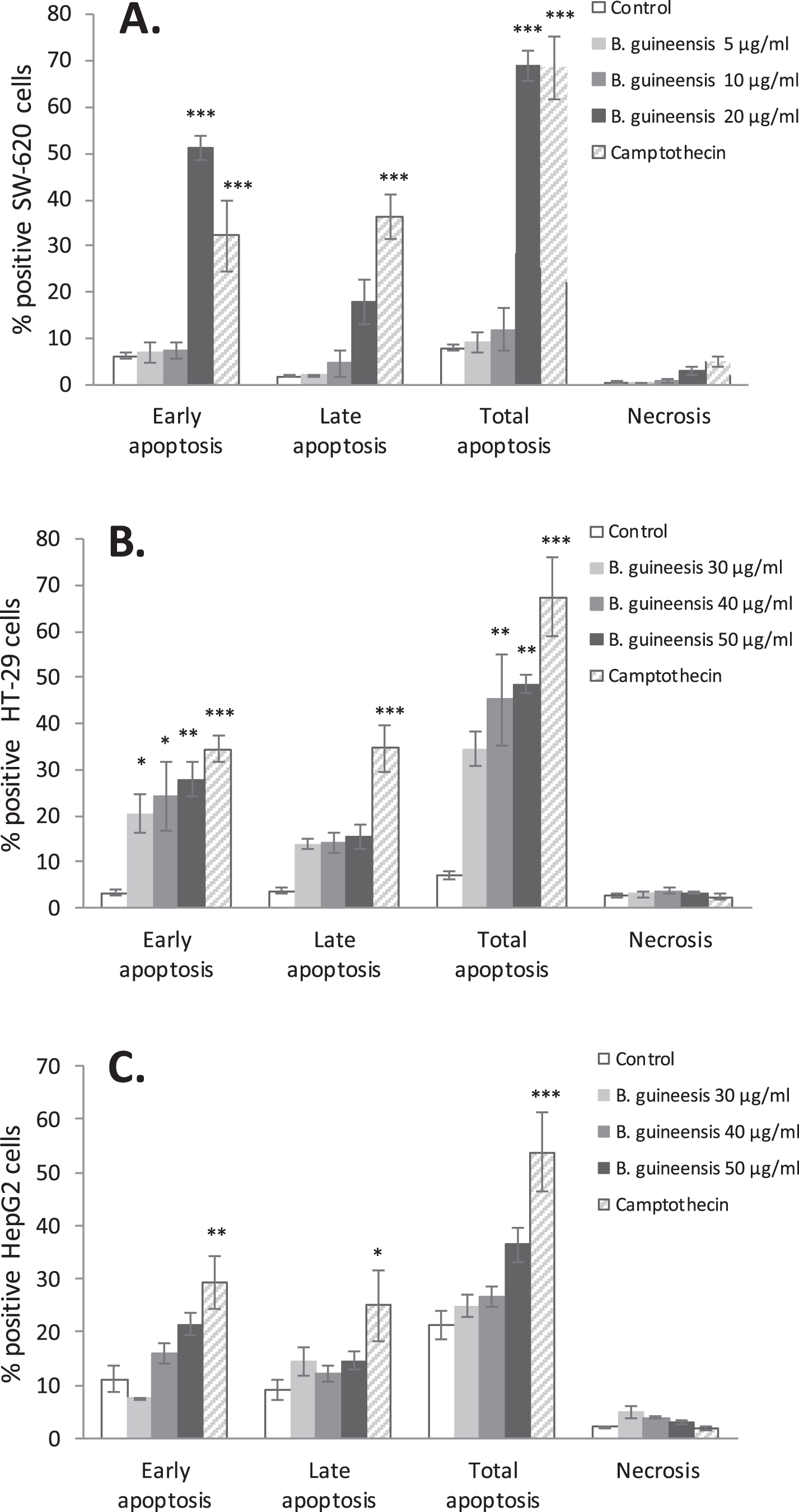
Apoptotic rates of SW-620 (A), HT-29 (B), HepG2 (C) cells determined by flow cytometry with Annexin V/PI staining. Three different concentrations of B. guineensis were evaluated and also camptothecin (10 μM) as positive control. Data of apoptotic rates are presented as the mean±S.E. of three independent experiments. *p < 0.05, **p < 0.01, ***p < 0.001 compared to control cells without extract.
A concentration of 20 μg/mL of B. guineensis caused a 51% of early apoptotic cells in SW-620 cells, and a concentration of 50 μg/mL caused a 28% of early apoptotic cells in HT-29 cells (Fig. 3). Other publications that evaluate early apoptosis of 50 μg/mL procyanidins grape seed extracts in colon carcinoma cells, reported values of 13% in HT-29 cells, 30% in SW-480 cells, 30% in LoVo cells, 45% in CaCo and 45% in HCT-8 cells [34, 37]. Comparison of these values denotes that the treatment of B. guineensis extract in SW-620 cells showed the highest levels of early apoptotic stimulation reported for the colon carcinoma cells treated with procyanidin extracts. Reports for procyanidin-rich extracts obtained from other samples denote non-apoptotic effects for 50 μg/mL of Japanese quince extract in HT-29 cells [354] and non-apoptotic effect for 100 μg/mL of cocoa procyanidins in Caco-2 cells [39].
Figure 3C showed a lower early apoptotic effect of 21% with 50 μg/mL B. guineensis extract in hepatocellular carcinoma (HepG2), however this increase is not significant compared to control cells. This lower apoptotic result suggests that the cytotoxic effect showed in Table 3 is likely to be caused by other signaling pathway rather than the apoptotic mechanism.
Based in previously reports, it could be suggested that the proapoptotic activity of B. guineensis is associated with the higher level of polymeric catechins and epi-catechins identified for this extract. A correlation between the proapoptotic effect and the presence of oligomeric and polymeric procyanidins has been previous demonstrated for grape seed extracts and Japanese quince extracts [35, 42]. The mechanism by which the B. guineensis extract is causing cytotoxicity and apoptosis in the cancer cell lines should be elucidated. It is also important to evaluate if the effect of this extract is only as intracellular ROS scavenger that promotes cell survival or if the extract can also activate intracellular signaling pathways that promote cancer defense. For other extracts, it has been demonstrated that procyanidins exert their action through inhibition of kinases and their subsequent pathways [40], inhibition of polyamine biosynthesis [39] or protection of alterations in the membrane fluidity [24, 25]. This last mechanism is a strong candidate to be involved given the fact of the inhibition of lipid peroxidation evidenced in this study.
The study of dietary phytochemicals is a promising area because of their few systemic toxic effects [42]. The extract of B. guineensis evaluated in this study showed an antioxidant activity evidenced in the radical scavenging activity, the intracellular ROS inhibition and also in the capacity to inhibit lipid peroxidation, providing protection to the membranes. The extract also demonstrated a strong cytotoxic activity against hepatic and both colorectal carcinoma cell lines with a marked selectivity when the IC50 is compared with non-tumor control cells. The major compound of B. guineensis extract are procyanidins, and according to Williamson and Manach [43], these molecules are poorly absorbed throughout the digestive tract due to their high molecular weight, however they reach the colon practically intact, where they interact directly with the cells.
Future assays will be required to determine B. guineensis polyphenols bioavailability. However, previous studies reported an uptake of procyanidins dimers, trimers and tetramers of 0.2 to 1.7% in Caco cells [44, 45]. Based on the lowest recovery rate of 0.2% reported for Caco cells, it could be suggested that to achieve the doses required for the antioxidant and the cytotoxic activity mentioned in our study, about 8 g of B. guineensis should be consumed. This amount of fruit is feasible considering that each fruit weights between 2 to 3 g without seed. Even more, being that the cell lines evaluated in this study correspond to gastrointestinal tract and procyanidins reach the colon practically intact as mentioned before [43], they could exert their biological activities.
Besides, it has to be considered that despite the low plasma values of procyanidins reached in human trial studies after consumption of procyanidin-rich foods, biological effects are observed and could be attributable to currently unidentified metabolites of the procyanidins, monomeric catechins or compounds produced by the microbiota [43].
To our knowledge, this is the first report on proapoptotic activities of B. guineensis polyphenol extract on human tumoral cell lines. However, in vivo and clinical studies should be performed to determine the therapeutic potential of this fruit.
Funding
This research was funded by Vicerrectoría de Investigación of Universidad de Costa Rica by the project number 422-B7-099.
Conflict of interest
The authors have no conflict of interest to report
