
Abstract
Artificial scaffolds play an important role in tissue engineering, which is used to mimic extracellular matrix (ECM) and provide a suitable microenvironment for cell growth. Many natural and synthetic biomaterials have been used to fabricate two dimensional or three dimensional scaffolds. However, missing electrical conductivity of these materials is one of the disadvantages. Recently, conductive polymers (CPs) and conductive nanomaterials (CNMs) have been chosen for doping into scaffolds to improve their conductivity. This review focuses on conductive scaffolds design, fabrication and application in tissue engineering for enhancing cell attachment and proliferation, promoting differentiation and maturation with and without electrical stimulations.
Introduction
In the 1990 s, tissue engineering was firstly emerged to create functional organs in vitro in order to replace damaged tissue [1]. To achieve this goal, tissue engineering has to combine many subjects such as engineering (material and mechanical), and life sciences (biology and physiology). The natural extracellular matrix (ECM) is a typical mixture of proteins and polysaccharides, which influences cell behaviors. Numerous research indicate that an appropriate scaffold plays one of the key roles for tissue engineering as they can mimic the ECM, and support cell proliferation and differentiation prior to implantation [2–9]. Many biocompatible and biodegradable materials have been used to synthesize scaffolds through electrospinning, lyophilization, etc. In addition, a number of studies showed that the cell behaviors were closely related on the stiffness and elasticity [10–12]. However, one disadvantage of current biomaterials is lacking electroactivity, which limits their applications in culturing a variety of tissues (e.g. cardiac, nerve, muscle, and skin). Therefore, the electroactive biomaterials lead the new avenue to the next generation of tissue engineering.
The conductive scaffolds are able to deliver electrical, electrochemical and electromechanical stimulation to cells directly, also promote multitude biological processes including cell signaling, nerve sprouting, cell division, angiogenesis, wound healing, etc. [13]. Due to its good electrical property and high conductivity/weight ratio, conductive polymer is able to be doped into biomaterial to form conductive composites for tissue engineering application [14, 15]. Besides, with the development of nanotechnology, conductive nanomaterials have become an alternative candidate to synthesize conductive scaffolds [16].
In this review, we will focus on using these conductive additives to generate conductive scaffolds for electroactive tissue engineering.
Conductive materials and its biocomposites
In general, the conductive scaffolds are fabricated by adding conductive additives into existed biomaterials. It can be divided into two major group according to conductive additives, conductive polymer based scaffolds, and conductive nanomaterial based scaffolds.
Conductive polymers
Conductive polymers (CPs) commonly have loosely held electrons in their backbones (Fig. 1). The atom along with the backbone forms a π bond, and the atom in the polymer chain is bonded through a σ bond, which is much stronger [15]. Based on this property, the neutral staged polymer can be oxidized or reduced to become either positively or negatively charged through the “doping” process. Without the doping, most conductive polymers will stay in non-conductive form. The doping process happens during synthesis through chemically or electrochemically via photodoping [17–19] as there is a charge transfer from dopant molecules to polymer chains, which causes charge carries, polarons and bipolarons to the conjugated chain. The conductivity of the CPs can be adjusted by the proportion between the dopant and CPs [20], however, different dopants could also affect the constructional properties (color, porosity, volume) of the polymer [17, 22]. Moreover, the doping process is also reversible, the dopant can leave or re-enter the polymer by applying an electrical potential [23]. In order to use CPs for biological applications, their inherent non-degradability becomes the major limitation. The in vivo CPs may induce chronic inflammation and need additional surgery to remove. To address the issue, the exist CPs are blended with suitable biodegradable materials to form composites [24].
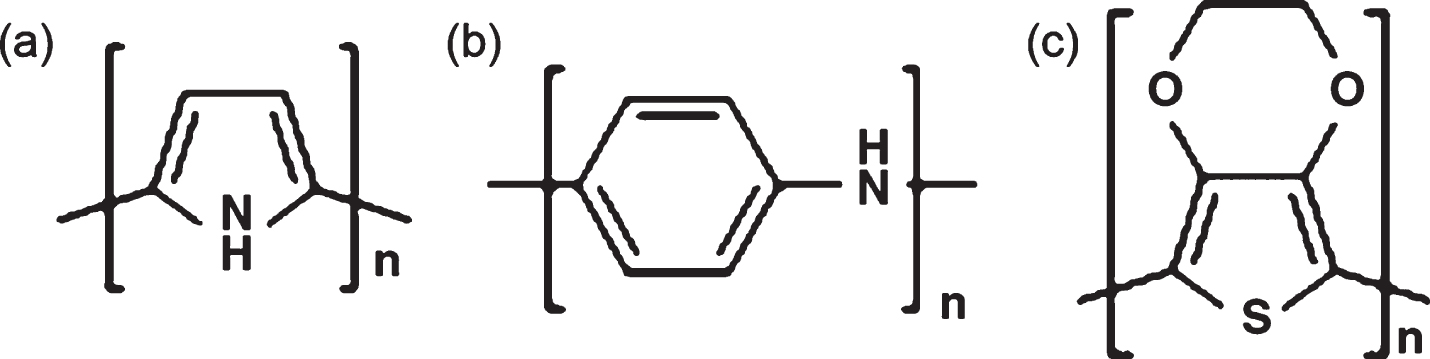
Molecular structure of conductive polymers, (a) Polypyrrole, (b) Polyaniline, (c) Poly(3,4-ethylenedioxythiophene).
Polypyrrole (PPy) possesses many excellent properties such as conductivity, biocompatibility, good chemical stability, etc. These make it become one of the most popular CPs which benefit in many applications, including fuel cells, computer displays, biosensor, drug delivery system, and tissue engineering [20, 26] The synthesis of PPy are extremely easy and flexible, which can be processed under room temperature with a wide range of solvents [25-27]. Moreover, PPy can be fabricated by doping into biomaterials to form electroactive scaffolds with a large surface area and variable porosities. Unfortunately, the synthesized PPy is very difficult to be further processed, as its molecular structure makes it non-thermoplastic and its mechanical property makes it brittle and insoluble [28–30].
Polyaniline
Polyaniline (PANI) is well known as aniline black [21, 31]. Depending on its oxidation level, PANI exists various forms of fully oxidized pernigraniline base, half-oxidized emeraldine base and fully reduced leucoemeraldine base [21]. As PPy, PANI is also easy to be synthesized, another notable property of PANI is the capability to switch between conductive and resistive states [32-36]. Unfortunately, the disadvantages of PANI including low processibility, non-biodegradability limit its biological application, moreover, it has been noted that PANI could cause chronic inflammation after implantation [23, 37]. However, more and more evidence have shown that the PANI in different states have sufficient biocompatibility for biomedical applications [38]. For instance, Guimarda et al. have assessed the in vivo responsing to different oxidation states of PANI implants. Their results shows that there is no significant inflammation at the implant site and no sign of muscle abnormal [17].
Poly(3,4-ethylenedioxythiophene)
Poly(3,4-ethylenedioxythiophene) (PEDOT) is another commonly used CPs, which is a polythiophene derivative compound. The heterocyclic ring structure of PEDOT decreases its band gap, potential reduction, and potential oxidation, which grants PEDOT better electrical, chemical and environmental stability, thus, it shows better conductivity and thermal stability comparing with PPy [39, 40]. There are studies show that synthesized PEDOT films have good biocompatibility for culturing various types of cells. Moreover, in vivo, PEDOT exhibits low intrinsic cytotoxicity and no significant inflammatory response after implantation, which makes it an ideal CPs for biomedical and biosensing application [41, 42].
Carbon based conductive nanomaterials
With the development of nanotechnology, three major groups of conductive nanomaterials (CNMs) have been found which are carbon-based nanomaterials metal-based nanomaterials; and silicon-based nanomaterials [16]. Due to their better conductivity compared with CPs, conductive nanomaterials have been widely used in electrical engineering and biomedical engineering.
Among carbon-based nanomaterials, carbon nanotubes (CNTs) are the most well-known and developed. CNTs mainly include the kinds of single-walled, double-walled and multi-walled type. CNTs have many impressive properties, such as tubular structure, electrical conductivity, and mechanical properties. According to the disciplined arrangement of their carbon atoms which linked together via strong sp2 bonds, CNTs exhibit superior electrical conductivity (104 S/cm2) and thermal conductivity (5000 Wm- 1K) [43]. Plus, with their small size and mass, carbon nanotubes have become famous nanomaterials for making functional nanocomposite in many materials science applications [44]. However, the cytotoxicity of CNTs has affected its biological application. Studies of CNTs cytotoxicity indicates that CNTs exhibits toxicity to cells in free suspension stage, while, the immobilized CNTs are non-toxic. Moreover, the immobilized CNTs is with great capability in improving cell adhesion, proliferation, and differentiation [45].
Unlike 3D CNTs, graphene consists of a monolayer of carbon atoms, which arranges in a 2D honeycomb lattice [46]. Besides, graphene has many excellent properties, such as flexibility, large surface area, thermal properties, electrical conductivity, high strength, stiffness, and biocompatibility [47]. Thus, graphene has become a very useful nanomaterial in biomedical applications [48-52]. More interesting is that due to its physicochemical properties, graphene has been utilized in stem cell-based tissue engineering with its great support for pluripotent stem cells proliferation and differentiation [50, 53–55].
Metal based conductive nanomaterials
Metal-based nanomaterials not only maintain the high conductivity, but also possess many nanoscale features including large surface areas and porous structure. Base on their shape and size, metal-based nanomaterials can be synthesized into nanoparticles, nanorods, and nanowires. For example, gold nanomaterials can be modified to functional reagents (drugs or proteins), for drug delivery, control release and photothermal therapy [56].
Silicon based nanomaterials
Silicon nanowires (SiNWs) also present good electrical conductivity, tunable dimensions, and convenient surface tailorability [57, 58]. Some studies demonstrates that SiNWs not only have good biocompability [59, 60], but also can be degraded into the form of Si(OH)4, which are metabolically tolerant in vivo [61, 62]. Thus, some researchers believe that SiNWs are favorable to in vivo applications, comparing with the other nonbiodegradable conductive nanomaterials.
Other conductive materials
Piezoelectric polymeric materials, such as poly (vinylidene fluoride) (PVDF), have also been used in tissue engineering [63]. For example, Tschoeke et al. have fabricated a PVDF composite scaffold to provide sufficient mechanical support during vascular composite graft [64]. Briefly, they have applied a two-step moulding technique to seed carotid myofibroblasts within PVDF scaffold to form a PVDF mesh in the wall of a fibrin-based vascular graft, and the artificial tissue structure demonstrated many similarities to the native tissue [64].
Melanin biopolymers are a class of naturally occurring conductive pigments [65]. The electrical conductivity of melanin is strongly dependent on temperature, physical form, and hydration state, for example, the dehydrated melanin films have conductivities of 10- 8 Scm- 1 and it reaches up to 10- 3 Scm- 1 in the hydrated condition. Even though the exact conduction mechanism of melanin is not known yet, there is enough evidence shows that their conduction mechanism is similar to CPs [66]. Bettinger et al. has evaluated the physical, chemical and electrical properties and biocompatibility of melanin film both in vitro and in vivo. They have found that the melanin could enhance proliferation in stem cells and neurite extension in PC12 cells in vitro. Moreover, the melanin implants are fully degraded after 8 weeks [67]. Kai et al. have fabricated melanin, poly(L-lactide-co-3-caprolactone) (PLCL) and gelatin composite nanofibrous scaffold through electrospinning for cardiac tissue engineering. Their studies have demonstrated that the conductive scaffold could promote human cardiac myocytes (HCM) attachment and enhanced cardiac-specific protein expression in vitro [68].
Conductive scaffolds design and synthesis
Due to the excellent electrical properties and biocompatibility, CPs and CNMs have become ideal candidates for tissue engineering. However, most of the CPs and CNMs cannot provide sufficient support to growing cells [17, 21]. Biomaterials are used for designing bioactive surface with particular interest, and living cells can interact with biomaterials via the interface or various bioactive ligands which are modified or immobilized on the surface of biomaterials [69]. It has become the most promising pathway for tissue engineering by making conductive biomaterials composites scaffolds. In addition, the aptamer technology opens a new field for binding cells to polymers [70]. As many attempts have been done to synthesize the composites scaffolds, it would be summarized and discussed the different fabrication and modification methods in this part.
CPs-based conductive scaffolds
Even though CPs can be easily synthesized, the preparation of clinically relevant CP-based scaffolds is still challenging. As we mentioned before, CPs need a doping process to become electroactive, but fortunately many components (e.g. collagen, chitosan, heparin, dextran sulfate, hyaluronic acid, growth factors, oligodeoxyguanylic acid, and ATP) can work as dopants during electropolymerization reactions [28, 71–73]. For instance, laminin peptides and collagen can be used to dope PEDOT with non-covalently incorporation, and improvement of PC12 cell adhesion on PEDOT film. Some ECM derivatives could also covalently incorporate with CPs during electropolymerization such as laminin peptides and hyaluronic acid [74–77]. The electropolymerization can not only be used to fabricate 2D CPs thin films, but also be enable to 3D substrates. For example, Peramo et al. have deposited PEDOT on a decellularized tissue network, and Wilks et al. have prepared electropolymerization of PEDOT in vivo [39, 78]. However, there are still some limitations of doping with biomolecules. Gomez et al. have found that the conductivity of PPy can be decreased by the physical properties such as surface roughness of the composites [79–81].
Electrospinning is a well-developed method for generating nanofibrous porous scaffolds with a tunable degree of fiber alignment, so many attempts have been made to electrospinning CPs-based biocomposites for tissue engineering. Firstly, CPs can be electrospun alone under specific condition such as organic solvent and chemical conditions (e.g. PANI doping and dissolution in hot sulphuric acid) which might be unsuitable for biological applications [33, 82–84]. Thus, electrospun CPs with biomaterials have become a promising approach to fabricate conductive biomaterials for biological applications. For example, the electrospinning composites scaffolds of gelatin and PANI can support cardiac myoblast cells (H9c2) attachment and proliferation in vitro, moreover, the additional PANI can also alter other properties of the composites including reducing fiber diameters and improving mechanical property [85]. Similarly, Ghasemi-Mobarakeh et al. have demonstrated that applying electrical stimulation could improve the proliferation and neurite outgrowth from nerve stem cells (NSCs) in vitro through fabricating PANI/gelatin/poly(ɛ-caprolactone) fibrous composites [86].
Surface modification of non-conductive fibrous biomaterials with CPs is an alternative method to fabricate conductive scaffolds. Abidian et al. have coated the surface of non-conductive poly(lactic acid-co-glycolic acid) with PEDOT via electropolymerization [87]. Xie et al. have found that the explanted dorsal root ganglia (DRG) could well adhere to the conductive core-sheath fibrous scaffolds to generate neurites across the surface (Fig. 2), and the length of neuritis can be increased by electrical stimulation [27].
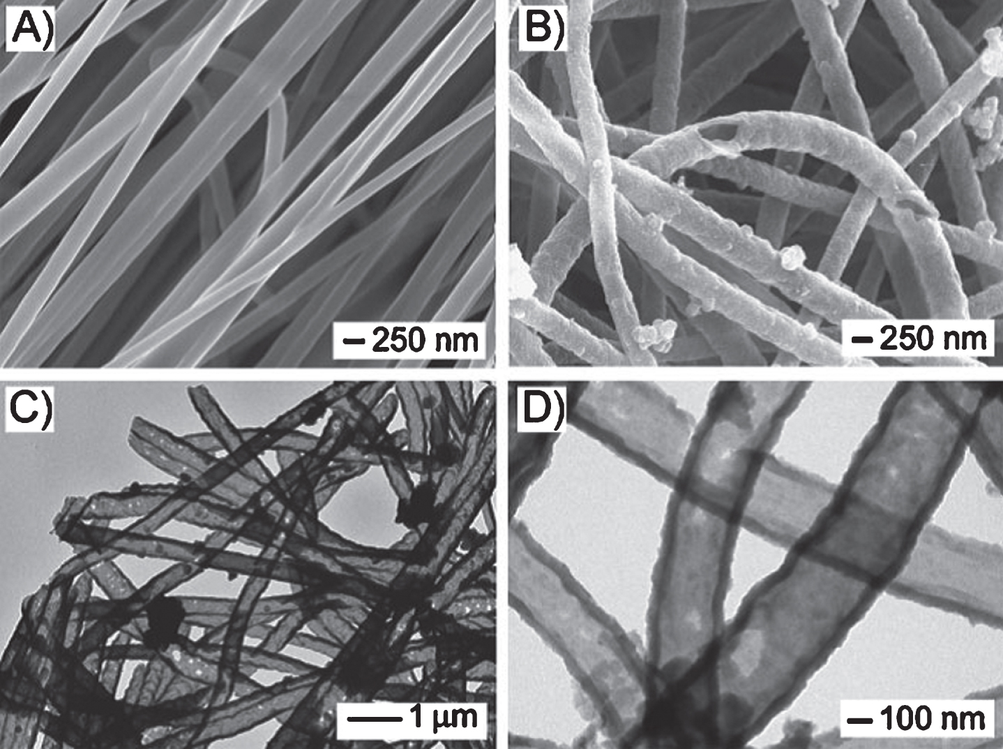
A,B) SEM images of PCL nanofibers and PPy nanotubes, respectively. C,D) TEM images of the PPy nanotubes. The nanotubes were obtained by soaking the PCL-PPy core–sheath nanofibers in DCM to selectively remove the cores. The polymerization was conducted with Fe3+ as an oxidant and Cl- as a dopant [27].
The conductive polymer can also polymerize inside hydrogel networks (e.g. PANI-PVP, PANI-polyacrylamide, and PPy/PANI-polyacrylamide hydrogels) [88, 89]. It is non-cytotoxic for the conductive hydrogels made by CPs and biocompatible hydrogels and it could be produced with a wide range of dimensions to mimic the ECM structure. This makes the conductive hydrogel to be an ideal material for biological applications such as the implantable biosensor, drug release devices, and deep brain stimulator [90].
Based on physical and chemical properties, The CNMs could be simply mixed with biomaterials to form conductive biocomposites. For example, CNTs have already been incorporated into numerous biomaterials including chitosan, collagen, poly-L-lactide (PLLA), polycarbonate-urethane (PCU), polycaprolactone (PCL), polystyrene (PS), etc. to form either 2D films or 3D scaffolds (electrospun or freeze dry). The CNTs cloud bring many useful properties to its nanocomposite such as enhanced mechanical properties, improved cell adhesion, and accelerated degradation which have been previously reviewed [44]. It makes CNMs easier to be applied with electrical stimulations by adding electrical conductivity to nanocomposites, which could better mimic the electrical properties of nerves and myocytes so such scaffolds are ideal to electrical stimulate developing tissues [91].
In general, graphene-based materials aggregate in the aqueous medium which contains salts, proteins or other ions, which require a chemical modification or functionalization to achieve desired properties [92]. Graphene and Graphene oxide (GO) are able to electrostatic or non-specific interact with proteins. Hence, functional graphene and graphene oxide can be formed by covalent interaction (through hydroxide, epoxy or carboxylic acid groups) or non-covalently interaction (through surface π electrons, hydrophobic or electrostatic interactions). In biomedical applications, GO is preferred while comparing with graphene, as GO presence of carboxylic, epoxy and hydroxide groups, which allow for a wide range of reactions and functionalization opportunities. For example, the carboxylic acid group of GO can be used to conjugate with the amino group of polyethylene glycol (PEG) and the nanocomposite of GO-PEG resulted is stable in various physiological solutions [92–94]. Similarly, GO can also form covalent conjugation via carboxylate group with the amide linkages of chitosan, and the functionalized GO could be used for drug and gene deliver [95, 96]. Moreover, Fan et al. and Depan et al. have fabricated graphene-chitosan thin films and GO-chitosan porous scaffolds respectively to improve the mechanical properties of the composite, and their cell culture is with good biocompatibility of the composites [97, 98].
Metal-based conductive scaffolds
Metal-based conductive nanomaterials have been recently developed in tissue engineering application. As shown in Fig. 3a, b, Dvir et al. have firstly synthesized alginate porous scaffold doped with electrical conductive bridge of gold nanowires (AuNWs), and the additional AuNWs have improved the mechanical properties of the composite. Moreover, AuNWs could deliver electrical stimulation through the scaffold which improves the interaction between cell to cell and cell to matrix [99]. Later, Dvir’ s group have investigated a gold evaporation method to coat gold nanoparticle (AuNPs) on the surface of existing scaffolds (Fig. 3c–f) (e.g. PCL electrospinning scaffold, decellularized matrix), and according to their reseach, the AuNPs films which on scaffolds’ surface could present high electrical conductivity [100–103].
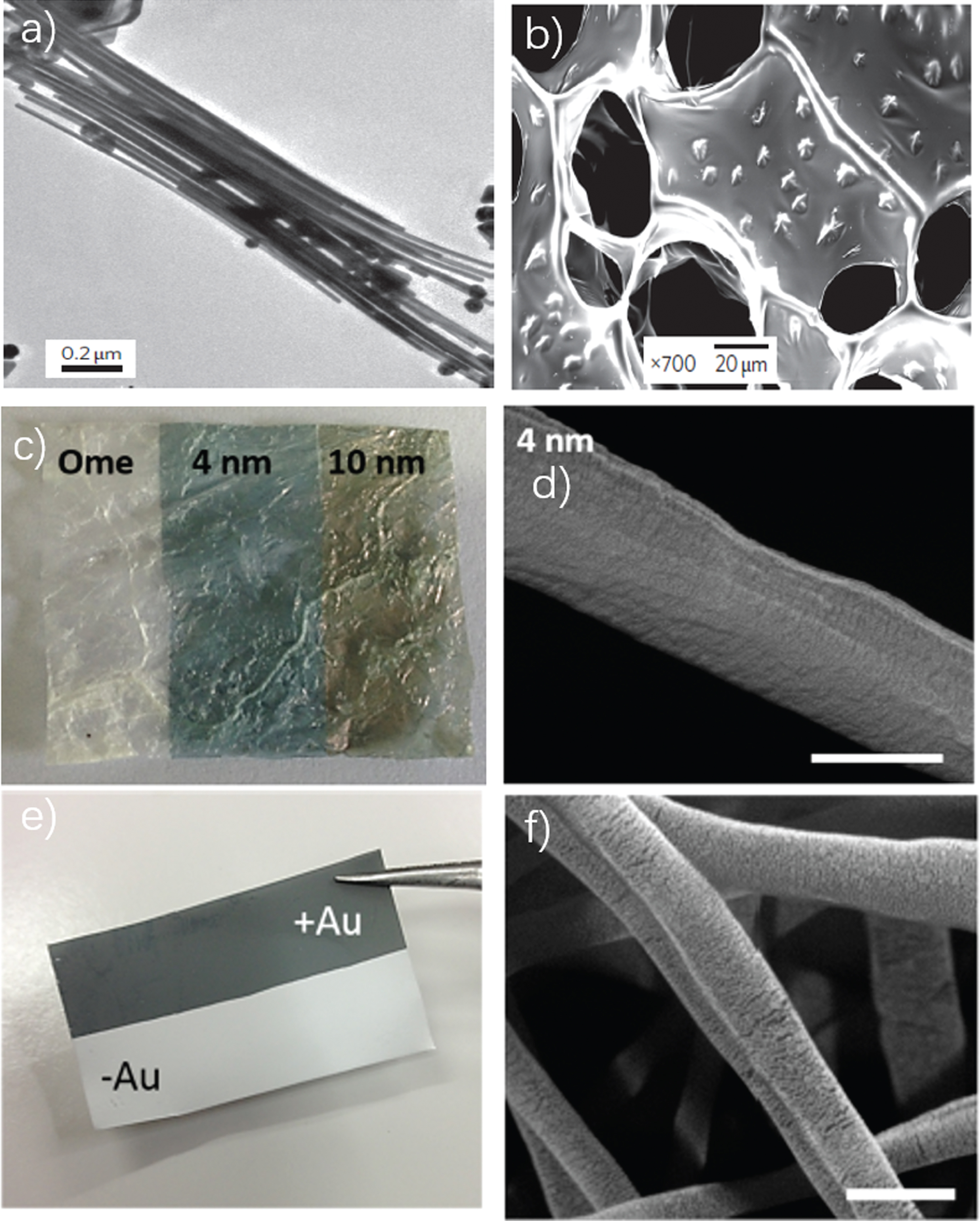
a) TEM image of the gold nanowire, b) SEM revealed AuNWs assembled within pore walls of the scaffold [99], c) AuNPs were deposited onto decellularized omentum scaffolds, the color changed due to the difference in NP dimensions. d) ESEM image of 4 nm AuNPs deposited scaffold, scale bar = 500 nm [102], e) AuNPs were evaporated on PCL nanofibrous scaffold surface, f) ESEM image of AuNPs on the surface of the fiber, scale bar = 5μm [103].
Tian et al. have firstly fabricated the nanoelectronic scaffolds (nanoES) through hybriding with synthetic or natural biomaterials. 3D macroporous nanoES could be alone or combined with other biomaterials as conductive scaffolds for a 3D culture of neurons, cardiomyocytes and muscle cells due to its robust electronic properties. Moreover, the hybrid conductive scaffolds shows a sensory capability of the nanoES by real-time monitoring of the local electrical activity and responding to the environment changing (drugs or pH) [104]. Similarly, Feiner et al. have integrated complex electronics with 3D nanofibrous scaffold for cardiac tissue engineering (Fig. 4), where the robust electrotonic properties were able to record cellular activates and employ electrical stimulation for synchronizing cardiac cells contraction [105]. Moreover, drug control release function has also been integrated within the hybrid scaffolds.
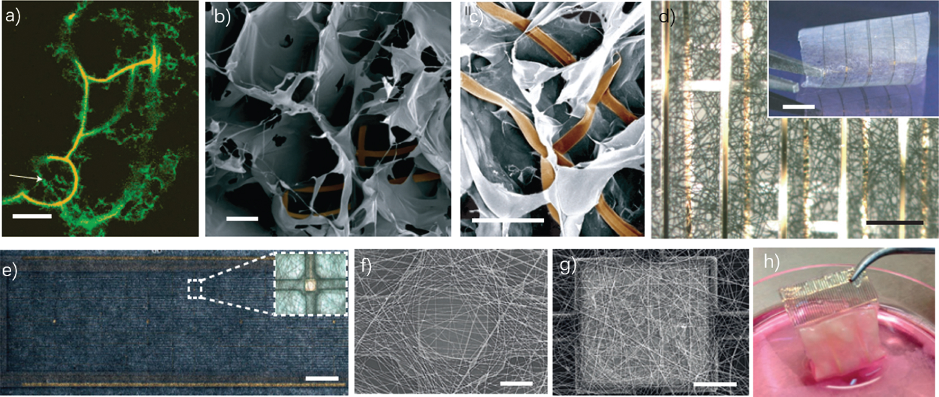
a-d hybrid macroporous nanoelectronic scaffolds. a) Confocal fluorescence micrograph of a hybrid reticular nanoES/collagen matrix. Green (fluorescein isothiocyanate): collagen type-I; orange (rhodamine 6 G): epoxy ribbons. The white arrow marks the position of the nanowire. Scale bar, 10μm. b, SEM images of a mesh nanoES/alginate scaffold, top (b) and side (c) views. The epoxy ribbons from nanoES are false-colored in brown for clarity. Scale bars, 200μm (b) and 100μm (c). d), A bright-field optical micrograph of the folded scaffold, showing multilayered structures of PLGA and nanoelectronic interconnects. The inset shows a photograph of the hybrid sheet before folding. A sheet of PLGA fibers with diameters of 1–3μm was deposited on both sides of the device. No damage or reduction of device yield was observed following this deposition. Scale bars, 200μm and 5 mm (inset) [99]. e-i Biomaterial–electronics hybrid. e) A stitched optical image of nanocomposite fibers of PCL–gelatin deposited on the electronic mesh. Scale bar = 1.5 mm. Inset, a single sensing/stimulating electrode covered with the nanofiber scaffold. f) SEM image of a recording/stimulating electrode covered by electrospun fibers. Scale bar = 20μm. g) The deposited electroactive polymer on the designated electrode was held by the nanocomposite fibers. Scale bar = 50μm. h) An image of the folded microECP after 7 days of cultivation with cardiac cells [105].
Engineered micro and nanoscale scaffolds have been used to mimic the ECM structure for supporting cells. Many studies demonstrates that the properties of scaffolds (e.g. topography, stiffness, mechanical properties) could affect cell behaviors such as adhesion, morphology, proliferation, differentiation, and migration [106–108]. However, the scaffolds made by biomaterials are commonly nonconductive, and electrical stimulation (ES) has presented many notable beneficial effects on biological applications especially for cardiac and neural tissues [21]. Thus, scaffolds composite with conductive materials have become an excellent candidate for tissue engineering because of their notable high efficiency of ES delivery.
Cardiac tissue engineering
Cardiac muscle is a significant electroactive tissue, which can transfer electrical signal and retain the beating heart [109]. The poor regeneration potential of cardiomyocytes leads to a very limit self-repair ability, in addition, the heart resource is even harder to be found. Thus, cardiac tissue engineering has become the most promising solution for repairing damaged heart tissue. Previously, many artificial scaffolds have been fabricated to support cardiomyocytes growth, and the corresponding designed scaffolds show some benefits for cells adhesion and beating function [110, 111]. However, the scaffold made by biomaterials usually exhibits high electrical resistance, which could cause cells to lose communications. The conductive scaffolds which could overcome this issue by allowing conductive polymers and conductive nanomaterials to synthesize composite scaffolds, could promote the electrophysiological communications between cells. So far, many attempts have been made for cardiac tissue engineering.
Table 1 has summarized several represented works which use conductive scaffolds in cardiac tissue engineering. For example, Kai et al. have used electrospun to synthesize PPy-PCL/gelatin scaffolds by balancing the concentration of PPy to maintain the other important properties (e.g. mechanical properties, biodegradability), and the cardiomyocytes cultured on this conductive scaffold have shown better adhesion and expression of cardiac-specific proteins [112]. Similarly, other CPs (PANI and Polyurethane) can also be used as electrospun with biomaterials (PLGA and PCL) to form conductive fibrous scaffolds, and the cardiac genes involved in contraction and relaxation (Troponin-T), cytoskeleton alignment (actinin-4) and gap-junction protein connexin 43 (Cx43) have been upregulated on conductive scaffolds (Fig. 5), moreover it is worthy of noting that the positive charge CPs have significantly improved the cell adhesion via electrostatic [35, 113].
Conductive scaffolds for cardiac tissue engineering
Conductive scaffolds for cardiac tissue engineering
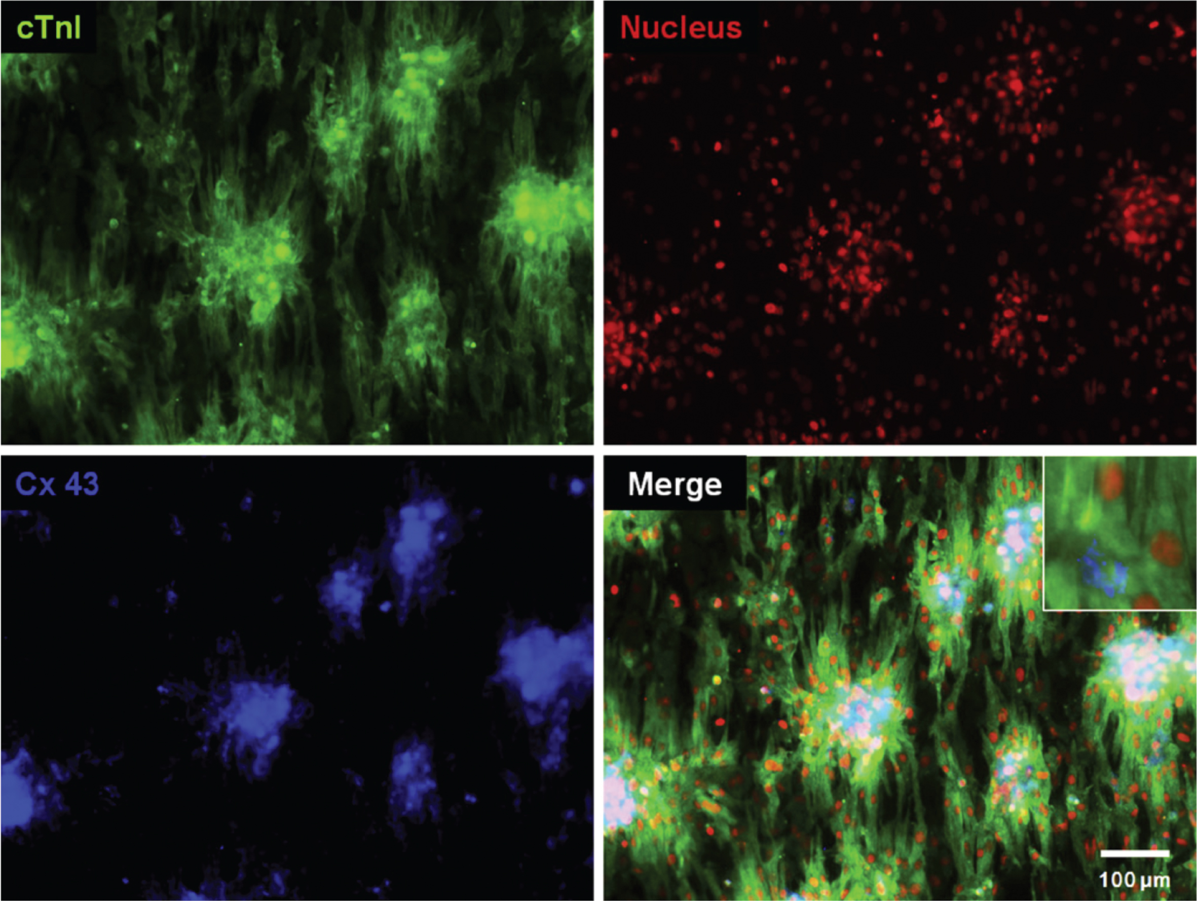
Fluorescence micrographs of the neonatal rat cardiomyocytes cultivated on PANI/PLGA mesh using the immunofluorescence staining for cardiac troponin I (cTnI, green), connexin 43 (Cx 43, blue), and nucleus (red). The inset in the panel shows a higher magnification image [35].
Compared with CPs, CNMs can not only provide conductivity but also improve 3D nano-environment in situ. Martinelli et al. have seeded cardiomyocytes on the substrate coated with CNTs film, which shows that the cardiomyocytes form tight contacts with CNTs. Moreover, they have observed the changes in the electrophysiological properties of cardiomyocytes, which reflects that CNTs are capable of promoting cardiomyocyte maturation [114]. CNMs biocomposites scaffolds also presents many benefits for cardiac tissue engineering. Through dopping CNTs into GelMA hydrogel, Shin et al. have discovered that the additional CNTs can enhance both mechanical and electrical conductivity properties of the hydrogel, and promote myocardial tissue attachment and proliferation. More interesting is comparing with pristine GelMA hydrogel, the spontaneous synchronous beating is 3 times higher than 50μm thick CNT-GelMA [115]. In addition, the constructs have been seeded with cardiomyocytes on CNT-GelMA gel which have presented homogenous Cx43 distribution and partial uniaxial alignment of sarcomeric structures [116]. Currently, GO-GelMA conductive scaffolds have also been synthesized for culturing cardiac tissue. The results indicate that GO could not only provide conductivity to the scaffolds but also affect many biological activities such as cell viability, proliferation, and maturation. Moreover, the cardiomyocytes has shown stronger contractility and faster spontaneous beating compared with GelMA [117]. Similarly, the nanofibrous CNTs-GelMA demonstrates suitable mechanical and conductive properties for cultivation cardiomyocytes with maintaining the viability and contractile activities. The cardiomyocytes can result in stronger spontaneous and synchronous beating behavior (Fig. 6) [118]. Martins et al. have found the increased expression of cardiac-specific genes involved in muscle contraction and electrical coupling on the carbon-chitosan porous scaffold without ES [119].
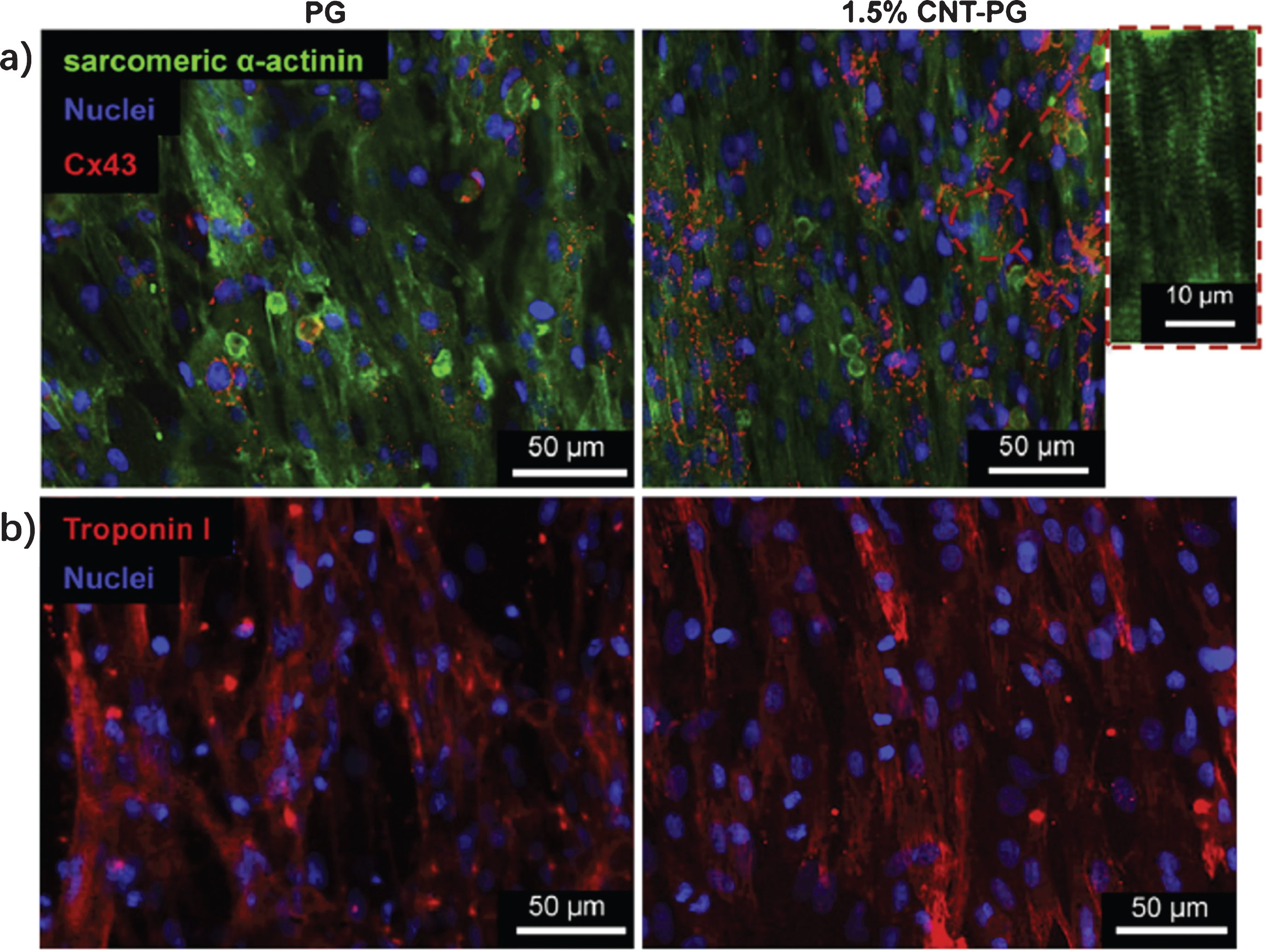
CM adhesion, viability, metabolic activity and maturation on CNTs-gelatin scaffolds. a) Sarcomeric a-actinin (green), Cx43 (red) and DAPI (blue), and b) Troponin I (red) and DAPI (blue) after 7 days of culture, revealing organized sarcomeres with a higher Cx43 expression on 1.5% CNT-PG than PG scaffold [118].
Recent studies have shown that the expression of Cx43 and reparative paracrine factors could be improved by adding graphene oxide (RGO) into mesenchymal stem cells (MSCs). In addition, RGO could adsorb ECM proteins (fibronectin) which affect the cell-ECM interaction. The implantation of RGO-MSCs hybrid spheroids into the infarcted myocardium could enhance cardiac repair and cardiac function restoration. Therefore, RGO has shown potential to enhance the therapeutic efficacy of MSCs for the treatment of myocardial infarction (MI) [120].
As the pioneers employing metal-based nanomaterials in cardiac tissue engineering, Dvir and his colleague have employed gold nanowires into the porous scaffold as a conductive bridge to transfer electrical signal through neighbor pore walls. With this approach, they have created homogeneous cardiac patches with stronger contractile properties (Fig. 7a–d) [99]. Later, they have investigated gold nanoparticles evaporation strategy for depositing AuNPs on the surface of scaffolds to improve conductivity. As shown in Fig. 7e, f, cardiac cells seeded on such scaffolds have presented good adhesion, proliferation, and maturation, moreover, the expression of electrical coupling proteins connexin 43 are significantly upregulated [100–102]. Similarly, the AuNPs formed inside hydroxyethyl methacrylate (HEMA) hydrogel also provide a new method to synthesize conductive scaffolds. The cardiomyocytes can increase the expression of connexin 43 on hybrid scaffolds with and without electrical stimulation [121].
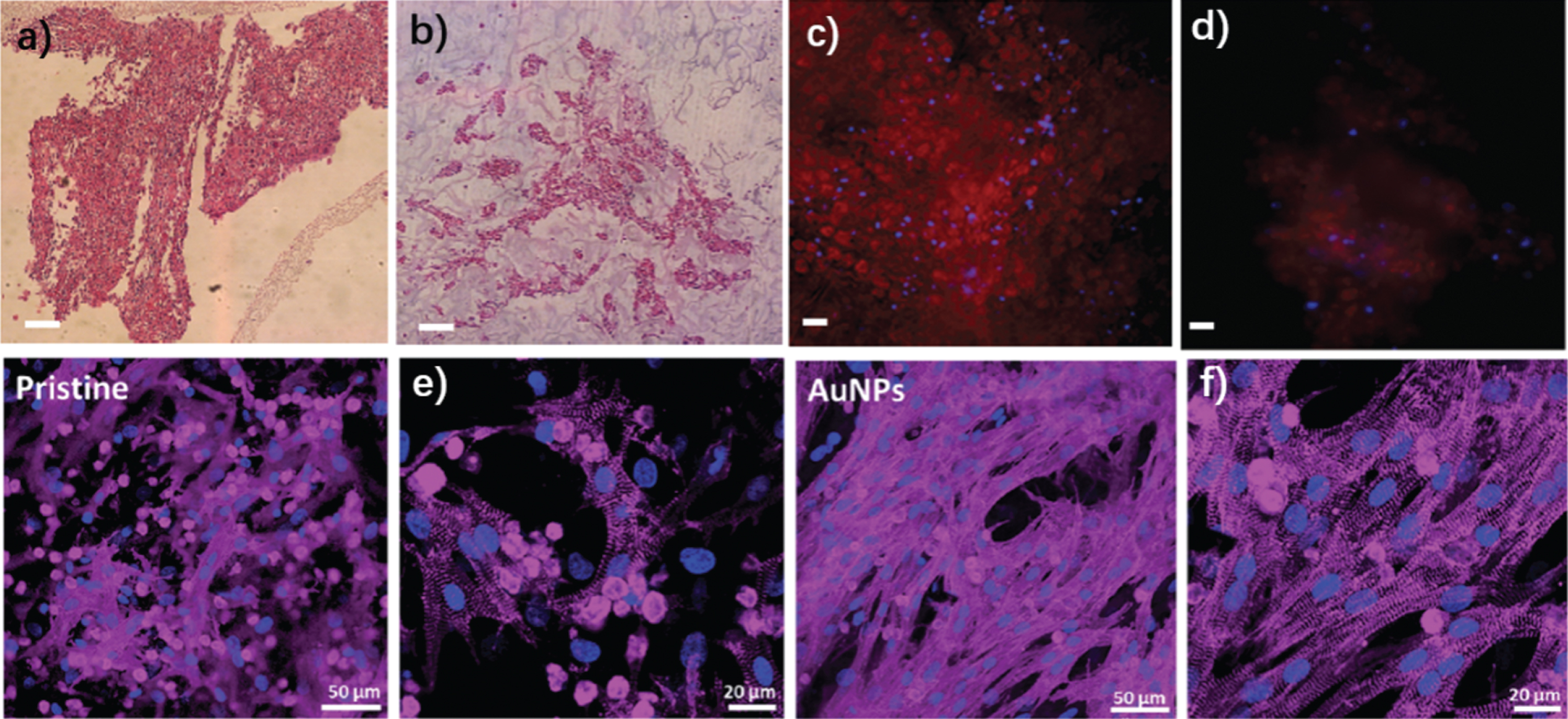
a-d cardiac cell organization within AuNWs scaffolds. a) H&E stained thin sections of the engineered tissues on day 8 revealed a thick tissue in the nanowire scaffold, b) the engineered tissue in the pristine scaffolds demonstrated non-continuous tissue separated by pore walls. c) Immunostaining of the cell-seeded scaffolds on day 8 revealed pervasive troponin I expression (red) within the Alg–NW scaffold. d) less staining was observed in the aggregates in the unmodified scaffolds [99]. e-f Cardiac sarcomeric actinin immunostaining on day 7. Actinin – pink, nuclei – blue. e) Cardiac tissue engineered within pristine PCL scaffolds. f) Cardiac tissue engineered within AuNPs-PCL scaffolds [101].
In another study, Tan et al. have shown the incorporation of conductive silicon nanowires in scaffold-free human could induce pluripotent stem cells (hiPSCs) to be derived into cardiomyocyte (CM) spheroids, where SiNWs form an electrically conductive network inside spheroids (Fig. 8). The CM spheroids which combined with SiNWs have presented strong synchronized contraction [122]. Moreover, an exogenous electrical stimulation has been applied to the nanowire hiPSCs-CM spheroids to further develop their functions. The results show that both SiNWs and ES can synergistically improve cell junction formation by increasing contractile properties and reducing endogenous spontaneous beating rate of hiPSCs-CM spheroids [123].
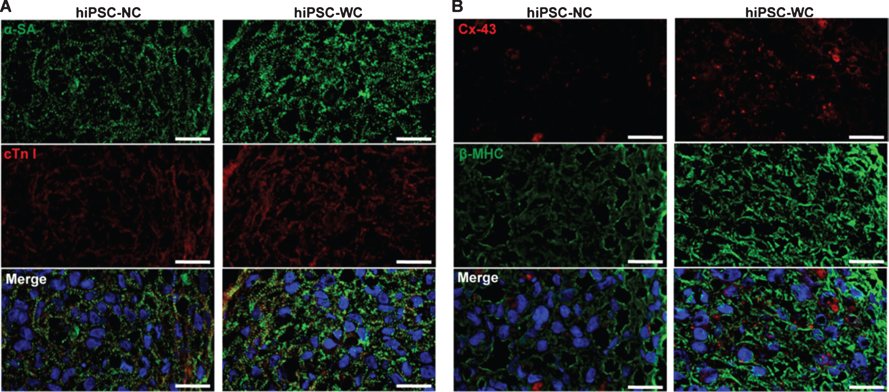
Structural analysis of hiPSC-derived cardiomyocyte spheroids. (A) Immunofluorescent staining of alpha sarcomeric actinin (α-SA) and troponin I (cTn I). (B) Immunofluorescent staining of connexin-43 (Cx-43) and beta-myosin heavy chain (β-MHC). hiPSC-NC=human induced pluripotent stem cell cardiac spheroids, no e-SiNWs, no stimulation; hiPSC-WC=human induced pluripotent stem cell cardiac spheroids, with e-SiNWs, no stimulation Scale bars = 20μm [122].
Central nervous system (CNS) plays an important role in the interaction between human biological processes and physiological processes. The injury of CNS could result in physical trauma, anoxia, hypoglycemia, diabetes, and viral infection. Even tough nerve axons can regenerate, it is limited to 5 mm and coaptation is usually necessary [124]. In terms of nerve regeneration and engineering, a number of studies demonstrates that scaffolds with optimized topographic, chemical and electrical cues could enhance neurocyte functions. Besides, neurocyte is a significantly electroactive tissue and its behaviors could be strongly affected by the electrical signals and electrical stimulations. Thus, electrical conductive scaffolds present a great potential in neural regeneration and engineering.
Prior conductive nanomaterials (CNMs), conductive polymers (CPs) are the best candidates for generating conductive scaffolds. Schmidt et al. have evaluated PPy film for the cultivation of PC-12 cell, and according to their research, PPy can enhance nerve cell interactions and it can significantly increase the neurite lengths with ES [125]. Electrospinning is the most favorable method for fabricating CPs nanofibrous composites scaffolds in tissue engineering. In last decade, CPs (PPy, PANI) have been mixed with various biopolymers (PCL, PLGA, gelatin, PLLA) and electrospun in conductive scaffolds to cultivate nerve cells (PC-12, NSCs, DRG). As shown in Fig. 9, the conductive scaffolds could enhance nerve cell interaction and proliferation, moreover, with applied electrical stimulation, the neurite outgrowth can be significantly increased to facilitate the regeneration of nerve [126, 127].
A-F Confocal microscopy micrographs at 24 h of PC12 cells cultured on different polymeric materials. The different scaffolds are A&B) Polycaprolactone fumarate (PCLF), C&D) PCLF-PPy naphthalene-2-sulfonicacidsodiumsalt (NSA), E & F) PCLF-PPy dodecylbenzenesulfonic acid sodium salt (DBSA), G-H Fluorescence microscopy images of neurite extension from DRG explants cultured on G) PCLF-PPy-NSA, H) PCLF-PPy-DBSA [127].
On the other hand, CNMs can mimic the hierarchical structure of the ECM, which promote selectively proteins adsorption and retain their bioactivity. The large surface area and high conductivity are favorable to nerve stem cells (NSCs) attachment and to establish the intercellular communication [128]. The electrical conductivity of CNMs is able to transfer ES, which may alter the local electric field of ECM proteins, improve attachment on scaffolds, and subsequently affect the neurite outgrowth [36].
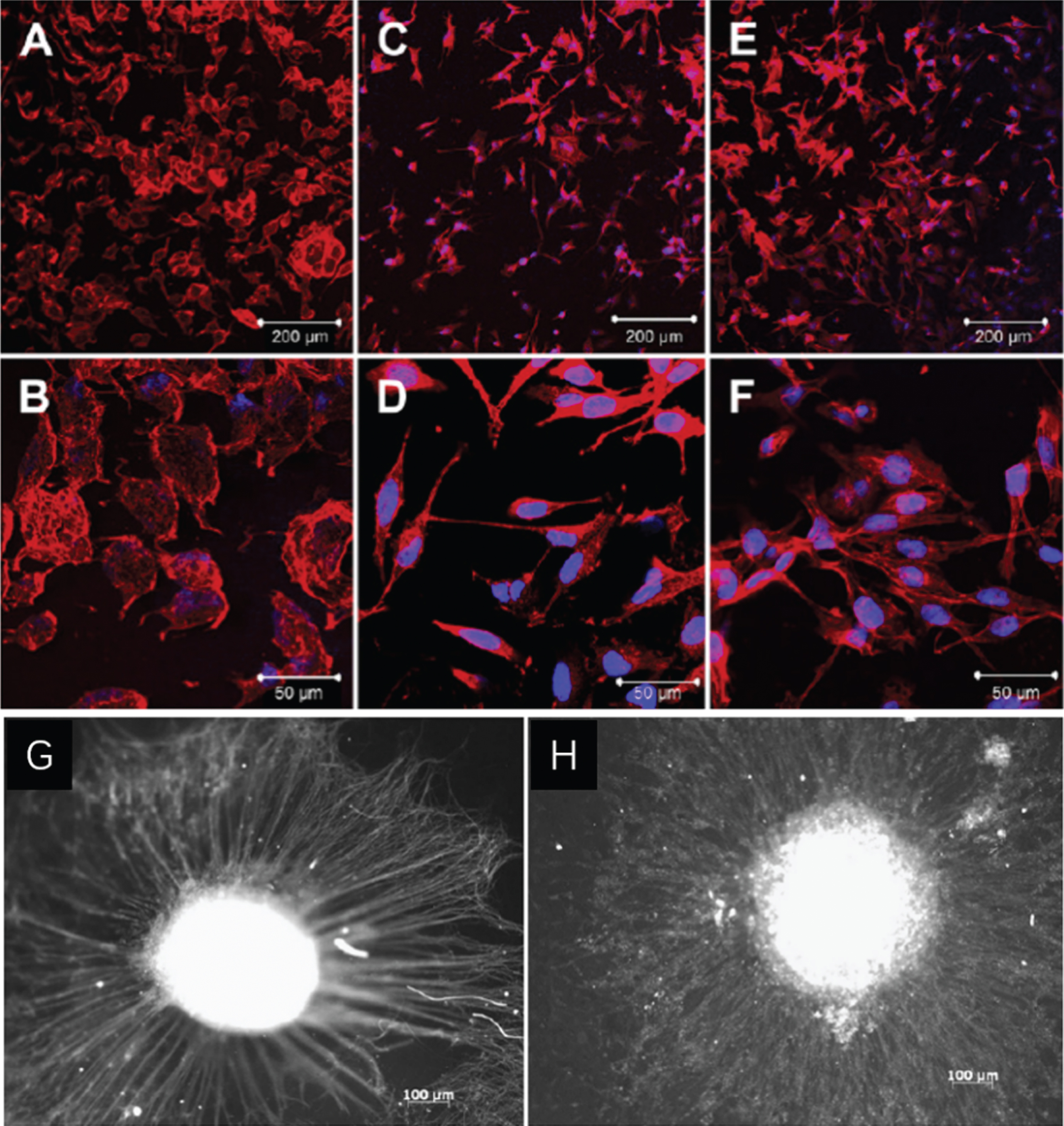
Due to its attractive architecture, physical, chemical and electrical properties, Carbon nanotubes/nanofibers have been employed in neural tissue engineering for promoting neuron growth and increasing the transmission signal among neurons [129]. Cellot et al. have proposed an “electrotonic hypothesis”, which can explain the physical interactions between the cell and carbon nanotube, and the mechanisms of carbon nanotubes affecting the collective electrical activity of cultured neuronal networks [130]. Base on single-cell recording and patch-clamp technique, it has been found that by interfacing to interface to neurons, CNTs can induce a potentiation of spontaneous synaptic activity in cultured neuron networks. A number of attempts have been made to use CNTs based scaffold in neural tissue engineering applications. For example, by seeding human bone marrow mesenchymal stem cells (hBMMSCs) on the carboxylated multiwall carbon nanotubes films, Chen et al. have found that CNTs can induce and maintain neural differentiation of hBMMSCs without any exogenous differentiation factors (Fig. 10) [131]. Similarly, Chao et al. have grafted CNTs with poly(methacrylic acid) (PMAA) to improve neuronal cells adhesion. Their results indicates that the grafted CNTs film scaffolds can provide a niche for human embryonic stem cells (hESCs) differentiation into neuronal cells [132]. By coupling with electrospun PLLA nanofibrous aligned scaffolds, CNTs can be synthesized and it presents the ability to enhance neural differentiation of embryonic stem cells (ESCs) and maturation with high expression of mature neuronal markers (Map-2) [133]. By interfacing on phosphate glass fibers scaffolds, Ahn et al. have synthesized CNTs which possesses good cell viability and neuronal interactions. Moreover, the corresponding in vivo experiments in rat sciatic injury model have demonstrated CNTs based scaffolds can be used at the interface between the nerve conduit and peripheral neural tissues [134].
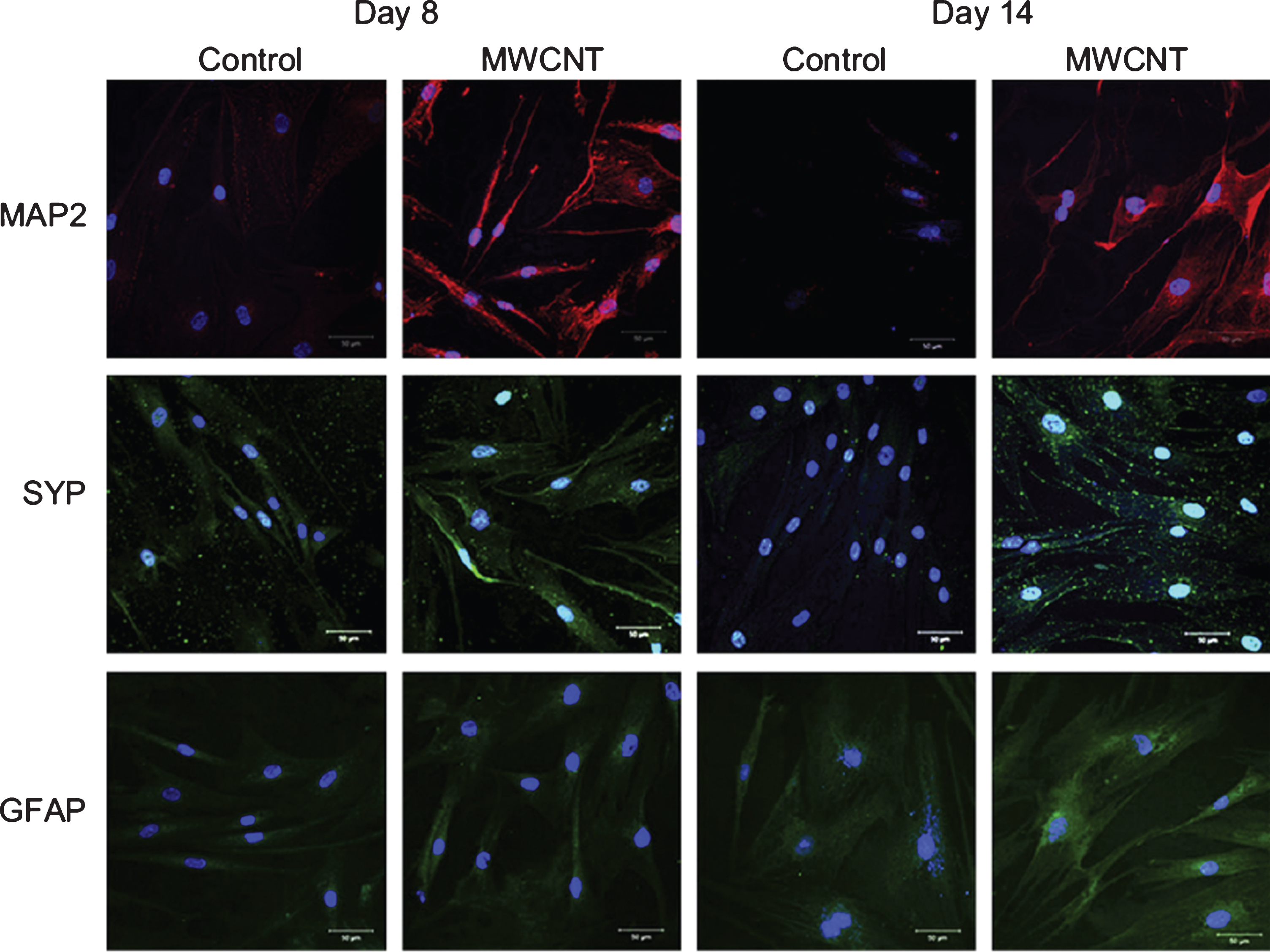
Immunofluorescence of hBMMSCs cultured on control (columns 1 and 3) and MWCNT groups (columns 2 and 4) in the basal medium. Neural protein markers including MAP2 (top), synaptophysin (SYP, middle) and GFAP (bottom) on days 8 (columns 1 and 2) and 14 (columns 3 and 4) were detected by confocal microscopy [131].
Similarly, as another carbon-based nanomaterial with 2D honeycomb lattice (sheet of CNTs), graphene and graphene oxide (GO) recently gain the most attention in biological applications. In neural tissue engineering, the conductive graphene and GO can act as the “brother” of CNTs, which could induce neuron differentiation and maturation. For instance, Park et al. have seeded human neural stem cells (hNSCs) on a graphene substrate, and their research shows that the grapheme can enhance the differentiation of hNSCs into neurons rather than glia during a long-term (Fig. 11a) [135]. The fluorinate modified graphene can demonstrate enhanced cell adhesion and proliferation of MSCs, which caused by a neuro-inductive effect via spontaneous cell polarization [136]. Besides, the fluorinated graphene has significant effects on cell morphology, cytoskeletal and nuclear elongation of MSCs. Another interesting finding which obtained by Solanki et al. shows that GO could also promote neuron differentiation of hNSCs as the extending axons begin aligning on the GO-based substrate. Even though the mechanism of governing axonal alignment has not been understood yet, only the GO-based substrate presented a great potential for central nervous system repair based on the neuron differentiation with self-aligned of axons (Fig. 11b, c) [55]. Tang et al. have shown that NSC differentiates structurally and functionally neural networks on graphene films, and the grapheme can enhance the network activity and the efficacy of neural signal [137].
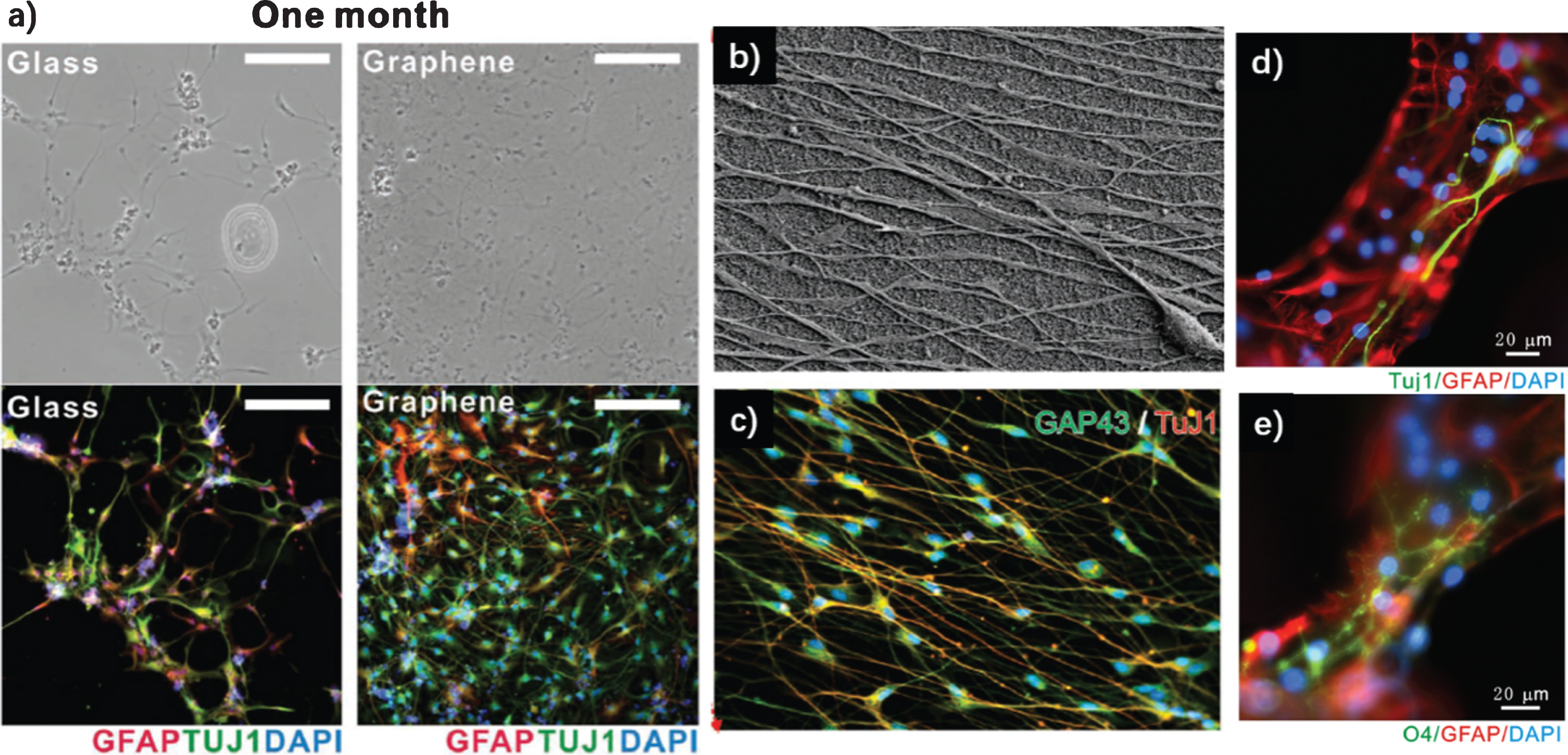
a) Bright-field (top row) and fluorescence (bottom row) images of hNSCs differentiated on glass (left) and graphene (right) after one-month differentiation. The differentiated hNSCs were immunostained with GFAP (red) for astroglial cells, TUJ1 (green) for neural cells, and DAPI (blue) for nuclei. Note that more hNSCs were adhered to graphene than to glass [135]. b-c Axonal Alignment of differentiated hNSCs on SiNP-GO on flexible and biocompatible substrates made from polydimethylsiloane (PDMS). b) SEM image of SiNP-GO on PDMS substrate showing highly aligned axons from hNSCs on Day 14. c) Immunocytochemistry results showing the expression of neuronal marker TuJ1 and axonal [55]. d-e Representative fluorescence images of differentiated NSCs under differentiation conditions, the cells were immunostained with Tuj-1 for neuron (green, d), GFAP for astrocyte (red, d&e), O4 for oligodendrocyte (green, e) and DAPI for nuclei (blue, d&e) [138].
A 3D graphene porous conductive scaffold has been fabricated by the Ni foam template. The NSCs on the 3D scaffold can not only keep a more active proliferation state with upregulation of Ki67 expression, but also enhance the NSCs differentiation to astrocytes and especially neurons (Fig. 11d, e) [138]. GO can also be coated on electrospun polymer nanofibers. For example, Feng et al. have fabricated the GO assembled on poly(vinyl chloride) nanofibrous scaffolds for cellular electrical stimulation, which have demonstrated an unprecedented accelerated growth and development of the primary motor neurons in a long-term culture period. In addition, the selective assemble GO on nanofibers extended the superior properties of graphene from traditional 2D nanoscales into 30 microscales architectural geometry [139]. More interesting is that the GO coated PCL nanofibrous scaffolds have shown selective differentiation of neural stem cells into mature oligodendrocytes without any inducers [140]. The selection is caused by merely changing the properties of the underlying biomaterial scaffold, and it is a valuable approach for tissue engineering by eliminating the use of exogenous differentiation inducers.
Recently, gold nanoparticles coated nanofibrous scaffolds (PCL-gelatin) have also been used in neural tissue engineering. The additional AuNPs on nanofibers can not only provide extra topographical properties of neuron cells adhesion and proliferation, but also improve the axonal elongation which leads to more complex neuronal networks [103]. Moreover, Orza et al. have found that the electrical conductive gold-coated collagen nanofibers could enhance the differentiation of placenta-derived mesenchymal stem cells into both myocardial and neuronal cells by investigating high expression of specific differentiation markers (atrium, natriuretic peptide, actin-f, and actin monomer, glial fibrillary acidic protein, and neurofilaments) [141].
In bone tissue engineering, the topographical cues of bone graft play an important role in cell adhesion, proliferation, differentiation, migration, and maturation [142, 143]. In addition, the bone tissue is also associated by electrical signals. Herein, employing electrical conductivity to existing scaffolds could significantly promote bone tissue behaviors.
Pelto et al. have studied how electrical stimulation (ES) affect the proliferation and osteogenic differentiation of human adipose stem cells (hASCs) on PPy-coated PLA scaffolds. According to their research, the conductive scaffolds can enhance hASCs proliferation significantly comparing with PLA scaffolds, and the alkaline phosphatase (ALP) activity has also increased [144]. By seeding Saos-2 cells on conductive scaffolds (PPy-PLLA), Meng et al. have found that that ES could modulate osteoblast activity through the conductive scaffold, including upregulation of osteoblast proliferation and expression and production of specific marker ALP (maturation) and OC (mineralization stage of bone formation) [145]. A 3D porous conductive scaffold has been synthesized by composing PEDOT-gelatin-bioactive glass through freeze-drying, and it can promote hMSC growth with high viability by adjusting the concentration of PEDOT. Even though ES has not been applied in this work, the PEDOT based scaffold has also shown great potential for bone healing [146].
By coating MWCNTs on the surface of 3D collagen scaffolds for culturing rat primary osteoblasts (ROBs) and implanting after 1 day, Hirata et al. have observed significantly higher alkaline phosphatase activity, calcium and osteopontin contents on CNTs based scaffolds, in addition, the ROBs differentiation have been earlier compared with nonconductive scaffolds. Even though a slight inflammation have been observed around CNTs after implantation, there have been more bone formation observed [147]. Li et al. have compared the effect of MWCNTs and graphite with the same dimension on human adipose-derived stem cells cultured in vitro and ectopic bone formation in vivo., and they have found that the MWCNTs can adsorb more proteins, which might not only improve cell adhesion and proliferation, but also promote the expression of osteogenic genes (ALP, cbfa1, and COLIA1), while graphite could not. The different effect of MWCNTs and graphite might attribute to the special surface structure. In addition, MWCNTs have also induced ectopic bone formation but graphite could not either. Therefore, CNTs have shown its capability in modulating downstream stem cellular response without exogenous growth factors [148].
Similarly, Nayak et al. have evaluated how graphene effect (hMSCs) on differentiation into bone cells. To minimize the stiffness and roughness of scaffold effected, graphene has been deposited on four different substrates (PDMS, PET, glass and silicon wafer). Its results indicate graphene on all substrates could promote hMSCs differentiation. More interesting, graphene have presented a remarkable accelerated rate for hMSCs differentiation into bone cells compared to the growth factor (BMP-2) [149]. GO-chitosan porous scaffolds which synthesized by covalently linking the carboxyl groups of GO with the amine groups of chitosan present a capability to modulate the biological response of osteoblasts, including cell attachment, proliferation, and growth [98].
Conclusions and perspectives
In summary, tissue engineering is the most promising pathway of repairing or replacing damaged organs. Studies have shown that the artificial scaffolds with proper topographical, physical, chemical and mechanical properties could mimic ECM, and provide a suitable microenvironment for cell adhesion, proliferation, and differentiation. In this review, we have chosen to focus on using electrically conductive materials (CPs and CNMs) to fabricate conductive scaffolds. The conductive composite scaffolds can not only possess all properties of the pristine scaffolds but also gain electrical conductive property which could significantly enhance cell to cell and cell to scaffold interaction. Even though most of the conductive polymers and conductive nanomaterials have been proved with low or no cytotoxicity, the non-biodegradability still has some limitation in vivo applications. The results of cell culture on conductive scaffolds both in vivo and in vitro have shown better adhesion and proliferation, moreover, the differentiation and maturation have been significantly promoted on the conductive scaffold with or without electrical stimulation. Therefore, the conductive scaffolds can be the most promising candidate for tissue engineering, especially for electrophysiological tissues including cardiac, neuron, bones, muscle and skin.
Moreover, different tissue cells cultured on conductive scaffolds have also shown highly maturation and function, which could be used for in vitro cell studies or fabricating complex biomimetic tissue-like structures for many biological and biomedical application, including organs-on-a-chip, disease model, drug screening and evaluation.
Conflict of interest
The authors have no conflict of interest to report.
Footnotes
Acknowledgments
This research was funded by the PhD development fund of Jiangxi University of Science and Technology (grant no. 3401223374); national nature science foundation of China (grant no. 21607151) and natural science foundation of Liaoning Province of China (grant no. 2018011534-301).