
Abstract
The conventionally synthesized nano-ferrite materials do not possess bulk properties, generally required for their use in mainstream industry. To make ferrite nanoparticles clinically applicable materials, it is important to have good control over morphology and optical properties of these materials. In this study, low-pressure microwave plasma was used to tailor the structural properties and surface chemistry of manganese ferrite nanoparticles. A facile sol-gel method was used to prepare cubic spinal structures of manganese ferrite nanoparticles. These nanoparticles were exposed to oxygen plasma sustained with a microwave source for improving their magnetic and photocatalytic activities. The techniques like XRD, SEM, PL, UV-Vis DRS, transient photocurrent response and EIS were used to characterize the samples. The plasma treated nanoparticles were used to degraded methyl blue (MB) dye in the solution. The photocatalytic activity showed 85% degradation of MB after 100 min of exposure of visible light. The second part of the paper studied the magnetic properties of the nanoparticles. The saturation magnetization decreased from 0.78 emu/g to 0.68 emu/g after plasma treatment of nanoparticles.
Introduction
Iron oxide nanoparticles have been extensively used in the fields of catalysis, pigments, biomedicals, electronics and data storage. These applications are ascribed to their exceptional optical, thermal and magnetic properties. Several studies reveal that physico-chemical traits of nanomaterials strongly depend on their morphology, size, shape, synthesis process and synthesis conditions [1–4]. Moreover, iron oxide nanoparticles with strong magnetic properties have previously been verified to be useful in biomedicine due to their strong chemical affinity, non-toxicity and biocompatibility while maintain their inherent magnetic properties [5]. Similarly, crystallite size of iron oxide nanoparticles strongly influences the magnetic properties as these nanoparticles become paramagnetic in nature below 15 nm while ferromagnetic aspect dominates for larger particle sizes [6–8].
The anticipated remediation of environmental pollution using a photocatalytic route has stimulated the intense research to design the efficient photocatalysts to meet the industrial standards. Iron oxide has been studied for photocatalytic application due to its appropriate stability, abundance, eco-friendliness and inexpensiveness. Iron oxide having a suitable optical photonic band gap of 2.1 eV has been proved to be effective material for photocatalytic applications [9, 10]. However, pristine iron oxide has been found to show insufficient photocatalytic results due to poor charge carrier potential, short exciton lifetime and small absorption coefficient, thereby rapid annihilation of charge carriers. Therefore, several strategies have been adopted to reduce the electron-hole recombination of iron oxide nanoparticles [11–14]. Doping of transition metals into metal oxides is one of the key strategies used for improving the lifetime of the excitons in metal oxide photocatalysts. Among transition metals, manganese (Mn) is 2nd highest transition metal element found in the earth and extensively used to suppress the electron hole recombination of semiconductor photocatalyst, like ZnO and TiO2 under light irradiation. Sing et al. [7] reported improved photocatalytic activities of Mn doped ZnO for degradation of different organic contaminates under light irradiation. The optimum Mn doped ZnO photocatalyst showed the maximum photocatalytic efficiency of 93% for congo red degradation due to effective electron-hole inhibition due to the presence of Mn+2 ions in the framework of ZnO. Similarly, Chauhan et al. [15] used sol-gel method to produce Mn doped TiO2 photocatalyst. They tested the photocatalytic activity of this photocatalyst by degrading MB under both UV and visible light irradiation. The optimum Mn doped TiO2 photocatalyst with 3 mol% of Mn demonstrated the highest red shift in the optical absorption and highest prolongation of charge carriers due to the effective role of Mn2+ to show better degradation efficiency under visible light irradiation than UV light 1 [6]. However, the selectivity of Mn as doping material to increase the magnetic and photocatalytic activity of iron oxide has never been reported. The introduction of Mn2+ into the framework of iron oxide will generate the intermediate energy levels between the VB and CB of iron oxide. The presence of these intermediate energy levels will effectively suppress the exciton recombination and consequently will increase the photocatalytic activity.
Majority of the conventional nano-ferrite materials do not own the bulk properties, generally required for their use in mainstream industry. The unprocessed and inactivated materials remain chemically inert and show low surface energy to different functional groups to interact with their surface. An effective way of making the ferrite nanoparticles clinically applicable materials is to modify their surface. However, most traditional approaches are not only energy intensive and costly but also harm the morphology of the material under treatment. Plasma discharges, on the other hand, are being used to produce energetic reactive species to tailor the surface chemistry and structural properties of almost all types of materials [17, 18]. In this study, we prepared Mn doped iron oxide ferrites using facile sol-gel method and then subjected to microwave plasma treatment in oxygen rich environment for improving the magnetic and photocatalytic properties. The photocatalytic activity of plasma modified Mn doped iron oxide was evaluated by degrading methyl blue under visible light irradiation. The plasma modified Mn doped iron oxide photocatalyst showed superior photocatalytic activity as compared to untreated photocatalyst.
Materials and methods
Sol-gel synthesis of nanoparticles
The manganese-iron oxide nanoparticles were produced using a simple sol-gel technique. Merck grade manganese nitrate hexahydrate and ferric nitrate nanohydrate were used in this synthesis. The apparatus was cleaned using deionized water contamination free production of the ferrite nanoparticles. Mn to Fe ratio was taken as 1:2. The metal salts were dissolved in deionized water and stirred and heated at 80°C using a magnetic hot plate. The citric acid was added dropwise under continuous stirring to produce sol from the solution. The sol was heated further to obtained gel. The gel settled down after some time. The heating was stopped at this point and the product was dried overnight in an oven at 90°C. the final product was grinded to obtain fine powder. The powder was sintered for 6 hours in a furnace at 700°C to induce better magnetic properties.
Plasma treatment of the product
The calcined manganese ferrite powder was exposed to microwave plasma of oxygen for 10 min in a partially evacuated chamber. Being a low-cost gas, oxygen was used to produce plasma discharge. A microwave source with a fixed input power of 900 W was used to perform the plasma procedure. Figure 1 shows the schematic of the microwave plasma setup. The ferrite powder was placed on a fixed holder in the plasma chamber, which was covered with a radiation shielding box. The chamber was evacuated using a vacuum pump and then oxygen gas was entered into the chamber in flow mode conditions. A microwave power source was used to break the molecular oxygen into ions, electrons, reactive oxygen radicals and UV radiations. There species interacted with the particle surface and changed its morphology and functionality through sputtering and oxidation reactions.
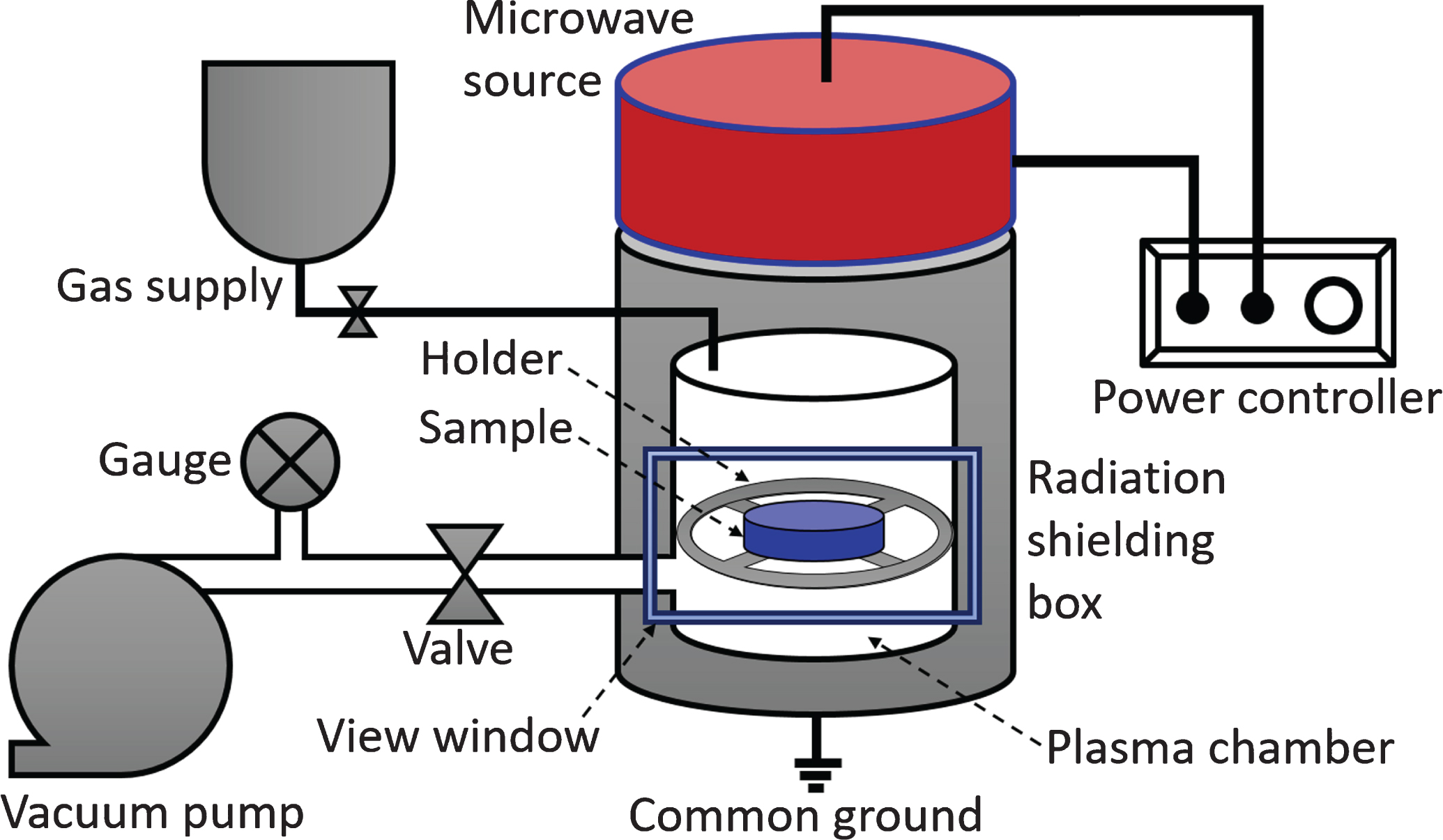
A schematic of the plasma setup used to treat ferrite powder.
The surface morphology, structures, crystallite size, band gap energy, photocatalytic activity and magnetization of the plasmas processed samples were studied using a set of analytical and quantitative techniques. Surface morphology was investigated using scanning electron microscopy. X-ray diffraction used to study the structural formation, crystallite size and phases of the ferrite nanoparticles. The XRD data was used in Scherrer’s formula for calculation of crystallite size. UV- visible and photoluminescence techniques were used to determine the band gap energy and absorbed wavelength of the nanoparticles. The magnetization of the ferrite nanoparticles was measured using vibrating sample magnetometer.
Photocatalytic activity
The photocatalytic activity of the prepared photocatalyst was evaluated in 250 mL quartz reactor for MB degradation under visible light illumination using 150 Xe lamp as irradiation source fitted with UV cutoff filter and kept at a distance of 12 cm from the reactor. The molecular structure of methyl blue compound is shown in Fig. 2. Typically, 0.15 g of photocatalyst was dissolved in aqueous solution of MB dye (25 mL, 25 mg/L) and left in the dark to attain the adsorption-desorption equilibrium. Then, reactor was exposed with the visible light to initiate the photochemical reaction. The eluent was withdrawn with 5 mL syringe after regular intervals for the analysis. The stability of as-prepared plasma treated Mn doped iron oxide was tested for five successive cycles by following the same process. After every cycle, the photocatalyst was washed, centrifuged and dried in the vacuum at room temperature before the starting of next cycle.
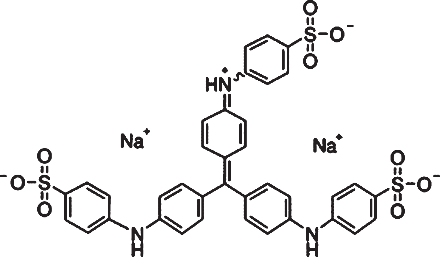
Molecular formula of methyl blue compound.
XRD analysis
XRD patterns of plasma treated and untreated ferrite nanoparticles are shown in Fig. 3. XRD peaks were compared with JCPDS card no.74-2403, which showed face-centered cubic structures of all manganese ferrite samples. The major XRD peaks of treated and untreated samples confirm the presence of same planes in the structure, which are (220), (311), (400) and (422). The plasma treatment did not alter the structure of the nanoparticles. Same phases and planes were found in both samples. However, XRD peak intensity of plasma treated samples was slightly higher than the untreated sample. The higher peak intensities reflect high purity and better crystallinity of the ferrite nanostructure. The crystallite size was calculated using a well-known Scherrer’s formula [19].
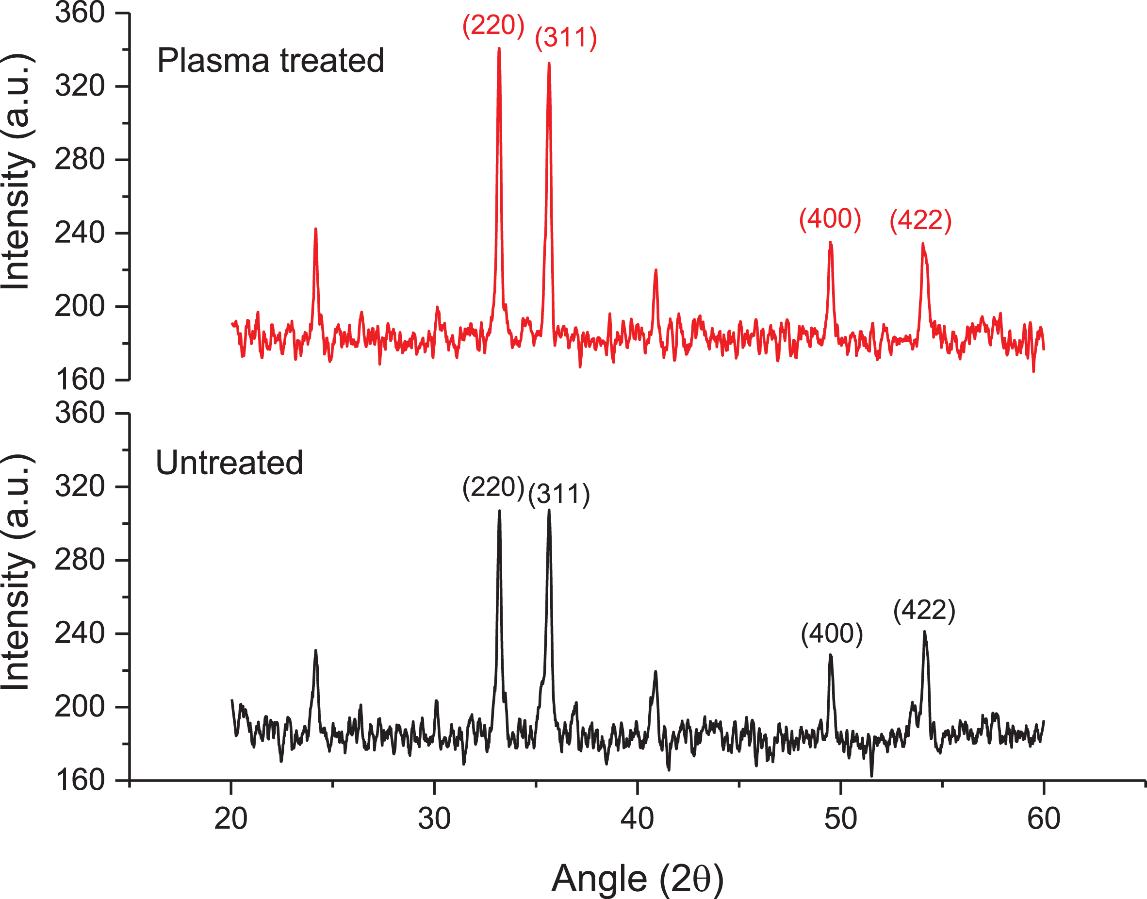
XRD spectra of plasma treated and untreated ferrite nanoparticles.
Where, D is crystallite size, θ is the Bragg angle, λ is the wavelength of X-rays and β is the full width at half maximum. The reactive species in oxygen plasma, including HO2-, O3, H2O2 and OH-, interact with the ferrite powder. As a result, the surface of the particles oxidized and some of the particles agglomerated into larger particles. The crystallite size of the plasma treated ferrite increased as compared to untreated ferrite. The crystallite size of untreated ferrite was measured about 35 nm. On plasma treatment for 10 min, the crystallite size changed to 39 nm. Similarly, the lattice parameter also increased from 8.35 Åto and 8.49 Åon plasma treatment of the ferrite, as shown in Table 1. A similar trend was observed in the volume of the unit cell.
Summary of XRD analysis of ferrite nanoparticles
The morphology of plasma treated and untreated Mn doped iron oxide ferrites were investigated by generating their SEM images. Figure 4 shows magnified SEM images of ferrites with and without plasma treatment. Since the samples were made up of some large clusters mixed with smaller clusters, defining the morphology from SEM images was difficult. Many of the clusters had unknown geometry. The plasma treated ferrite had few clusters of larger sizes as compared to untreated ferrite. The size of these clusters, however, was substantially greater than that of the untreated clusters. The increased size of the clusters may be attributed to agglomeration of the particles into larger clusters during plasma treatment. Also, the surface of the plasma treated clusters was more porous as compared to untreated clusters. The pores form during plasma treatment due to an interaction of reactive species with the clusters. The reactive species, on interaction with cluster surface, transfer their energy to the atoms of the surface. As they gain energy, these atoms become excited and leave the particle by causing surface sputtering. The pore size grows as the microwave powder supply to the discharge increases. Plasma species with higher energies are more detrimental to the target surface. The removal of impurities during plasma exposure is another reason of pore formation in the sample surface. XRD peak intensity increases with a decrease in impurity level, as revealed in XRD analysis of the plasma treated and untreated ferrite samples.
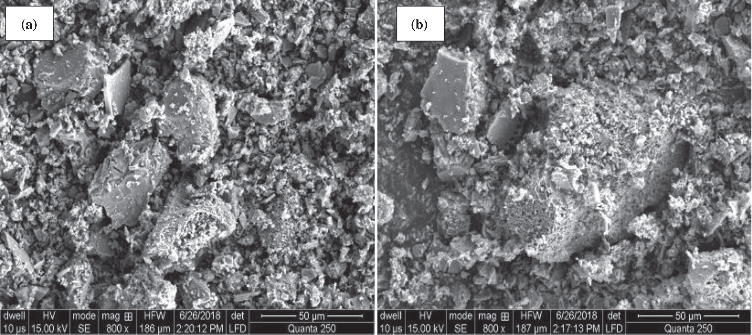
SEM images of (a) untreated and (b) plasma treated manganese ferrite samples.
UV-visible absorption spectra of Mn doped iron oxide ferrite nanoparticles with and without plasma treatment are shown in Fig. 5. It is evident that plasma treated iron oxide ferrite nanoparticles showed higher absorbance as well as increased optical absorption towards distributed wavelength than untreated ferrite samples. The untreated ferrite absorption peak was observed at 579 nm, while plasma treated ferrite adsorption peak was observed at 596 nm. The band gap energy of the untreated ferrite sample was 5.14 eV, which reduced to 5.08 eV on microwave plasma treatment. The decrease in the band gap can be due to creation of intermediate states between the valence and conduction band of iron oxide nanoparticles by doping of Mn2+ ions, which lowered the Fermi level to narrow the band gap [19].
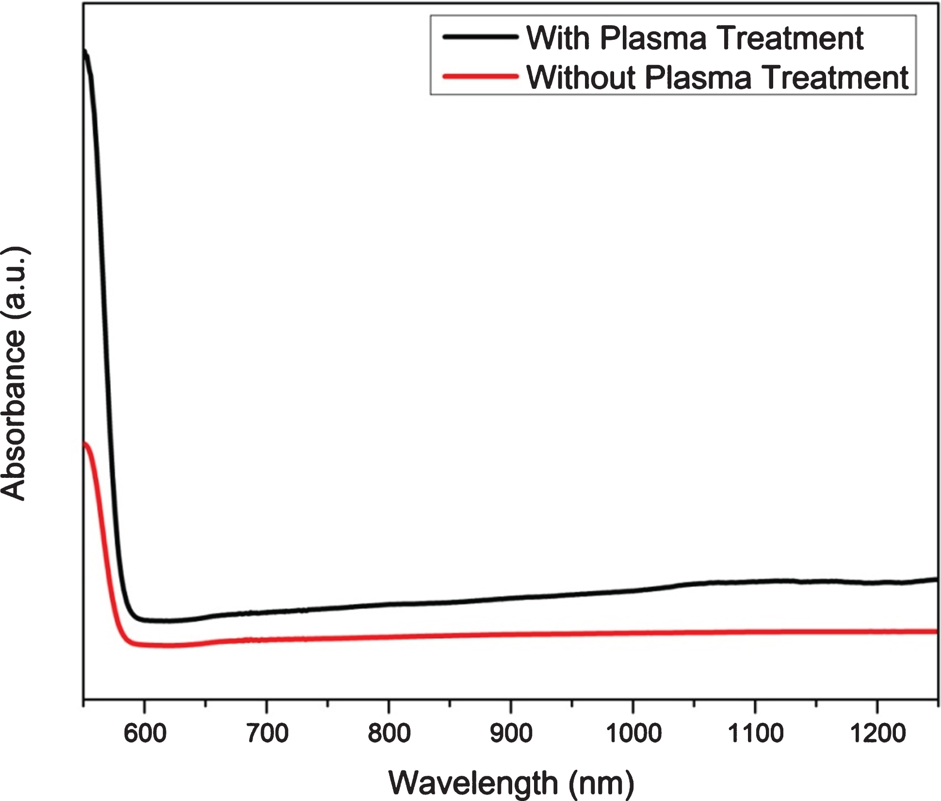
UV-Vis spectra of untreated and plasma treated manganese ferrite nanoparticles.
In order to study the recombination rate of the excitons, the room temperature photoluminescence (PL) spectra at exciton of 325 nm of as-synthesized Mn doped iron oxide ferrite nanoparticles was studied with and without plasma treatment and the results are shown in Fig. 6. It can be observed that ferrite nanoparticles showed one band of luminescence at wavelength of 370 nm, which is consistent with previous studies [20]. Moreover, the fluorescence intensity of plasma modified Mn doped iron oxide ferrite nanoparticles is less than untreated ferrite nanoparticles. Since lower PL intensity indicates the inhibited recombination of the photoinduced charge carriers [21]. Therefore, plasma modified Mn doped iron oxide nanoparticles demonstrated the retarded recombination of photoinduced charge carriers. This suppressed recombination of the excitons is very significant for improved photocatalytic activity.
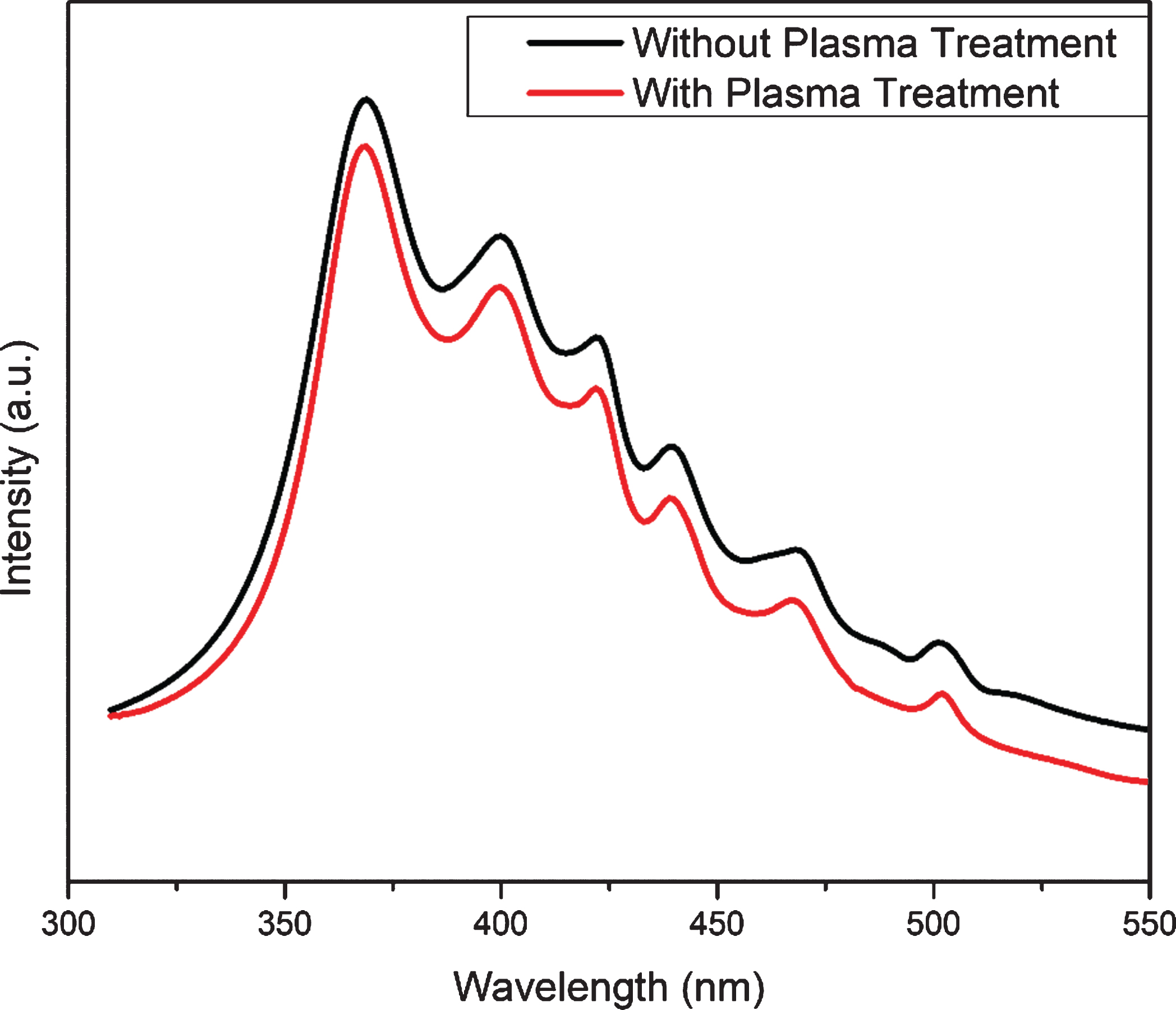
PL spectra of untreated and plasma treated Mn doped iron oxide ferrite nanoparticles.
To further study the recombination rate and electron migration ability of as-synthesized Mn doped iron oxide ferrite nanoparticles with and without plasma treatment, transient photocurrent response and EIS were studied and the results are reported in Fig. 7. It is evident from the transient photocurrent response that there was significant increase in the transient current response for plasma modified Mn doped iron oxide ferrite nanoparticles as compared to untreated ferrite nanoparticles. This increase in current over plasma modified Mn doped iron oxide ferrite nanoparticles confirmed the prolong separation of charge carriers.
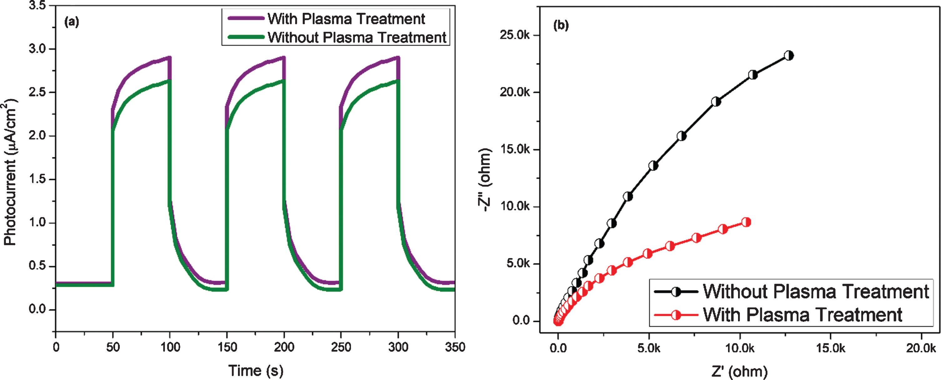
(a) Transient photocurrent and (b) EIS Nyquist plots of untreated and plasma treated ferrite nanoparticles.
The further ascertain was brought through EIS Nyquist plot of the photocatalyst. The smaller arc radius of EIS Nyquist plot reflects the higher separation of photo-induced charge carriers [22]. Therefore, plasma modified Mn doped iron oxide photocatalyst showed higher lifetime of charge carriers and hence this photocatalyst will be suitable to show the high photocatalytic activity. Figure 8 schematically illustrates the transition of electrons from valance band to the conduction band of the manganese ferrite and the formation of electron-hole pairs for initiation of oxidation and reduction reactions in the dye solution. The reactions derive the photocatalytic degradation of MB blue into water and carbon dioxide along with some by products [25–27].
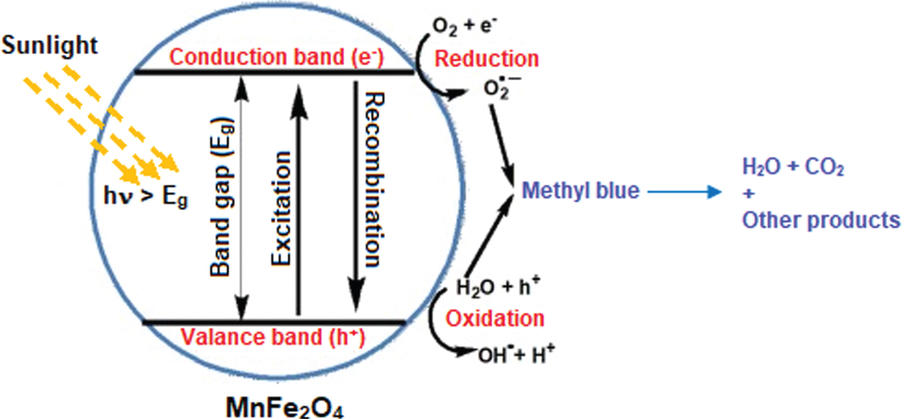
Illustration of photoactivation of ferrite catalyst and formation of electron-hole pairs.
The photocatalytic activity of manganese ferrite nanoparticles with and without plasma treatment was carried out for the degradation of MB dye under visible light illumination and the results are shown in Fig. 9. The photolysis did not show any significant degradation rate. The degradation was measured about 4.2%. Similarly, blank experiment in the absence of light and presence of the catalyst also did not demonstrate any appreciable photocatalytic degradation activity. When degradation of MB was conducted in the presence of untreated ferrite nanoparticles, it showed a significant increase in the photocatalytic activity under visible light illumination. The degradation rate reached to 72% within 100 min of light illumination. The plasma modified ferrite nanoparticles showed further increase in degradation efficiency. The degradation of dye with plasma treated photocatalyst increased to 85% within 100 min of visible light illumination. The apparent increase in MB degradation activity of plasma modified ferrite nanoparticles was due to increase in the optical absorption towards longer wavelength and inhibited electron-hole recombination rate as confirmed by UV-Vis DRS and EIS studies [23]. The k value of plasma modified ferrite nanoparticles was four times higher than untreated ferrite nanoparticles under identical experimental conditions. This rise in value of k is in good agreement with the rising trend of the photocatalytic degradation of MB.
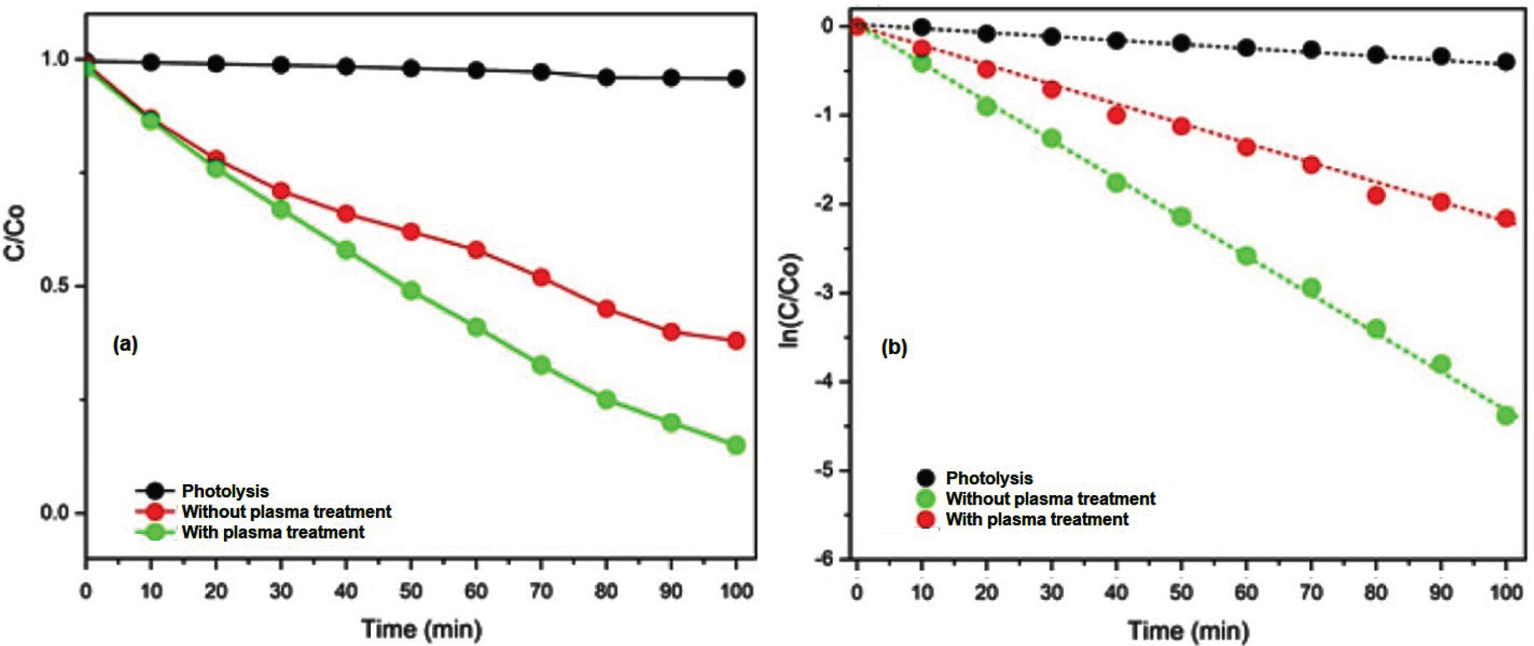
(a) photocatalytic degradation rate of MB and (b) apparent reaction constant k values.
The recyclability of the photocatalyst is also important for the industrial application. Stable photocatalyst did not show significant loss in the photocatalytic activity after repeated experiments. As shown in Fig. 10, plasma modified ferrite nanoparticles did not show any significant loss in degradation efficiency within five successive cycles of MB degradation experiments. The catalyst retained 97% efficiency after five cycles.
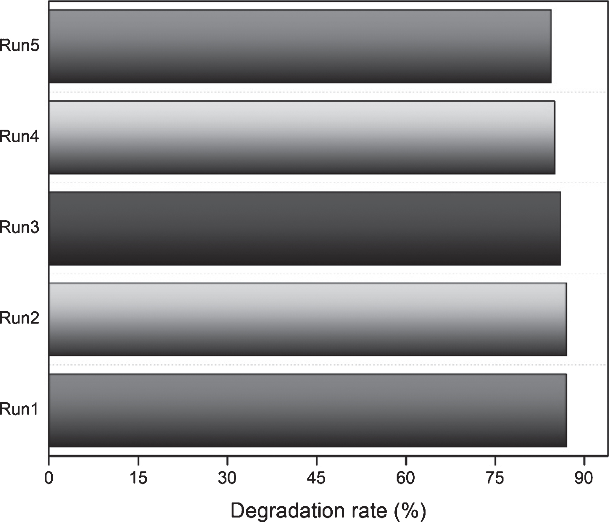
Photocatalytic stability of plasma treated ferrite nanoparticles after five successive cycles of dye degradation.
The magnetic properties of both plasma treated and untreated manganese ferrites were investigated by creating hysteresis loops for them. Figure 11 depicts the hysteresis loops, which revealed that both samples exhibited ferromagnetic activity. The hysteresis loops observed are caused by the formation of soft ferrite nanoparticles. The size of the crystallites grew larger after plasma treatment. It is discovered that the size of the magnetic domain grows with the size of the crystallites. Strong magnetization resulted from an increasing number of atomic spins synchronized with the magnetic field [28]. The microwave plasma treatment may also raise the temperature of the sample upto 300°C. It means, the plasma treatment not only reform the surface morphology but also alters the magnetic character of the sample due to calcination effect. The plasma calcination of ferrites effectively improves their magnetic properties even at lower temperatures due to an interaction of reactive species with the surface atoms. The B-H curves revealed an increase in magnetization of manganese ferrite on plasma treatment [29].
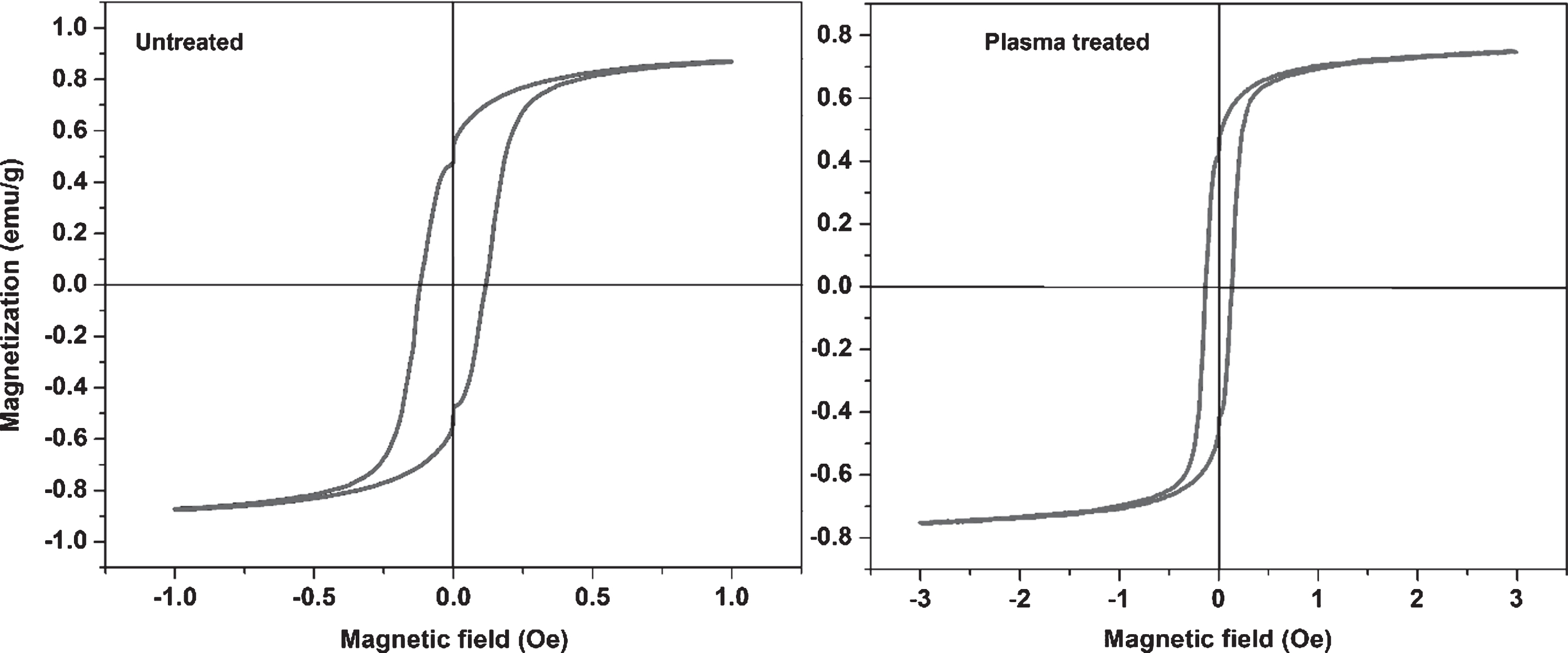
Magnetization curves of plasma treated and untreated manganese ferrites.
The saturation magnetization of untreated ferrite sample was higher than that of the plasma treated ferrite sample. The sample’s low saturation magnetization under plasma exposure demonstrates the well-ordered structures of a ferromagnetic material [20]. The saturation magnetization of iron oxide ferrite nanoparticles of untreated and plasma treated ferrites was measured about 0.78 emu/g and 0.68 emu/g, respectively. After plasma procedure, the coercivity was also reduced. The coercivity of manganese ferrite decreased from 0.3 kOe to 0.1 kOe on microwave plasma treatment. The magnetization response of manganese ferrite nanoparticles depends on oxidation of manganese and iron cations. The magnetization also depends on the location of these cations in the spinel crystal lattice. The divalent cations, like Zn2+ and Mn2+, are mostly found in tetrahedral sites while trivalent cations (Fe3 +) are located in octahedral sites in the spinel crystal lattice. Such arrangements ensure high magnetization of manganese doped iron oxide ferrite nanoparticles. It is assumed that Mn2+ cations may oxidize to Mn3 + cations during synthesis process and Fe3 + may reduce to Fe2 + . These redox reactions cause a change in cations’ arrangement in the sub-lattices. The Fe2+ cations partially transfer to tetrahedral sites while Mn3 + cations partially transfer to octahedral sites of the spinel crystal lattice. This change in occupancy of sites sometimes has negatively impact on magnetic properties of the ferrite nanoparticles. However, it is worth mentioning here that the oxidation of Mn2+ into Mn3 + takes place at higher temperatures (900–1000°C). In our case, the sintering of the manganese ferrites was conducted 700°C, therefore changes of oxidation Mn2+ were very low.
This study was focused on magnetic and photocatalytic properties manganese ferrite nanoparticle with and without conducting microwave plasma. The prepared samples were characterized using XRD, SEM, UV-Vis DRS, PL, transient photocurrent response and EIS study. The band gap of the plasma treated nanoparticles decreased to support the photocatalytic activity of the ferrite nanoparticles against methyl blue. The PL and EIS study showed effective separation and prolong lifetime of the photo-induced charge carriers. The photocatalytic activity of the samples was tested for the degradation of methyl blue dye under visible light illumination and the plasma treated nanoparticles showed highest degradation efficiency of 85% after 100 min of visible light illumination. The saturation magnetization nanoparticles with and without plasma treatment was measured to be about 0.68 emu/g and 0.78 emu/g, respectively. The coercivity of the nanoparticles also decreased from 0.3 kOe, to 0.1 kO e on plasma treatment.
Footnotes
Acknowledgments
Authors would like to acknowledge the support of the Deputy for Research and Innovation- Ministry of Education, Kingdom of Saudi Arabia for this research through a grant (NU/IFC/INT/01/010) under the institutional Funding Committee at Najran University, Kingdom of Saudi Arabia).
Conflict of interest
Authors declare no conflict of interest regarding this submission.