






Abstract
OBJECTIVE:
To evaluate the diagnostic accuracy and utility of bedside ultrasound (US) by neonatology residents to confirm position of umbilical venous catheter (UVC), umbilical arterial catheter (UAC), and peripherally inserted central catheter (PICC).
METHODS:
In this prospective study, we included neonates who required UVC, UAC or PICC insertion. Two neonatology residents performed all bedside US examinations after a short period of training. Plain radiograph was taken as gold standard. Time taken for confirmation of catheter position by US and radiograph was recorded.
RESULTS:
We recruited 71 neonates for UVC and UAC, and 40 neonates for PICC. Sensitivity and specificity of US in identifying a malpositioned catheter was good for UVC (94% and 66.7% respectively) and UAC (86.7% and 94.5%). Agreement between radiograph and US was good for UVC [0.718 (0.512, 0.861); p < 0.001] and UAC [0.857 (0.682, 0.953); p < 0.001]. Sensitivity (47.8%) of US in identifying a malpositioned PICC was low, though specificity (82.4%) was good. Agreement between radiograph and US in identifying PICC position was poor [0.25 (–0.084, 0.545); p 0.024]. This was due to incorrect interpretation of catheter position on radiograph in some infants, which was confirmed by the radiologist. The median time taken for US was significantly less than time taken for radiograph in confirming the position of UVC (50 vs. 155 minutes; p < 0.001)), UAC (45 vs. 128 minutes; p < 0.001), and PICC (60 vs. 136 minutes; p < 0.001).
CONCLUSION:
US examination byneonatology residents has good diagnostic accuracy in confirming the position of UAC and UVC, and possibly PICC in neonates. The time taken to confirm catheter position by US is significantly less than radiograph.
Keywords
Background
Placement of central vascular catheters (CVC) such as umbilical venous catheter (UVC), umbilical arterial catheter (UAC), and peripherally inserted central catheter (PICC) is an integral part of neonatal intensive care. CVC placement is a blind procedure and length of insertion is decided based on birth weight or shoulder-umbilical length for UVC and UAC, and on measurement of length on the surface marking of the vein from the point of insertion to heart for PICC [1, 2]. The estimate is often incorrect and results in malpositioning of the CVC, which in turn leads to untoward complications affecting the heart, lungs, and liver [3–6]. Hence, it is imperative to confirm the CVC tip position soon after placement.
Plain antero-posterior radiograph of chest and abdomen, including the limb of catheter insertion for PICC, is the most widely used method to confirm catheter tip position in neonatal intensive care units (NICUs). Though previous studies have demonstrated the utility of bedside ultrasound examination (US) to confirm the position of CVCs in neonates [7–9], US is still not widely used. This could be due to concerns that US examination requires an expert radiologist and that it is time-consuming. A recent pilot study showed that US could easily be learnt by neonatology consultants and residents, and time taken by neonatologists to confirm catheter position was three minutes for UAC and UVC and seven minutes for PICC [10]. We conducted this study to evaluate the diagnostic accuracy and utility of bedside US examination by neonatology residents to confirm the position of UVC, UAC, and PICC in neonates.
Methods
This was a prospective diagnostic evaluation study, done between July 2014 and February 2016, in level three NICU of a tertiary care teaching hospital in India. The Institutional Ethical Committee and Review Board approved the study before commencing recruitment (Institutional Review Board, Christian Medical College, FG/2014/07/008943). Informed written consent was obtained from the parents before enrolment.
We included neonates of any gestational and postnatal age who required UVC, UAC, or PICC. We excluded babies who were too unstable to undergo sonography and those where we could not obtain parental consent. We used umbilical catheters (Vygon) 5, 6, or 8Fr for UVC and 3.5 or 4Fr for UAC. We used 28 G (1Fr) catheter (Vygon; Premiecath) or 24 G (2Fr) catheter (Vygon; epicutaneocava) for PICC. We used weight-based formula to estimate the depth of insertion of UVC (2×weight + 5) and UAC (3×weight + 9), and surface marking-based measurement to estimate the depth of insertion of PICC [11].
Two neonatology residents (MPS and TA) performed all bedside US examinations using Vivid 6 (GE healthcare) machine and 13 MHz linear probe. The residents had neonatology training for 18 months prior to the study and had been doing ultrasound examination of brain and heart regularly. They underwent a short period of training to evaluate CVC position by US; each resident performed US for 5 to 10 babies for each UVC, UAC, and PICC under the guidance of the radiologist (SG).
Plain radiograph was taken as the gold standard. Radiograph of chest and/or abdomen, with inclusion of the limb for PICC, was taken bedside using portable radiograph machine. Both US examination and radiograph were done with baby in supine position, head in neutral position, and limbs in semi-abduction (30–45 degrees) and complete extension. The investigators were blinded to radiograph until US was completed. Repositioning of malpositioned catheters was done after both radiograph and US were completed. Both radiograph and US reports were taken into consideration while deciding repositioning.
We aimed to do both US and radiograph within four hours from the time of insertion. Call for portable radiography was made soon after CVC insertion. Time taken for confirmation of CVC position by US and radiograph from the time of completion of CVC insertion was recorded.
Interpretation of catheter position in radiograph and US
UVC
Catheter tip at inferior vena cava (IVC) - right atrium (RA) junction was considered as the optimal position for UVC [12]. On US, IVC-RA junction was visualized directly and the tip position in relation to IVC-RA junction was recorded. On the radiograph, UVC tip at or just above the level of diaphragm was considered to correlate with IVC-RA junction. UVC was considered to be malpositioned, if the tip was found inside the liver, inside the cardiac chambers or if the catheter was taking a deviant course and turning into other vessels such as portal veins or splenic vein (Fig. 1).
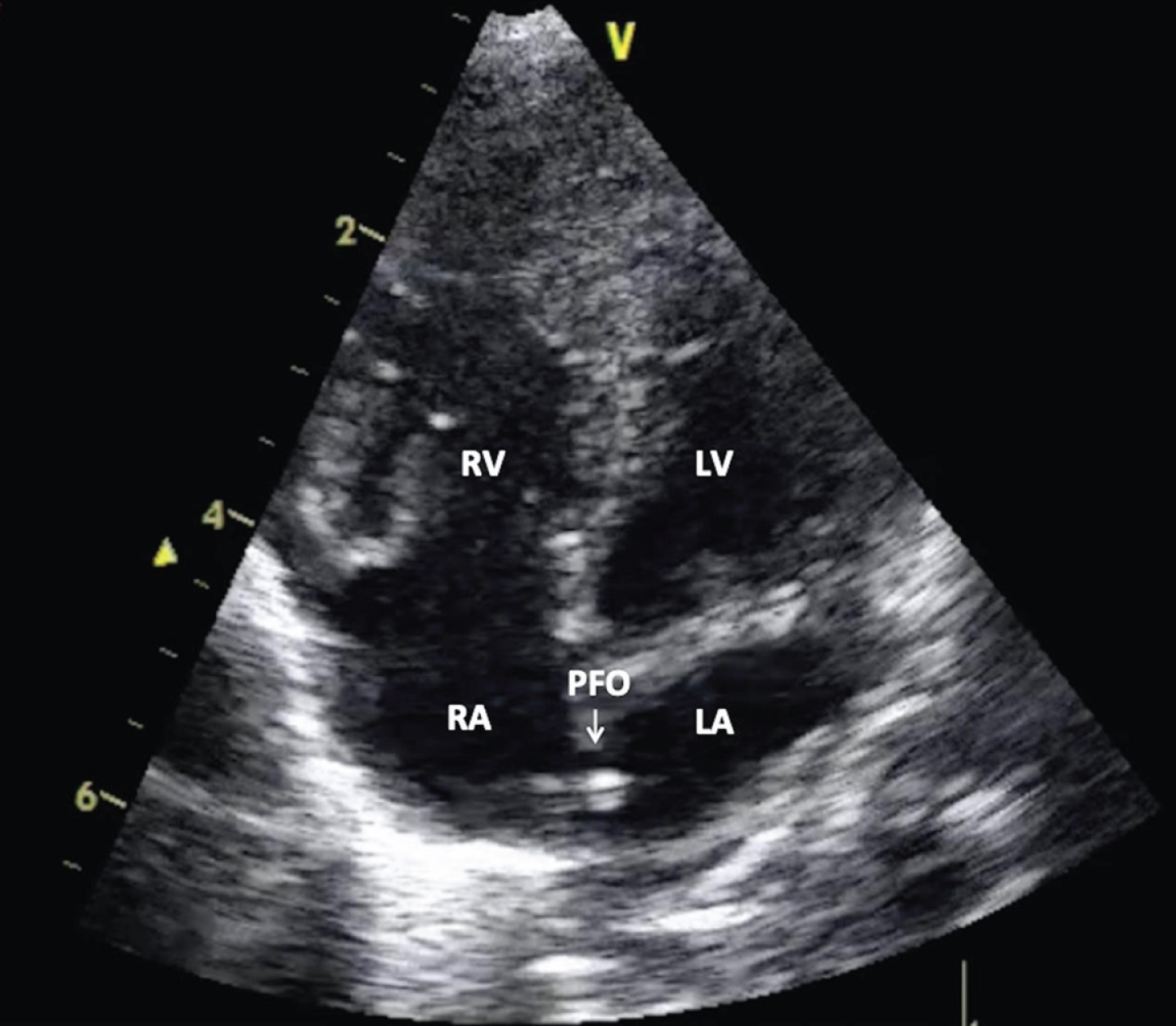
US image of UVC; malpositioned catheter with tip in left atrium (LA; left atrium, LV; left ventricle, PFO; patent foramen ovale, RA; right atrium, RV; right ventricle).
The position of UAC was judged based on vertebral level on both radiograph and US; catheter tip at T6-T10 level was considered optimal [13]. UAC was considered to be malpositioned if the catheter tip was found above T6 or below T10. On US, the vertebra just above the origin of coeliac artery from the aorta was taken as T12 and vertebrae were counted upwards till the catheter tip position (Fig. 2). On the radiograph, the vertebral level corresponding to the UAC tip was recorded.
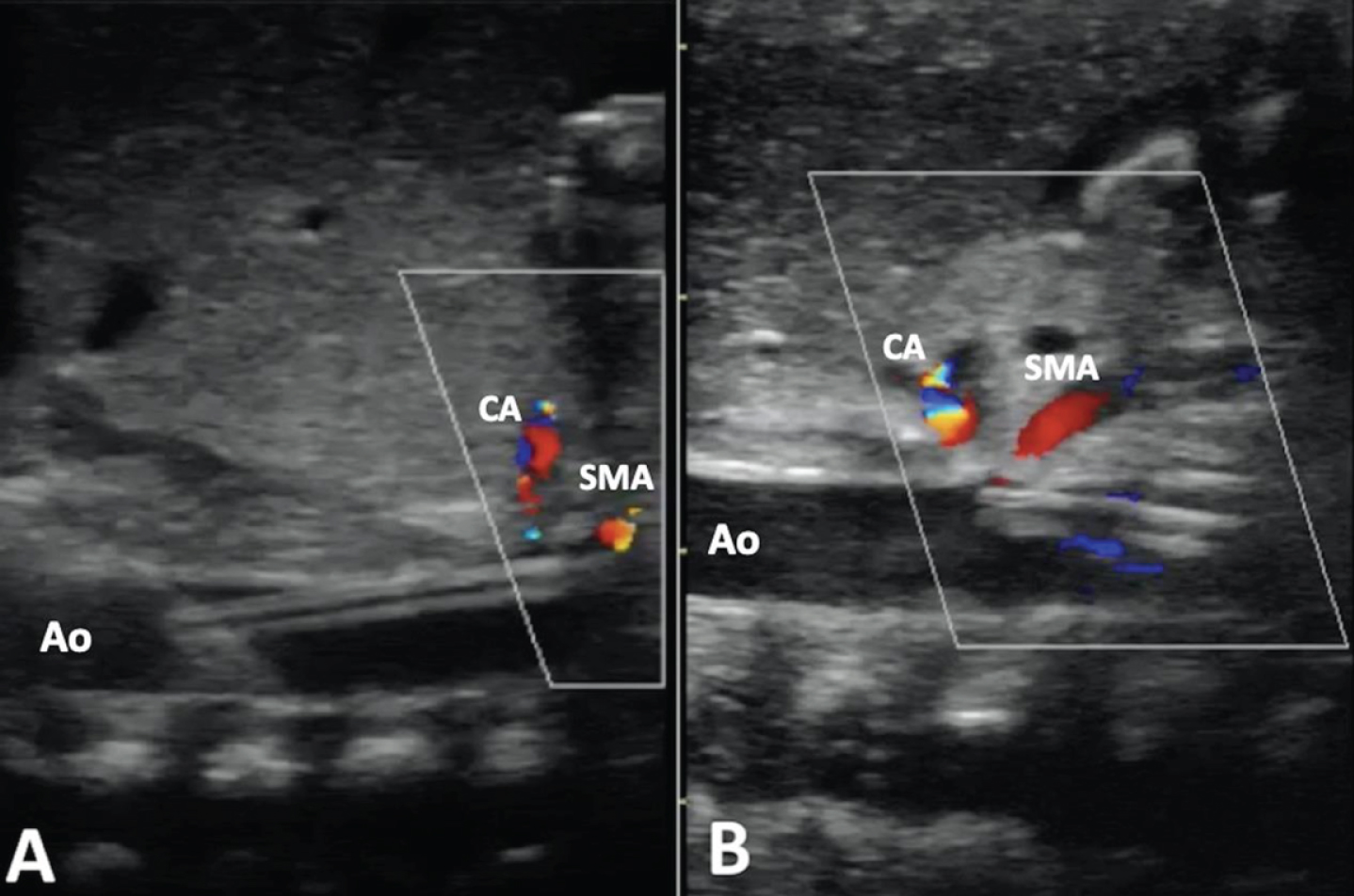
US images of UAC with colour Doppler of coeliac and superior mesenteric arteries. A-Appropriately positioned UAC tip at T9 level; B- Low and malpositioned UAC with tip at L1 level near the origin of coeliac and superior mesenteric arteries (Abbreviations: Ao; Aorta, CA; coeliac artery, SMA; superior mesenteric artery).
The catheter tip at SVC-RA junction or in superior vena cava (SVC) was considered ideal for an upper limb PICC [14]. On US, the SVC and SVC-RA junction was visualized directly in right parasternal axis and subcostal views and catheter tip position was recorded (Fig. 3). On the radiograph, T2-T3 intervertebral disc was considered to correlate with SVC-RA junction. Catheter tip beyond the first rib and before the IVC-RA junction was considered to correlate with SVC [15].
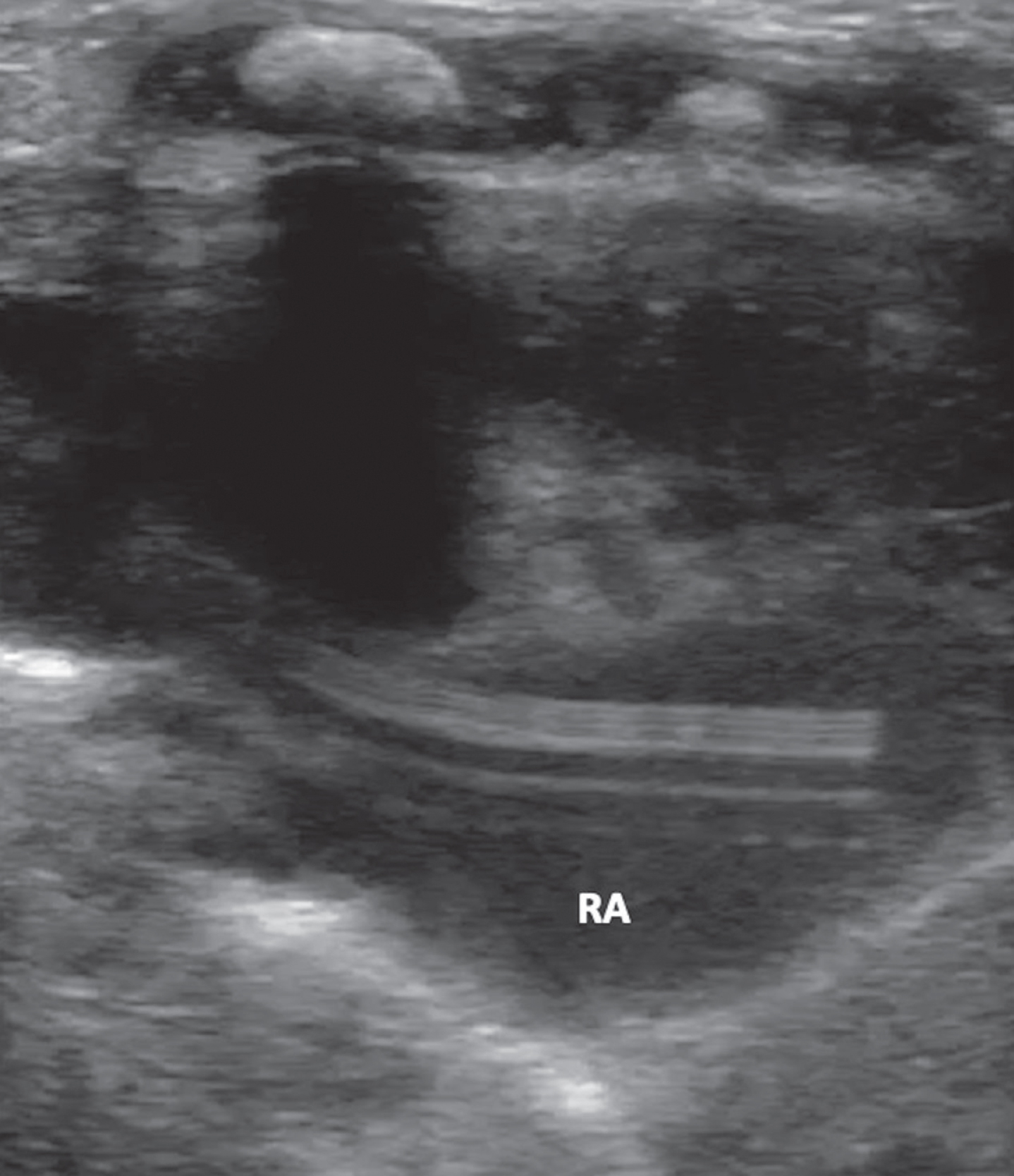
US image of PICC; catheter tip in right atrium (RA; right atrium).
The catheter tip at IVC-RA junction or in IVC was considered ideal for a lower limb PICC [14]. On the radiograph, catheter tip at or just above the level of diaphragm was considered to correlate with IVC-RA junction; and catheter tip in the midline of the abdomen above L5 was considered to be positioned in IVC.
Sample size for UAC and UVC was calculated based on a hypothetical sensitivity of 90% for US. For 90% sensitivity, 7% precision, 80% power and 5% alpha error, we required 71 infants for each UVC and UAC. For PICC, a convenient sample size of 40 was taken.
Statistical methods
Descriptive statistics are presented as median and interquartile range (IQR) or number and percentage as appropriate. Diagnostic accuracy is reported using sensitivity, specificity as well as positive and negative predictive values. Agreement statistics is reported using Cohens’s Kappa and Prevalence-Adjusted and Bias-Adjusted Kappa (PABAK). Mann-Whitney U test was used as the test of significance to compare time taken for the radiograph and US, since the data was not normally distributed. All statistical analyses were done using SPSS 16.0 and Dx test software. A p value < 0.05 was considered statistically significant.
Results
We recruited 71 neonates requiring UVC, 71 requiring UAC, and 40 neonates requiring PICC in the study. Table 1 shows the baseline characteristics of study infants.
Baseline characteristics
Baseline characteristics
*Median (Inter-quartile range); #n (%). Abbreviations: PICC: peripherally inserted central catheter; UAC: umbilical arterial catheter; UVC: umbilical venous catheter.
Among the 71 UVCs, 50 were shown to be malpositioned on radiograph; 35 malpositioned inside the heart, 11 inside the liver, and four catheters were found to be deviating and turning into other vessels. Of the 35 UVCs shown to be malpositioned inside the heart on radiograph, two (5.7%) were found to be in ideal position on the US and hence inappropriate repositioning, which would have resulted in a peripherally placed UVC, was avoided. Of the 11 UVCs shown to be malpositioned inside the liver on radiograph, one (9%) was found in ideal position on US. In four (5.6%) neonates in whom the UVC was found to have a deviant course on radiograph, we could not identify the catheter position on US. Among the 21 UVCs shown to be in ideal position on radiograph, 7 (33%) catheters were found to be malpositioned inside the heart on US and required repositioning.
Among the 71 infants with UAC, the catheter tip was shown to be at T10 on radiograph in three infants; however, the tip was found to be at T11 near the origin of coeliac artery on US. These catheters were pulled down to lower position at L3-L4. On the other hand, the catheter tip was erroneously reported to be at T6 on US in two infants; the tip was shown to be at T4-T5 on radiograph.
Of the 40 infants with PICC, 12 PICC were shown to be malpositioned on radiograph. Five catheters were erroneously reported on radiograph to be deviating from the normal course and turning into other vessels based on some vague linear shadows; they were found to be in ideal position on US. The radiologist reviewed the radiograph and US images of all these infants and confirmed this finding, and unnecessary removal of the catheters was avoided. In the remaining seven infants, radiograph showed the catheter tips inside heart. However, they were found to be in correct position on US. Three catheters that were shown to be in ideal position on the radiograph were found to be malpositioned inside the heart on US and they were repositioned. On the other hand, the catheter could not be visualized on US in two (5%) infants with PICC in upper limbs; in both the infants the catheter was found to be deviating from the normal course and turning into other vessels on the radiograph.
Table 2 shows the results of US versus radiograph in identifying a malpositioned catheter. Table 3 shows the measures of diagnostic accuracy of US in identifying malpositioned CVCs, taking radiograph as the gold standard. US had good diagnostic accuracy in identifying a malpositioned UVC and UAC. The sensitivity and predictive values of US in identifying malpositioned PICC were low. The agreement between radiograph and US was good for UVC and UAC and poor for PICC (Table 4).
Sonography vs. radiography to identify a malpositioned catheter
Abbreviations: PICC: peripherally inserted central catheter; UAC: umbilical arterial catheter; US: ultrasound; UVC: umbilical venous catheter.
Measures of diagnostic accuracy of US in identifying malpositioned catheters
Abbreviations: NPV: negative predictive value; PICC: peripherally inserted central catheter; PPV: positive predictive value; UAC: umbilical arterial catheter; UVC: umbilical venous catheter.
Agreement between radiograph and US in identifying catheter position
Abbreviations: IQR: interquartile range; PICC: peripherally inserted central catheter; UAC: umbilical arterial catheter; UVC: umbilical venous catheter.
The time taken for the US was significantly less than the time taken for the radiograph in confirming the catheter position in all three CVCs (Table 5).
Time taken to confirm catheter position by radiograph and US
Abbreviations: IQR: interquartile range; PICC: peripherally inserted central catheter; UAC: umbilical arterial catheter; UVC: umbilical venous catheter.
Our study shows that neonatology residents could promptly confirm the position of UVC, UAC, and possibly PICC by bedside US examination after a short period of training. Though many previous studies have shown that US is equivalent to or superior than radiograph in confirming CVC tip position in neonates, most studies have evaluated only radiologist-performed US [7, 8]. Pulickal et al. showed that neonatologists could identify the position of UVC by bedside US [16].
US was found to be superior to radiograph in confirming UVC tip position in previous studies [7, 16]. Michel et al. reported a sensitivity and specificity of 93% and 95% for US and 66% and 63% for radiograph to determine the UVC position [8]. Radiograph is less accurate in confirming UVC position because of the indirect ways used to determine the IVC-RA junction, based on either the vertebral level (T8-9) or the diaphragm [17]. In our study, we used the diaphragmatic or cardiac silhouette method to determine the IVC-RA junction on the radiograph since it was found to be better than the vertebral level method [17].
US is definitely better than radiograph in identifying the UVCs that are malpositioned inside the cardiac chambers. Pulickal et al. showed that 27% of the UVCs that were judged to be in correct position on radiograph required repositioning since they were found to be malpositioned inside the heart on US, the results being similar to our study (33% in our study) [16]. However, in case of UVCs malpositioned inside the liver, it may be difficult for a novice neonatology resident to identify the catheter tip. This is because the hyperechoic walls of blood vessels inside the liver mimic the catheter on US and may mislead the resident. Nevertheless, in our study we could identify the catheter tip in all 11 infants with UVCs malpositioned inside the liver. It is also difficult to identify the catheter tip on US if the UVC is deviating and turning into other vessels such as portal veins or splenic vein. We could not identify the catheter tip position in four infants with UVC having a deviant course. These fallacies and difficulties could potentially be overcome by neonatology residents with experience. The number of appropriately positioned UVCs was low in our study. This was because we considered the catheter position as appropriate only if the tip was at IVC-RA junction. All other positions were considered as ‘malpositioned’.
In our study, we could confirm the UAC position in all infants, similar to previous studies [10, 18]. US imaging of the intra-thoracic descending aorta can be challenging for neonatology residents, since it has to be seen beyond the diaphragm and behind the heart. We could not trace the catheters beyond T6 in two infants with UAC at T4-5. Furthermore, it is difficult to identify the UAC tip when it is very low, because of bowel gas interference.
US had low diagnostic accuracy and poor agreement with radiograph in identifying PICC position in our study. This was mainly due to incorrect interpretation of catheter position on radiograph rather than on US, which was confirmed by the radiologist. Though some studies have used radiograph as the gold standard, we found the radiograph to be less accurate in determining PICC position [10, 19]. Jain et al. found US to be better than radiograph to confirm PICC position; radiograph had a low sensitivity and specificity of 64% and 55% respectively [20]. It is difficult to trace the thin PICC catheters on radiograph. Lateral radiograph and injection of a contrast agent are other methods used for better visualization of PICC, but they have the disadvantages of increasing the radiation exposure and adverse effects of the contrast respectively. US is better in identifying the PICCs that are malpositioned inside the heart chambers, as in UVC. However, visualizing the tip of PICC in the lower IVC and in the cervical vessels above SVC on US is difficult because of interference by bowel gas and excess soft tissues respectively [21].
Since neonatology residents are routinely doing bedside US for head and heart nowadays, they can easily learn US techniques for CVC position. The time taken to confirm CVC position by US was significantly less when compared that of radiograph in our study. With the ready availability of bedside US machine in most NICUs, neonatology residents could promptly confirm the position of CVCs. This would reduce the duration of malpositioned CVCs and their untoward complications [3–6].
The major advantage of using US to confirm CVC position is avoidance of radiation exposure. Though radiation dose from a single radiograph is minimal, cumulative radiation exposure during the NICU stay was found to be significantly high especially in sick neonates requiring CVCs [22]. The other advantage is direct visualization of the catheter in relation to the heart and blood vessels on US, compared to the indirect method of inferring the heart and blood vessels based on soft tissue shadows and bony landmarks on a radiograph. US can be used to reposition the catheter, thus repeat radiographs to confirm position after repositioning can be avoided [19, 20]. US can be used for repeated assessments of catheter position until removal to identify catheter migration over time. US is also used real-time to guide the placement of CVCs in neonates [15, 24].
The limitation of the study was that radiologist did not interpret all the US and radiograph images of study infants. The reliability of interpretation of radiograph and US by neonatologists may be questionable. However, this is more pragmatic since the radiographs and US for CVCs are interpreted only by neonatology residents and/or consultants in most centers across the world. Though we considered plain radiograph as gold standard, it was found to be erroneous in some infants. Hence the calculations of diagnostic accuracy markers of US became less accurate. Another limitation was the small sample size in the PICC group.
To conclude, US examination by the neonatology residents has good diagnostic accuracy in confirming position of UVC and UAC, and possibly PICC. The time taken to confirm the CVC position by US is significantly less when compared to that of radiograph.
Funding source
The study was supported by the research funds of Christian Medical College Vellore and the department of Neonatology. This research received no specific grant from any funding agency in the public, commercial or not-for-profit sectors.
Potential conflicts of interest
The authors have no conflicts of interest relevant to this article to disclose.