






Abstract
Objectives:
Small clinical trials reported that repetitive sessions of tDCS could improve naming abilities in post-stroke aphasia. However, systematic meta-analyses found no effect, but all of these analyses pooled data from both single and repetitive sessions at the group level. The aim of this paper was to perform a meta-analysis based on individual patient data to explore the effects of repetitive tDCS sessions on naming in post-stroke aphasia and in prespecified subgroups.
Methods:
We searched for published sham-controlled trials using the keywords “aphasia OR language” AND “transcranial direct current stimulation OR tDCS” AND “stroke”. We computed an active and sham improvement ratio by dividing the difference between naming scores after and before the active or sham sessions, respectively, by the total number of picture items. Because of heterogeneity (I2 = 66%, p: 0.002), we used random-effects models to estimate the standardized mean difference (SMD) for the naming outcome. We then analyzed subgroups according to number of sessions, polarity, side/location of the active electrode, post-stroke delay, aphasia severity and comprehension disorders.
Results:
Seven eligible studies were identified, including 68 chronic stroke patients. tDCS was beneficial on naming ability (35% ±34% in the active vs. 25% ±37% in the sham condition). An SMD of 0.8 (95% CI: 0.27–1.33) was found for the naming outcome. Additionally, there was a dose-dependent effect (5 vs. >5 sessions). We also demonstrated a prevalence of anodal vs. cathodal condition and left vs. right targeting electrode. Finally, repetitive sessions were beneficial regardless of the severity of aphasia, comprehension disorders or post-stroke delay.
Conclusion:
Repetitive sessions of tDCS are likely to be valuable in enhancing naming accuracy in post-stroke aphasia.
Introduction
Transcranial direct current stimulation (tDCS) is a non-invasive brain stimulation technique that has garnered increasing interest due to its ability to induce changes in cortical excitability. tDCS influences the firing rate of tonically discharging neurons likely by shifting the resting membrane potential of the neurons underneath the active electrode. Indeed, anodal stimulation depolarizes the resting potential of the neuronal membrane whereas cathodal stimulation induces hyperpolarization (Nitsche et al., 2000). In addition, tDCS can also induce aftereffects that involve changes in N-methyl-D-aspartate (NMDA) receptor efficacy and non-synaptic mechanisms based on changes in the neural membrane (Nitsche et al., 2003). Due to its acceptable safety record, low cost and potential use in outpatients, this technique has opened new areas to experimental treatment and is increasingly being evaluated in pivotal clinical trials in a wide range of neurological diseases. For example, in stroke survivors, a single session of tDCS can be used to understand the involvement of a specific area with a given symptom, whereas repetitive sessions of tDCS have been used to promote clinical recovery (Fregni et al., 2007; Rosso et al., 2014).
Aphasia in stroke survivors is one of the most studied deficits because of its frequency and social impact (Pedersen et al., 2004). Neuromodulation with tDCS has been introduced to increase the efficacy of speech and language therapy using two classical approaches: (i) applying anodal tDCS over the left hemisphere language regions to enhance perilesional area activities; or (ii) applying cathodal stimulation over the right hemisphere to downregulate the homologous regions (Monti et al., 2013). However, anodal tDCS has also been applied over the right hemisphere with the hypothesis that the non-dominant hemisphere could contribute to language recovery (Floël et al., 2011). Recent meta-analyses and reviews have tended to investigate the ability of tDCS to enhance naming accuracy in stroke survivors after a left hemispheric infarct, but conflicting results have been published, and tDCS efficacy is still controversial (Elsner et al., 2011; Elsner et al., 2015; Sandars et al., 2016; Holland et al., 2012). Indeed, confounding factors include (i) the fact that data obtained from single and repetitive sessions of tDCS has been pooled together, and (ii) several studies were not sham-controlled. In addition, there are still pending questions that need to be addressed including the optimal tDCS parameters that should be used (polarity, side and location of the target area) and a description of the population who may benefit from tDCS (such as aphasia severity level).
The primary aim of this study was to determine the potential of repetitive sessions of active tDCS to improve naming abilities in post-stroke aphasia patients with respect to sham stimulation by performing a meta-analysis based on individual patient data. The secondary aims were to determine whether tDCS-induced aftereffects were related to (i) the number of sessions, (ii) polarity, side and location of the active electrode and the relationship between these variables, (iii) post-stroke delay, or (iv) aphasia severity and comprehension disorders. To that purpose, we performed an individual patient data (IPD) assessments from studies using repetitive sessions of tDCS aimed at improving naming accuracy in post-stroke aphasia.
Methods
Search strategy
Electronic searches were performed in the following databases: MEDLINE (Pubmed), Google scholar database, Cochrane Collaboration Central Register of Clinical Trials (CENTRAL) and ELSEVIER (Embase). According to Medical Subject Headings (MeSH), the search keywords were: « transcranial direct current stimulation » (OR « tDCS»), « stroke », « aphasia»(OR«language») with the Boolean operator « AND ». In addition, references of the selected articles and of the recent Cochrane database reviews (Elsner et al., 2011; Elsner et al., 2015) were reviewed to maximize the identification of relevant papers. Language restriction was applied to identify trials published in English only. The research process was performed in agreement with the guidelines proposed by Preferred Reporting Items for Systematic Reviews and Meta Analyses (PRISMA). The PRISMA checklist is available in the supplementary material.
Study selection
The following inclusion criteria were applied: (i) sham-controlled design (with both sham and active conditions), (ii) repeated (n > 1) sessions of tDCS, (iii) aphasia due to left-hemisphere stroke, (iv) naming as an endpoint (action, picture), (v) more than 3 patients, and (vi) available individual patient data reported. Studies were excluded if they had a parallel group design (as we needed within-subject comparisons to extract relevant data) or a risk of multiplicity. Two independent reviewers (C.R. and C.D.) searched and evaluated the literature at the same time for eligible studies based on their titles and abstracts. If relevant, the full article was obtained to confirm eligibility and to assess its methodological quality. Any uncertainties regarding inclusion were clarified through discussion.
Among the 88 studies identified from the initial literature search, 21 full texts were assessed for eligibility, of which 11 were excluded for not meeting the eligibility criteria or not providing IPD (n = 4) despite several attempts to contact the corresponding authors of the papers. Three additional studies were excluded because of the use of a parallel design (n = 2) (Polanowska et al., 2013; You et al., 2011) or the risk of multiplicity (n = 1); this study (Marangolo et al., 2013) had patients also enrolled in Fiori et al. (2013) (Fiori et al., 2013). We ultimately included 7 studies (Baker et al., 2010; Fiori et al., 2013; Floël et al., 2011; Kang et al., 2011; Marangolo et al., 2016; Volpato et al., 2013; Wu et al., 2015). The common dataset consisted of 87 active-sham comparisons from 68 patients (see Fig. 1 for a flow chart). Indeed, two studies had a double crossover design, including one investigating both polarities on the same group (anodal, cathodal and sham) (Floël et al., 2011) and a second one targeting both Broca’s and Wernicke’s regions with the active electrode (Fiori et al., 2013). In all trials, authors reported that the order of the sessions (active or sham) was randomized, and no effect of the order was found. All included studies reported that a written informed consent was signed from participants and that ethics committees approved the research.
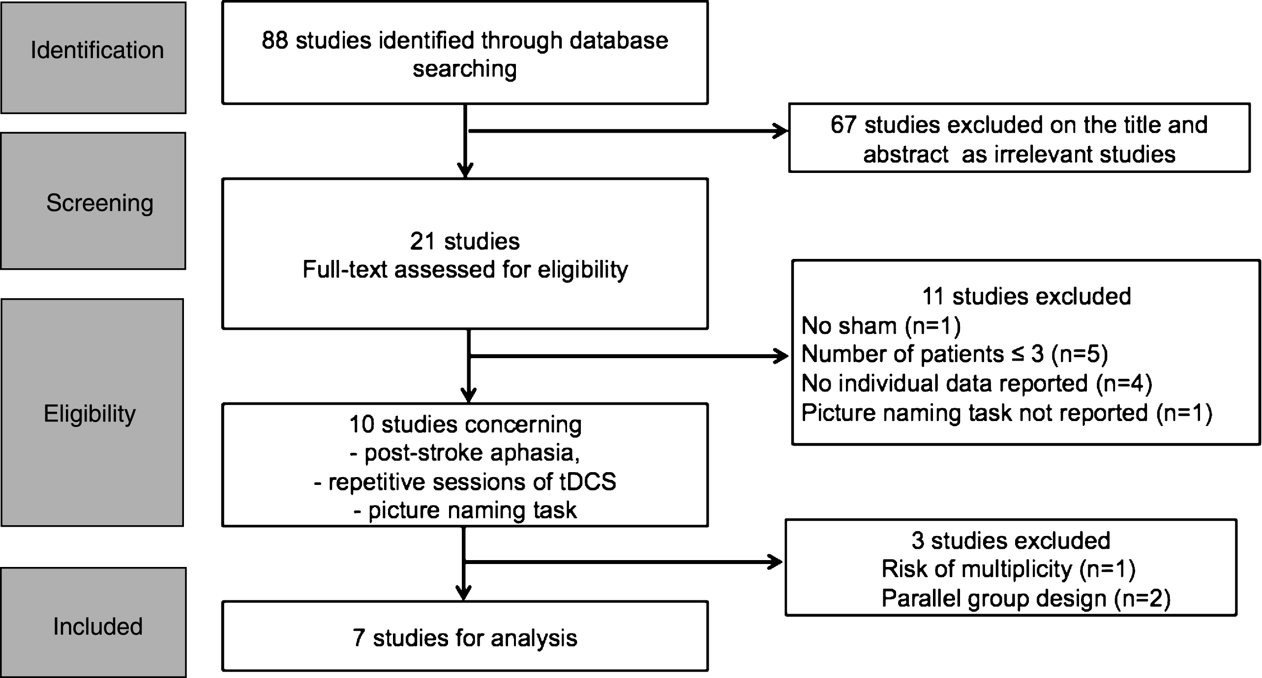
Flow chart of the study.
Picture (object) naming accuracy was the primary endpoint of all studies. The naming accuracy score was assessed on the number of items named accurately, as this was the most consistent and available outcome in all studies. These scores were collected before (pre-) and immediately after (post-) active and sham tDCS interventions. For the active condition, each pre- and post-score was normalized to the number of pictures (total number of items), i.e., (Nactive – Npre)/Nitems, and we refer to this as the “active improvement ratio” or AIR (where Nactive is the number of items correctly named after the active condition, Npre is the number of items correctly named at baseline and Nitems is the total number of items used in each study). For the sham condition, the same ratio was computed and referred to as the “sham improvement ratio” (SIR). From these two normalized ratios, we computed the treatment effect as the difference between AIR and SIR.
In three studies, the difference between post-active (or post-sham) and pre-scores (Nactive – Npre) was reported instead of raw scores (Baker et al., 2010; Kang et al., 2011; Marangolo et al., 2016). These differences were still normalized based on the total number of items to directly determine the treatment effect.
Recorded variables
To characterize the population, we systematically extracted the following data using a standardized data-extraction form: age, gender, time elapsed between stroke and tDCS sessions (i.e., “post stroke delay”), presence or not of comprehension disorders, and aphasia severity score. For the latter, we used a 3-point aphasia severity-scale (1: mild aphasia; 2: moderate aphasia; 3: severe aphasia). For each patient, we derived a severity score from explicitly reported severity (Volpato et al., 2013), Aphasia Quotient (AQ) scores (Baker et al., 2010; Kang et al., 2011), BDAE Aphasia severity rating scale (Wu et al., 2015), or clinical descriptions and quantitative data reviewed by an experienced speech therapist (and only in case the patient profiles were sufficiently documented) (Floël et al., 2011; Marangolo et al., 2013). We chose a 3-items scoring (mild, moderate, severe) because a normalized score (in % of the maximum score) was impossible to compute due to the heterogeneity of the provided information. For Fiori et al. (Fiori et al., 2013), we could not extrapolate any score of aphasia severity because of the lack of clinical descriptions and insufficient quantitative data. Patients were considered to have comprehension disorders whenever authors reported pathological scores on one or more of their comprehension subtests (including different versions of the Token Test, Aachener Aphasie Test-AAT, and Western Aphasia Battery-WAB, subtests). When information was not available, or when we did not have normative values for reported scores (Wu et al., 2015), we classified some of the patients as having or not having a comprehension disorder according to their aphasia profile (global and Wernicke patients: yes, anomic patients: no, and no classification for other reported subtypes). When available, we recorded aphasia profile and location of the stroke lesion in the frontal/parietal/temporal lobes.
For the parameters of the tDCS interventions, we extracted the polarity (anodal or cathodal), the location and side of the active and reference electrodes, and the number of sessions. Concomitant speech therapy was also recorded.
Data analysis
Each variable was tabulated, cross-checked for errors by two investigators (C.R. and C.D.) and compared to the original publication of the respective trials. Individual patient data were then merged to create a common dataset.
tDCS efficacy was assessed by paired samples t-tests between the AIR and SIR values. The standardized mean difference (SMD) (Sedgwick et al., 2013) was extracted for each study and for the common dataset by comparing AIR vs. SIR. The impact of pre-specified subgroups was first tested using paired samples t-tests between the AIR and SIR in each subgroup and then by a direct comparison using independent sample t-tests on the between AIR and SIR. These pre-specified subgroups analyses were performed according to the number of sessions, polarity, side and location of the active electrode, post-stroke delay, aphasia severity level and presence/absence of comprehension disorders. Moreover, we aimed to disentangle the interaction between the SIDE of the active electrode (left or right), POLARITY (anodal or cathodal) and LOCATION of the electrode (frontal or temporoparietal targets). However, as no study reported cases with left cathodal montages, it was not possible to compute a classic 3-way ANOVA. Therefore, we only computed two-way ANOVAs to investigate the interactions between (i) LOCATION of the target electrode and POLARITY and (ii) LOCATION of the target electrode and SIDE. Statistical significance was assumed if p < 0.05. No data were input for missing values. All statistics were performed using MedCalc Statistical Software version 12.7.5 (MedCalc Software BVBA, Ostend, Belgium; http://www.medcalc.org; 2013).
Results
Characteristics of the studies and of the common dataset
Table 1 summarizes the characteristics of the tDCS parameters, and Table 2 the profile of the patients of the seven selected studies (n = 68 patients for 87 observations). In the common dataset, the mean±SD age of the patients was 56±11 years. Time post-onset was 49±56 months. Aphasia profiles were as follows: Broca’s aphasia (35%, n = 24), anomia (19%, n = 13), non-fluent aphasia (24%, n = 16), conduction aphasia (3%, n = 2), global aphasia (6%, n = 4), mixed aphasia (3%, n = 2), motor transcortical (3%, n = 2), sensory transcortical (1%, n = 1), Wernicke’s aphasia (4%, n = 3) and not classified (1%, n = 1). Comprehension disorders could be assessed in 64 patients, and 72% of them showed comprehension impairments (n = 46/64). The anatomical location of the lesion was available in 65 cases and involved the frontal (62%, n = 40), temporal (75%, n = 49) and parietal (63%, n = 41) lobe. Concomitant therapy was administered in 6 out of the 7 studies (n = 79/87 observations). Eligible studies were assessed for selection, detection and attrition bias (see supplementary Table 1) (Viswanathan et al., 2013).
Characteristics of the tDCS parameters for the included studies
Characteristics of the tDCS parameters for the included studies
N is the number of subjects. IFG is the inferior frontal gyrus. L is left, R is right.
Characteristics of the included patients
AAT: Aachen aphasia test; AQ: aphasia quotient; BDAE: Boston diagnosis aphasia examination, EL: esame del linguaggi;, NA: not available.
The primary outcome on naming accuracy was positive, with a mean significant improvement of 35% (±34%) in the active vs. 25% (±37%) in the sham condition, relative to the total number of items (p < 0.0001). This result was still significant when the study of Marangolo et al. (Marangolo et al., 2016), which used dual tDCS, was removed (p < 0.0001). One interesting finding was that the higher the improvement in the sham condition, the higher the improvement in the active condition (r = 0.993, 95% CI: 0.896–0.954, R2: 87%, p < 0.0001). Figure 2 shows the plot of the SMD across studies. As the measure of statistical heterogeneity, I2 was 66% (p: 0.002), the random fixed-effect approach was chosen to calculate the global SMD (mean: 0.802, 95% CI: 0.273–1.333). In addition, the difference between the AIR and SIR was higher for more than 5 sessions of tDCS compared to 5 sessions only (p: 0.02) suggesting a dose-dependent effect (Table 3).
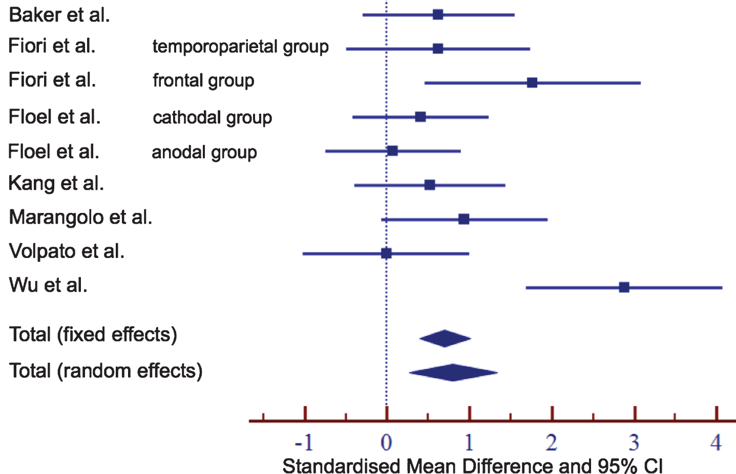
Standardized mean difference (and 95% confidence interval-CI) of active vs. sham improvement ratios for each study and for the common dataset.
Efficacy in pre-specified subgroups
Anodal and cathodal polarities were beneficial (p < 0.0001 and p: 0.02; respectively). However, anodal stimulation was more effective, as the difference between active and sham was higher in anodal than in cathodal conditions (p: 0.004).
Improvements in naming accuracy were also greater after left vs. right stimulation (p: 0.005), although both montages statistically differed from the sham conditions (p < 0.0001 and p: 0.002; respectively). There was only a trend for improved naming abilities when comparing temporoparietal vs. frontal locations of the active electrodes (p: 0.08). Both locations showed benefits in AIR vs. SIR (p < 0.0001 and p: 0.001; respectively).
However, there was a significant interaction between SIDE and LOCATION (F(3, 83): 4.03, p: 0.04). Indeed, when the active electrode targeted the left temporoparietal lobe (n = 19), naming ability was more improved than when the electrode was applied over the left frontal lobe (n = 34) (mean±SE difference: 21% ±3 vs. 7% ±3, p: 0.002). Conversely, when the active electrode was positioned over the right hemisphere, there was no difference between frontal (n = 10) and temporal (n = 24) stimulation locations (p: 0.61) (Fig. 3). No significant interactions between POLARITY and LOCATION were found (F(3,83): 2.7, p: 0.12) (Fig. 3).
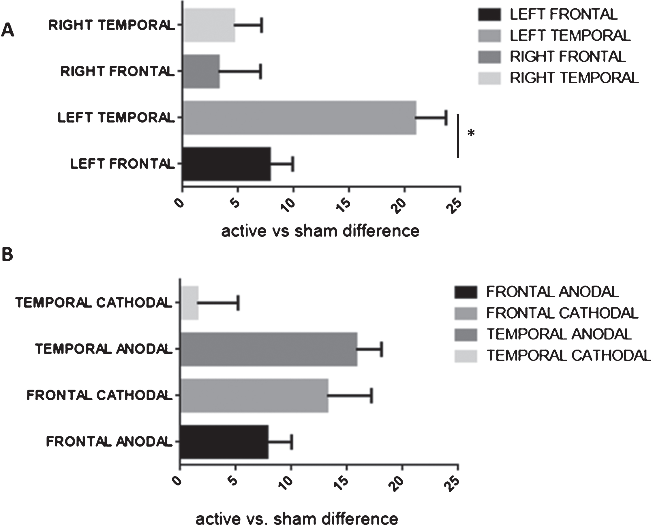
Bar graphs of the mean±standard error of the mean of the difference in the active minus sham improvement ratios in patients with frontal or temporoparietal target electrodes. A: based on the side of the active electrode (left vs. right). B: based on the polarity (anodal vs. cathodal). *p < 0.05.
Finally, for the severity of aphasia (mild, moderate, severe), active stimulation led to better improvements than sham stimulation (p: 0.01, p: 0.002, and p: 0.001, respectively), without a preference for one of these subgroups (p: 0.40). There was no difference in the tDCS effect on patients with stroke ≤ or >1 year (p: 0.79), nor in patients with or without comprehension disorders (p: 0.42).
Results of this IPD meta-analysis confirmed that repetitive tDCS sessions could enhance naming accuracy in chronic stroke survivors. Additionally, there was a dose-dependent effect (5 vs. >5 sessions). We also demonstrated a prevalence of anodal vs. cathodal conditions and left vs. right targeting electrode. Although there was no overall effect of the location of the target, an effect did appear when assessing protocols stimulating the left hemisphere. In these cases, the temporoparietal location led to better improvements than the frontal location. Finally, we found that repetitive tDCS sessions were beneficial regardless of the severity of aphasia.
In contrast, with our findings, recent Cochrane database reviews (Elsner et al., 2011; Elsner et al., 2015) concluded that there was no evidence of the efficacy of tDCS on naming accuracy. However, it is worth noting that these reviews did not focus on repetitive sessions of tDCS only but rather pooled data from studies using both single and repetitive session(s) of tDCS. This confounding factor likely accounts, at least in part, for the difference in our observations. Indeed, it has been hypothesized that the short-lasting effects induced by a single tDCS session will accumulate with repetitive sessions and temporarily create a state that promotes relearning of language (Holland et al., 2012). The effect of repetitive vs. single session(s) is also supported by our findings showing that tDCS aftereffects were more marked in studies in which more than 5 sessions were delivered, demonstrating a dose-dependent effect. One may consider that more sessions extend incrementally the therapeutic window of the consolidation processes in which concomitant speech therapy reinforces the networks that were previously facilitated by tDCS neuromodulation (Hamilton et al., 2011). However, it must be stressed that it has been reported that the enhancing effects of anodal tDCS may reverse inhibitory effects (Fricke et al., 2011) according to the rules of metaplasticity if the inter-session interval is too short.
The benefit of anodal stimulation was higher in our meta-analysis than that of cathodal stimulation. We only found one study with a left cathodal montage (Monti et al., 2008), but because of the lack of both repetitive sessions and individual data, this study was not included in our meta-analysis. Anodal stimulation to the left hemisphere is delivered under the assumption that it enhances the function of the perilesional areas (Schlaug G., 2008). The after-effects of anodal stimulation are relatively consistent in the literature, i.e., they drive the target area into an excitatory state through a mechanism of membrane potential depolarization (Liebetanz et al., 2002). However, both the after-effects of cathodal stimulation and the role of the right hemisphere in picture naming accuracy in post-stroke aphasia are still matter of debate. Cathodal stimulation has been proposed on the right hemisphere under the assumptions that (i) the contralesional hemisphere is responsible for excessive inhibition of left perilesional areas through transcallosal pathways, and (ii) cathodal stimulation usually leads to inhibition. This approach is simplistic, and contrasts with other studies that demonstrated that the right hemisphere could be beneficial in aphasic patients (Hillis AE, 2006; Meltzer et al., 2013), especially in the more severe cases (Bradnam et al., 2012). In this context, it is conceivable that cathodal tDCS may not induce the expected effects. In the debate of the beneficial or detrimental role of the right hemisphere in aphasia recovery, it is conceivable that the volume of the lesion in the left hemisphere affects the results of right-based tDCS experiments. Indeed, the right hemisphere may be the sole source of recovery when damage to the left hemisphere is extensive enough that no language regions have survived (Anglade et al., 2014). Future research should assess the extent and location of the damage of critical regions.
Regarding the targeted area, we found, for the left hemisphere, a stronger effect when the active electrode was positioned over the temporoparietal region compared to the frontal lobe. This finding can be explained by the underlying anatomy of language fasciculi (Hickok et al., 2004). Indeed, the arcuate fasciculus, which constitutes the main tract of the dorsal route, travels from a region in the posterior sylvian fissure to frontal regions through the temporoparietal junction. The ventral route, which is represented by the inferior fronto-occipital and the inferior longitudinal fasciculi, also travels through this temporoparietal junction. In other words, the temporoparietal junction is a crossroad area containing both dorsal and ventral streams. Damage to this area has been associated with poor aphasia recovery (Rosso et al., 2015). This can be explained by the fact that this simultaneous disruption of both streams precludes the compensation of one route by the other, as is also suggested by the Lichtheim 2 model of Ueno et al (Ueno et al., 2013). In this model, when virtual lesions are produced at different loci, Lichtheim 2 developed aphasic syndromes mimicking human symptoms. In the “conduction aphasia” simulation, the recovery-related phenomena were underpinned by an increased activation of the ventral pathway, confirming that aphasia recovery may be hampered if the infarct lesion damages both the dorsal and the ventral pathways. By integrating the fact that the temporoparietal junction is a major language fasciculi crossroad, we can argue that the stimulation of this area by tDCS may affect the entire language network through both routes, from the temporoparietal junction to the frontal areas via the dorsal route and from the temporoparietal junction to occipital, temporal and the frontal pole through the ventral route. This activation could strengthen and facilitate the potential of picture naming circuit.
Finally, a benefit of tDCS was found regardless of the severity of aphasia, and even for severe aphasic patients (as it has already been shown for anomia treatment without tDCS) (Nickels, 2002). Although results must be interpreted with caution, as they were based on a small number of studies, this is the first time that a meta-analysis on tDCS and aphasia classified the patients according to their severity from data extrapolated from specific scores used in each study. The other tDCS/TMS meta-analysis failed to subgroup the results by aphasic severity degree (Ren et al., 2014). Our results demonstrated that tDCS was valuable even for severe aphasic patients. This contrasts with the use of tDCS in motor stroke patients. Indeed, a recent meta-analysis revealed a statistically significant benefit of tDCS in nine studies who demonstrated mild/moderate impairments but not those classified with moderate/severe impairments (Marquez et al., 2015).
Our study has limitations. Given that most of patients suffered from ischemic strokes (59/68, 88%), the extrapolation of our findings to hemorrhagic strokes is limited and should be done with caution. Second, no conclusions can be drawn for the acute and subacute phases at this time. Although we did not restrict our web search to the chronic phase, six out of the seven studies enrolled patients after 6 months (Baker et al., 2010; Fiori et al., 2013; Floël et al., 2011; Kang et al., 2011; Marangolo et al., 2016; Volpato et al., 2013). This limited the applicability of our results to earlier phases of stroke. Third, the reliability of our results should be interpreted with caution since four studies could not be included because of a lack of IPD. It is also important to note that no study presented sample size calculations or estimations. These omissions could impact the validity of the results by introducing a “study selection bias”. Selection biases cannot be perfectly controlled for since studies with positive results are more likely to be published as papers than studies with negative conclusions. Finally, the heterogeneity of stroke patients’ lesions and profiles, pre-stroke language dominance implied that our meta-analysis is an early attempt albeit an important one at bringing order to a small and very mixed body of literature.
Conclusion
This meta-analysis provides clues that repetitive sessions of tDCS are likely to enhance picture-naming accuracy in post-stroke aphasia. This opens up the perspective of harnessing tDCS as a therapeutic treatment to promote language recovery in clinical routines. However, tDCS use is worth further explorations to confirm these preliminary results.
Conflicts of interest
The authors declare no conflicts of interest
