






Abstract
Background:
Replantation of the avulsed nerve root has been proposed for the treatment of severe brachial plexus injury for several decades. However, due to the complexity of the technique and limited functional improvement, practical applications are yet to be implemented.
Objective:
In the present study, we investigated the effect of pretreatment with resveratrol on nerve autografts used for replantation surgery in a rat model of nerve root avulsion.
Methods:
Resveratrol pretreatment was performed using an explant culture technique. Two surgical procedures were performed. During the first surgery, Sprague-Dawley rats were subjected to left C6 nerve root avulsion, and nerves were harvested for autografting. The harvested grafts were explant-cultured for 1 week. A second procedure was performed to replant the C6 nerve root using the explant-cultured nerve graft 1 week after the first procedure. Histological and immunohistochemical analyses were performed 8 weeks after the second procedure. We first compared findings between explant-cultured nerve grafts and fresh nerve grafts, following which we compared findings between explant-cultured grafts pretreated with and without resveratrol. Changes induced within nerve grafts by 1 week of explant culture with or without resveratrol were investigated in vitro.
Results:
There was no significant difference in outcomes between 1 week-explant-cultured and fresh nerve grafts. Addition of resveratrol to the explant culture medium resulted in a significant increase in the number and myelin thickness of regenerated axons, and in the number of regenerating motor neurons in the C6 spinal cord segment. In vitro analyses revealed that nerve grafts pretreated with resveratrol exhibited significant increases in glial cell line-derived neurotrophic factor (GDNF) expression and the number of dedifferentiated Schwann cells.
Conclusions:
Resveratrol may promote axonal regeneration following replantation surgery for the treatment of nerve root avulsion injury; however, further studies are required to verify these findings in humans.
Introduction
Due to disruptions in the connections between the spinal cord and peripheral nerves, complete surgical repair of nerve root avulsion injuries has been considered impossible. Two decades ago, replantation of the avulsed nerve root into the spinal cord was reported as a clinically promising surgical option (Carlstedt, Grane, Hallin, & Noren, 1995). However, such treatment resulted in limited functional recovery of the shoulder and elbow joints in several adult patients. Subsequent research revealed that, while functional recovery of the hand was also achieved in a pediatric patient, recovery remained limited (Carlstedt, Anand, Htut, Misra, & Svensson, 2004). Limitations in the number of axons regenerated from the avulsed motor neuron into the replanted nerve have been considered among the most important factors impeding good functional outcomes following nerve replantation surgery (Carlstedt et al., 2000; Htut, Misra, Anand, Birch, & Carlstedt, 2007).
In nerve root avulsion injury, the avulsed nerve root gradually shrinks following detachment from the spinal cord and retraction from the spinal vertebrae. Moreover, the stump of the avulsed nerve root is sometimes severely damaged by traction force, necessitating resection of the damaged segment. Therefore, during replantation surgery, a nerve graft is usually required to bridge the spinal cord and the stump of the avulsed nerve root. In general, better outcomes are observed for autograft bridging than for either artificial conduits or allograft bridging. Therefore, pretreatment of nerve autografts may further improve axonal regeneration into the replanted nerve (Chu et al., 2009).
Glial cell line-derived neurotrophic factor (GDNF), a member of the transforming growth factor β superfamily (Lin, Doherty, Lile, Bektesh, & Collins, 1993), has been reported to promote the migration of Schwann cells, axonal regeneration (Bergerot, Shortland, Anand, Hunt, & Carlstedt, 2004; Chu et al., 2009; Chu et al., 2012), and myelin thickening following replantation surgery for nerve root avulsion injury. However, because direct treatment using neurotrophic factors may exert adverse effects on the human body, such treatment should be avoided when possible (Sendtner, Pei, Beck, Schweizer, & Wiese, 2000).
Resveratrol is a non-flavonoid polyphenol with anti-oxidant, anti-inflammatory, and anti-cancer effects (Zhang, Liu, & Shi, 2010) that has been reported to promote the release of GDNF from glial cells in vitro (Zhang et al., 2012). In the present study, we investigated the effect of resveratrol pretreatment on nerve autografts for replantation surgery in a rat model of nerve root avulsion injury.
Materials and methods
Animals
Eight-week-old male adult Sprague-Dawley (SD) rats (Nippon SLC Inc., Hamamatsu, Japan) weighting approximately 280 g (250–300 g) were used for the present study. All animals were housed under a 14-h light/10-h dark cycle (lights on at 7:00 am) with ad libitum access to food and water. Prior to surgery, rats were deeply anesthetized via an intraperitoneal injection of sodium pentobarbital (40 mg/kg). This study was approved by the Animal Research Committee of the Graduate School of Medicine at Kyoto University (approval number: MedKyo 14530), and all procedures were performed in accordance with the guidelines of the Animal Research Committee.
First surgery: C6 nerve root avulsion and C5 nerve root dissection
A midline skin incision was made on the posterior side of the neck. The left paravertebral muscles of the cervical spine were unilaterally dissected from the spinous processes, laminae, and anterior articular processes (lateral mass) at the level of C3 to T2. The C5-C6 vertebral arch was confirmed in reference to the T2 spinous process, the most prominent spinous process in rats. The transverse processes and lateral third of the C4, C5, and C6 lateral masses were carefully removed using a surgical bur (NSK-Nakanishi Co, Ltd., Tochigi, Japan). The C6 spinal nerve was exposed and gently avulsed using Adson forceps without teeth (nerve root avulsion). Successful nerve root avulsion was confirmed when the bifurcated stump of the avulsed nerve root was observed in conjunction with the leakage of spinal fluid from the vertebral foramen. The C5 spinal nerve was dissected just outside of the intervertebral foramen from which it emerged (nerve root dissection), following which the muscle and skin were sutured in layers.
Harvesting of the nerve autograft
Two different types of nerve autografts were used in the experiments of the present study. In Experiment 1, the peroneal nerve was used. The left peroneal nerve was exposed from the mid-thigh to the middle one-third of the lower leg using a posterior approach. A 5-mm length of the peroneal nerve was then harvested. However, due to the complexity and invasive nature of the procedure in rats, C5 nerve root autografts were used for Experiment 2. When C5 nerve roots were harvested during C6 nerve root avulsion, no substantial increases in the extent of paralysis were observed relative to that following C6 nerve root avulsion alone. C5 spinal nerves were harvested at the greatest lengths possible from immediately outside of the intervertebral foramen to the point at which the C5 and C6 ventral rami merged.
Explant cultures of the harvested nerve grafts
The harvested nerve grafts were incubated in Schwann cell medium (ScienCell Research Laboratories, Carlsbad CA, USA) at 37°C in a humidified atmosphere containing 5% CO2 for 1 week. Half of the medium was exchanged with fresh medium every 3 or 4 days. One week after explantation, the incubated nerves were used as grafts during the second surgery.
Second surgery: Replantation of the nerve grafts
The second surgery was performed 1 week after the first surgery. The stump of the avulsed C6 nerve root was identified via the same posterior approach. A few millimeters of the stump were resected to eliminate the portion that may have been damaged by the forceps during the first surgery, following which the remainder was sutured with the proximal stump of the nerve graft using 10-0 nylon sutures. A few millimeters were also trimmed from each stump of the nerve graft prior to neurorrhaphy. Dorsal hemilaminectomy was performed to expose the dura at C5. The dura and arachnoid mater were partially excised at the sixth myelomere, and a small incision was made on the lateral side of the spinal cord. Then, the other stump of the same nerve graft was inserted into the spinal cord incision. The connection between the grafted nerve and the spinal cord was covered with fibrin glue, following which the muscle and skin were sutured in layers.
Tissue preparation
The rats were administered a lethal dose of sodium pentobarbital and transcardially perfused with 200 mL of phosphate-buffered saline (PBS, pH 7.4), followed by 350 mL of 4% (w/v) paraformaldehyde (PFA) in 0.1 M phosphate buffer (PB, pH 7.4). We then harvested cervical spinal cord segments (sixth myelomere) as well as the left C6 spinal nerve (cut 2 mm distal to the neurorrhaphy site). The harvested C6 spinal cord segments were post-fixed overnight in perfusion fixative at 4°C and cryoprotected for 24 h in 20% (w/v) sucrose in PB (4°C). The segments were cut into serial transverse sections (thickness: 40μm) using a cryostat. The harvested left C6 spinal nerve were fixed in 1% glutaraldehyde and 1.44% PFA, post-fixed in 1% osmic acid, and embedded in epoxy resin. The harvested spinal cord segments were used for motor neuron counting, while the harvested spinal nerves were used for histological analysis. For immunohistochemistry experiments, the harvested left C6 spinal nerves were fixed in 4% PFA overnight at 4°C, cryoprotected in 20% sucrose for 48 h at 4°C, and cut into transverse sections (thickness: 20μm) using a cryostat.
Counting of spinal motor neurons
For spinal motor neuron counting, all sections of the C6 spinal cord segment were stained with 0.5% (w/v) cresyl violet (Nissl staining). The numbers of surviving motoneurons on the lesioned side of the spinal cord were counted in all sections and compared with those on the unlesioned side, as described in our previous report (Noguchi, Ohta, Kakinoki, Kaizawa, & Matsuda, 2013). Large multipolar cells with abundant cytoplasm larger than 30μm in the ventral horn were counted as viable motor neurons. To avoid double counting, the number of surviving motor neurons was corrected using Abercrombie’s formula(Clarke & Oppenheim, 1995). No cell loss was expressed as 100%. Cell counting for all serial sections was performed by two observers who were blinded to the groups.
Retrograde labeling of the bilateral C6 spinal motor neurons
Rats were deeply anesthetized with intraperitoneal pentobarbital 53 days after the second surgery. A transverse incision was made on the anterior portion of the upper chest to split the pectoralis major muscles, exposing the bilateral upper trunk of the brachial plexus. Three microliters of 3% hydroxystilbamidine (Life Technologies, Eugene OR, USA) was injected into each upper trunk of the brachial plexus using a Hamilton syringe. Three days after the injection, rats were administered a lethal dose of sodium pentobarbital and intracardially perfused with PFA, following which the C6 spinal cord segments were harvested as previously described in section 2.6. The harvested C6 segments were post-fixed overnight in perfusion fixative at 4°C and cryoprotected for 24 h in 20% (w/v) sucrose in PB (4°C). The segments were cut into serial transverse sections (thickness: 40μm) using a cryostat. The number of labeled motor neurons on the lesioned side of the ventral horn was counted using confocal fluorescence microscopy and compared with that on the unlesioned side.
Histological analysis of the nerve root
Serial transverse sections (thickness: 1μm) were prepared 1 mm distal to the site of neurorrhaphy between the grafted nerve and the original C6 nerve root. The sections were also located approximately 5 mm distal to the autograft replantation site. The prepared sections included only axons that had regenerated a distance of at least 5 mm from the replantation site. The sections were stained with 0.5% (w/v) toluidine blue solution and examined via light microscopy (ECLIPSE 80i, Nikon, Tokyo, Japan). The total number of myelinated axons and the axon diameter were determined using ImageJ software (Ver. 1.51, National Institute of Health, Bethesda, MD, USA), as previously described (Kaizawa et al., 2016). Briefly, the entire neural area of each specimen was calculated for a single image. Four or five fields were chosen at random, such that the area analyzed would represent >20% of the entire neural area of each specimen. The number of myelinated axons was calculated for each field at a final magnification of 200×. The numbers of myelinated axons and neural areas of each field were then added together. The total number of myelinated axons in each specimen was estimated as follows: total number of myelinated axons = number of myelinated axons in each field×(entire neural area/neural area of each field). To assess nerve-fiber diameters (NFDs) and axon diameters (ADs), ultrathin sections of the same tissues stained with uranyl acetate and lead citrate were examined using transmission electron microscopy (TEM: Model H-7000; Hitachi High-Technologies, Tokyo, Japan). Fifteen fields of the specimen were randomly chosen at a magnification of 2000×. Myelin thickness (MT) and G-ratio were calculated according to the formulas (NFD - AD)/2 and AD/NFD, respectively.
Experiment 1: Fresh nerve autograft vs. explant-cultured nerve autograft
Sixteen rats were randomly divided into the following two groups: the fresh nerve autograft group (fresh group, n = 8) and the explant-cultured nerve autograft group (explant group, n = 8). All rats underwent C6 nerve root avulsion (Noguchi et al., 2013). In the explant group, the left peroneal nerve was harvested for nerve grafting on the day of C6 root avulsion surgery, following which the harvested nerve was incubated in Schwann cell medium for 1 week. One week after the first surgery, replantation surgery was performed using the explant-cultured nerve graft. In the fresh group, the left peroneal nerve was harvested immediately prior to replantation surgery. Thus, replantation surgery was performed using fresh peroneal nerve grafts.
Experiment 2: Explant-cultured nerves pretreated with resveratrol vs. untreated explant-cultured nerves
Twenty-four rats were randomly divided into following two groups: those that received C5 nerve grafts that had been pretreated with resveratrol for 1 week in explant cultures (resveratrol group, n = 12), and those that received C5 nerve grafts that had been cultured without resveratrol (control group, n = 12). During the first surgery, left C6 nerve root avulsion was performed, and the C5 spinal nerve was harvested for autografting. In the control group, the harvested nerve graft was incubated in Schwann cell medium for 1 week. In the resveratrol group, the harvested nerve graft was incubated in Schwann cell medium with 100 U/mL of resveratrol (Sigma-Aldrich, St Louis, MO, USA) for 1 week. The second replantation surgery was performed 1 week after the first surgery using the explant-cultured nerves.
Experiment 3: In vitro examination of explant-cultured nerve grafts
Immunohistochemistry
Bilateral C5 spinal nerves were harvested from 15 SD rats for immunohistochemistry experiments. Each of the harvested nerve roots was regarded as one sample. The 30 total nerves were randomly assigned to the following three groups: the naïve group (n = 10), the control group (n = 10), and the resveratrol group (n = 10).
In the naïve group, tissue preparation procedures were performed immediately after nerve harvesting. In the resveratrol and control groups, the harvested nerves were incubated in Schwann cell medium with or without 100 U/mL of resveratrol for 1 week, respectively. Following preparation of the tissue samples, immunohistochemistry procedures were performed using the following primary antibodies: chicken polyclonal anti-glial fibrillary acidic protein (GFAP) antibody (1:1000; Abcam plc, Cambridge, UK) and mouse monoclonal anti-CD68 antibody (1:50; Bio-Rad, Hercules, CA). The sections were pretreated with 0.3% (v/v) H2O2 in PBS and preincubated with 2% (v/v) normal goat serum in 0.2% (v/v) TritonX-100 in PBS at 28°C. The sections were then incubated overnight with the primary antibodies at 4°C, following which they were treated with secondary biotinylated antibody [CF™488 (for anti-GFAP antibody) and CF™543 (for anti-CD68 antibody) (Biotium, Richmond, CA)] for 1 h.
For all serial sections, the numbers of immunopositive cells were counted by two observers blinded to the groups. Results were expressed as the mean number of immunopositive cells per section.
Real-time polymerase chain reaction analyses
Preparation of nerve samples. Six SD rats were used for polymerase chain reaction (PCR) analyses in the present study. The bilateral partial brachial plexus emerging from C5, C6, C7, and C8 and 15 mm lengths of the sciatic nerve were harvested under deep anesthesia with a lethal dose of sodium pentobarbital administered via intraperitoneal injection. The unilateral cervical nerves and unilateral sciatic nerves harvested from a single rat were each regarded as one sample, respectively. The 12 cervical nerves and 12 sciatic nerves were randomly assigned to the following three groups: naïve group (n = 8), control group (n = 8), and resveratrol group (n = 8). In the naïve group, the nerves were snap-frozen and immediately stored at –80°C following harvesting until further use. In the control group, the nerves were incubated in Schwann cell medium for 1 week, snap-frozen, and stored at – 80°C. In the resveratrol group, the nerves were incubated in Schwann cell medium with 100 U/mL resveratrol for 1 week, snap-frozen, and stored at – 80°C.
Real-time PCR
Total RNA was extracted from the prepared nerves using TRI Reagent (Molecular Research Center Inc., Cincinnati OH, USA), chloroform (Sigma-Aldrich), and isopropanol (Kishida Chemical Co. Ltd, Osaka, Japan). Two hundred micrograms of total RNA was transcribed to cDNA using ReverTra Ace qPCR RT Master Mix (Toyobo, Osaka, Japan). The cDNA was analyzed via real-time PCR using Light Cycler 1.5 (Roche, Basel, Switzerland), and the standard curve was created using THUNDERBIRD SYBR qPCR Mix (Toyobo). The primers for GDNF and β-actin were designed based on methods described in a previous report (Pedersen, Jacobsen, Mollerup, & Gjerstad, 2010) (Table 1). Data were expressed relative to the value obtained for β-actin.
List of primers used for real-time reverse-transcriptase polymerase chain reaction
List of primers used for real-time reverse-transcriptase polymerase chain reaction
GDNF: glial cell line-derived neurotrophic factor.
Preparation of cervical nerve roots and hind limb peripheral nerves. Fourteen SD rats were used for enzyme-linked immunosorbent assay (ELISA) experiments in the present study. The bilateral partial brachial plexus emerging from C5, C6, C7, and C8 as well as the bilateral sciatic, peroneal, and tibial nerves were harvested at the maximum possible lengths under deep anesthesia with a lethal dose of intraperitoneal sodium pentobarbital. The bilateral cervical nerves and whole hind limb nerves harvested from a single rat were each regarded as one sample. The 14 cervical nerves and 14 hind limb nerves were randomly assigned to the following three groups: the naïve group (n = 8), the control group (n = 10), and the resveratrol group (n = 10). In the naïve group, the nerves were snap-frozen and stored at –80°C on the day of harvesting until further use. In the control group, the nerves were cut into 5 mm segments, incubated in Schwann cell medium for 1 week, snap-frozen, and stored at – 80°C. In the resveratrol group, the nerves were also cut into 5 mm segments, incubated in Schwann cell medium with 100 U/mL resveratrol for 1 week, snap-frozen, and stored at – 80°C.
ELISA
To quantify the amount of GDNF expressed in the explant-cultured nerves, all 28 nerve samples were dissected without thawing and resuspended in 1 mL of NP-40 extraction buffer containing 150 mM NaCl, 50 mM Tris-HCl (pH 8.0), 0.1% Tween-20 (Sigma-Aldrich), 1% Nonidet P40 substitute (Sigma-Aldrich), protease inhibitor PMSF (Sigma-Aldrich), and 0.2 mM EDTA-2Na (Sigma-Aldrich). The samples were then further homogenized with a vortex mixer for 30 s and centrifuged (15000 rcf, 30 min, 4°C), following which the supernatant was collected. The concentration of GDNF was measured using an ELISA kit (Raybiotech, Norcross, GA, USA) in accordance with the manufacturer’s instructions, and the total GDNF content within each nerve sample was calculated.
Statistical analysis
All data were expressed as the mean±standard error. The results of Experiments 1 and 2 and ELISA findings for Experiment 3 were compared using unpaired t-tests following confirmation of a normal distribution (JMP 12, SAS Institute, Cary, NC, USA). Other data for Experiment 3 were compared using one-way analyses of variance (ANOVA) followed by Tukey’s test. The level of statistical significance was set at P < 0.05.
Results
Experiment 1: Fresh nerve autograft vs. explant-cultured nerve autograft
Motor neuron counting
The percentage of surviving motor neurons in the lesioned ventral horn was 72.4±3.79% in the fresh group and 72.5±2.95% in the explant group (Table 2). Thus, no significant difference was observed between the two groups (P = 0.98).
Results of Experiment 1: Fresh nerve autografts vs. explant-cultured nerve autografts
Results of Experiment 1: Fresh nerve autografts vs. explant-cultured nerve autografts
Fresh group: replantation surgery performed with fresh nerve autografts; explant group: replantation surgery performed with explant-cultured (1 week) nerve autografts.
The total number of regenerated myelinated axons at 1 mm distal to site of distal neurorrhaphy was 1,328.13±114.88 in the fresh group and 1,529.88±252.52 in the explant group, and there was no significant difference between the two groups (P = 0.48). Furthermore, there were no significant differences in NFD, AD, or MT between these two groups.
Experiment 2: Explant-cultured nerves pretreated with resveratrol vs. untreated explant-cultured nerves
Motor neuron counting
The percentage of surviving motor neurons in the lesioned ventral horn was 69.22±3.44% in the control group and 67.23±3.60% in the resveratrol group (Fig. 1). Thus, there was no significant difference between the two groups (Table 3).
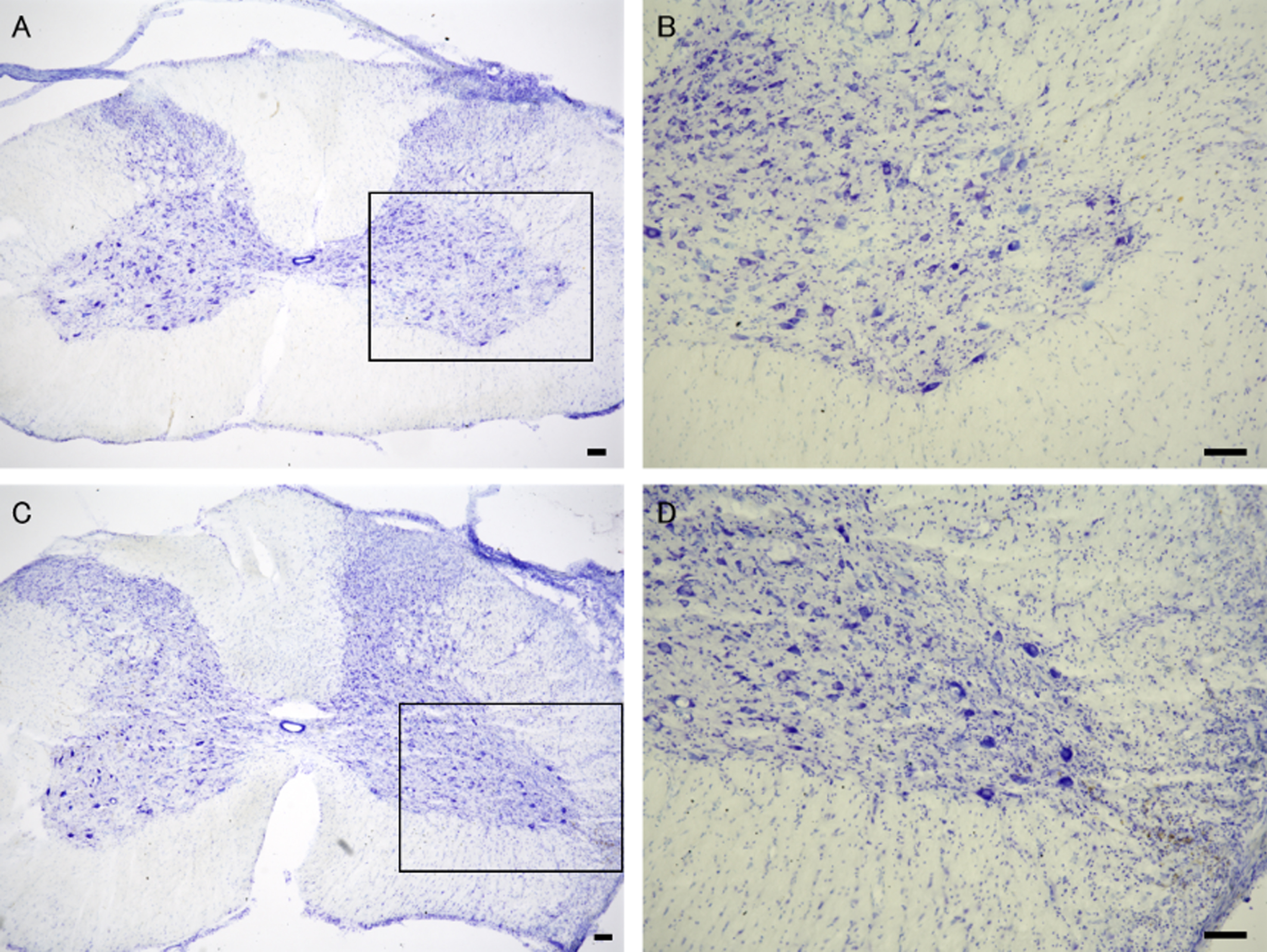
Nissl staining of the C6 ventral horn in the control (A, B) and resveratrol groups (C, D). B and D are high-magnification images of the boxed area in the control (A) and resveratrol groups (C), respectively. Bar: 100μm.
Results of Experiment 2: Explant-cultured nerves pretreated with resveratrol vs. untreated explant-cultured nerves
Control group: replantation surgery performed with explant-cultured (1 week) nerve autografts without resveratrol pretreatment; resveratrol group: replantation surgery performed with explant-cultured (1 week) nerve autografts pretreated with resveratrol.
The total number of regenerated myelinated axons at 1 mm distal to the site of distal neurorrhaphy was significantly higher in the resveratrol group than in the control group (P = 0.0046, Fig. 2A–2 C). MT was also significantly greater in the resveratrol group than in the control group (P = 0.0079, Fig. 2D and 2E), although no significant differences in NFD or AD were observed.
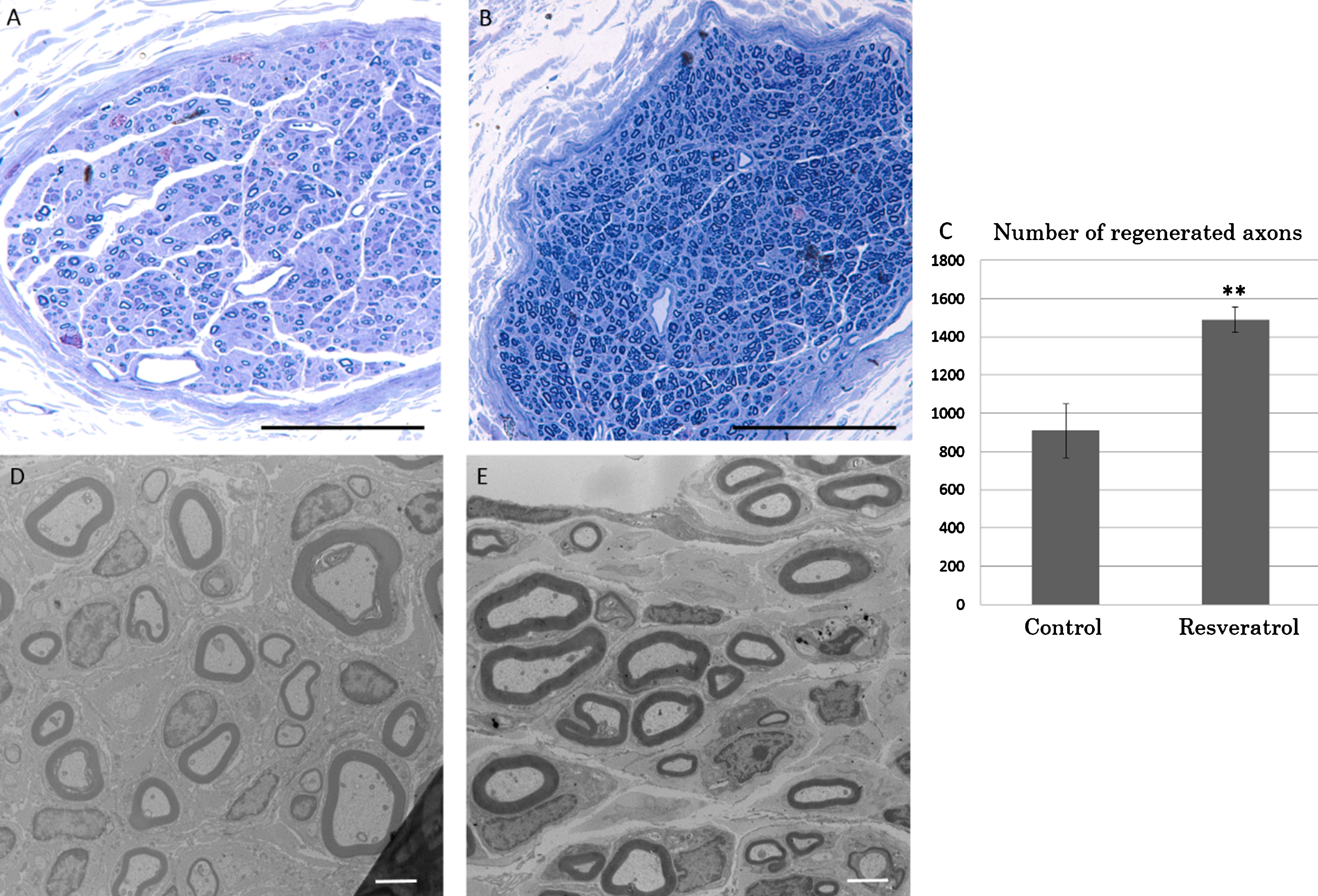
Toluidine blue staining and transmission microscopic images of the replanted C6 root. The replanted C6 roots were harvested at 1 mm distal to the site of distal neurorrhaphy. Significantly more axons were regenerated in the resveratrol group than in the control group (A, control group; B, resveratrol group. Scale bar: 100μm). Axonal regeneration was significantly higher in rats that had received nerve grafts pretreated with resveratrol (resveratrol group) than in those that had received grafts cultured without resveratrol (control group) (C). Transmission microscopic images (D, control group; E, resveratrol group. Scale bar, 2μm) of the replanted C6 roots at the site of toluidine blue staining. The myelin sheath was significantly thicker in the resveratrol group than in the control group. **P < 0.01.
The number of labeled C6 motoneurons was significantly higher in the resveratrol group than in the control group (P = 0.037, Fig. 3).
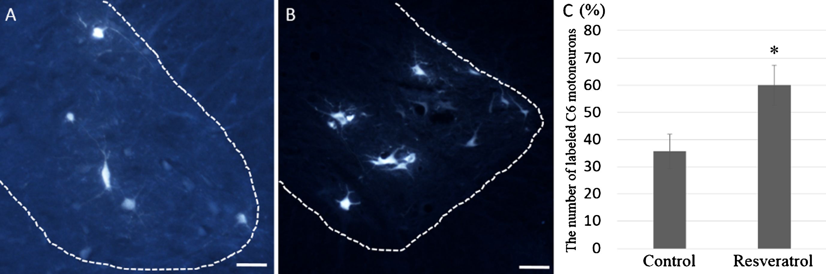
Labeled motor neurons in the C6 ventral horn in control group (A) and resveratrol group (B). Significantly more motor neurons were labeled in resveratrol group (C). Bar: 100 m, *P < 0.05.
Immunohistochemistry
The number of CD68-positive cells did not significantly differ among the three groups (naïve group: 14.9±3.03; control group: 18.5±2.71; resveratrol group: 24.6±3.82; P = 0.721 between naïve and control groups, P = 0.126 between naïve and resveratrol groups, and P = 0.400 between control and resveratrol groups) (Fig. 4A–4D).
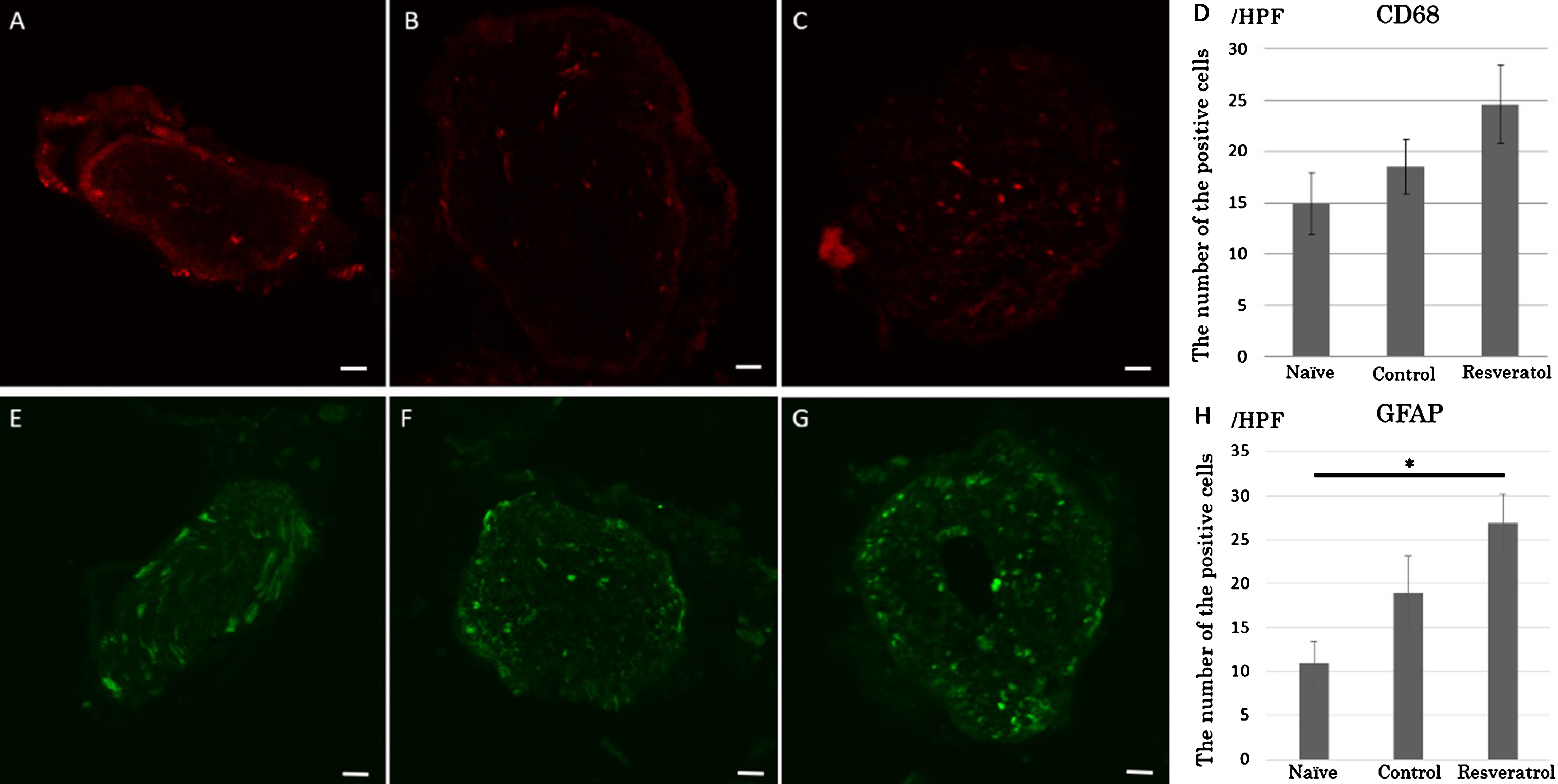
Immunohistochemistry results for CD68 and glial fibrillary acidic protein (GFAP). Immunohistochemistry results for CD68 in non-pretreated nerve grafts (A: naïve group), nerve grafts explant-cultured without resveratrol (B: control group), and nerve grafts explant-cultured with resveratrol (C: resveratrol group). The number of CD68-positive cells was higher in the resveratrol group than in the other two groups, but no significant differences were observed among the three groups. (D). Immunohistochemistry results for GFAP in non-pretreated nerve grafts (E: naïve group), nerve grafts explant-cultured without resveratrol (F: control group), and nerve grafts explant-cultured with resveratrol (G: resveratrol group). A significant difference in the number of the GFAP-positive cells was observed between the naïve group and the resveratrol group (H). Scale bar: 100μm. *P < 0.05.
The number of GFAP-positive cells was significantly higher in the resveratrol group than in the naïve group (resveratrol group: 26.9±3.34; naïve group: 10.9±2.43; P = 0.015) (Fig. 4E–4 H). There was no significant difference in the number of GFAP-positive cells between the naïve group and the control group (control group: 19.0±4.16; P = 0.25), or between the control group and the resveratrol group (P = 0.26).
GDNF mRNA expression was significantly higher in the resveratrol group than in the control group (resveratrol group: 0.910±0.14; control group: 0.412±0.051; P = 0.0013) (Fig. 5A). In addition, GDNF mRNA expression was significantly higher in the resveratrol and control groups than in the naïve group (naïve group: 0.00658±0.0038; P = 0.0078 between the naïve and control group, and P < 0.0001 between the naïve and resveratrol group).
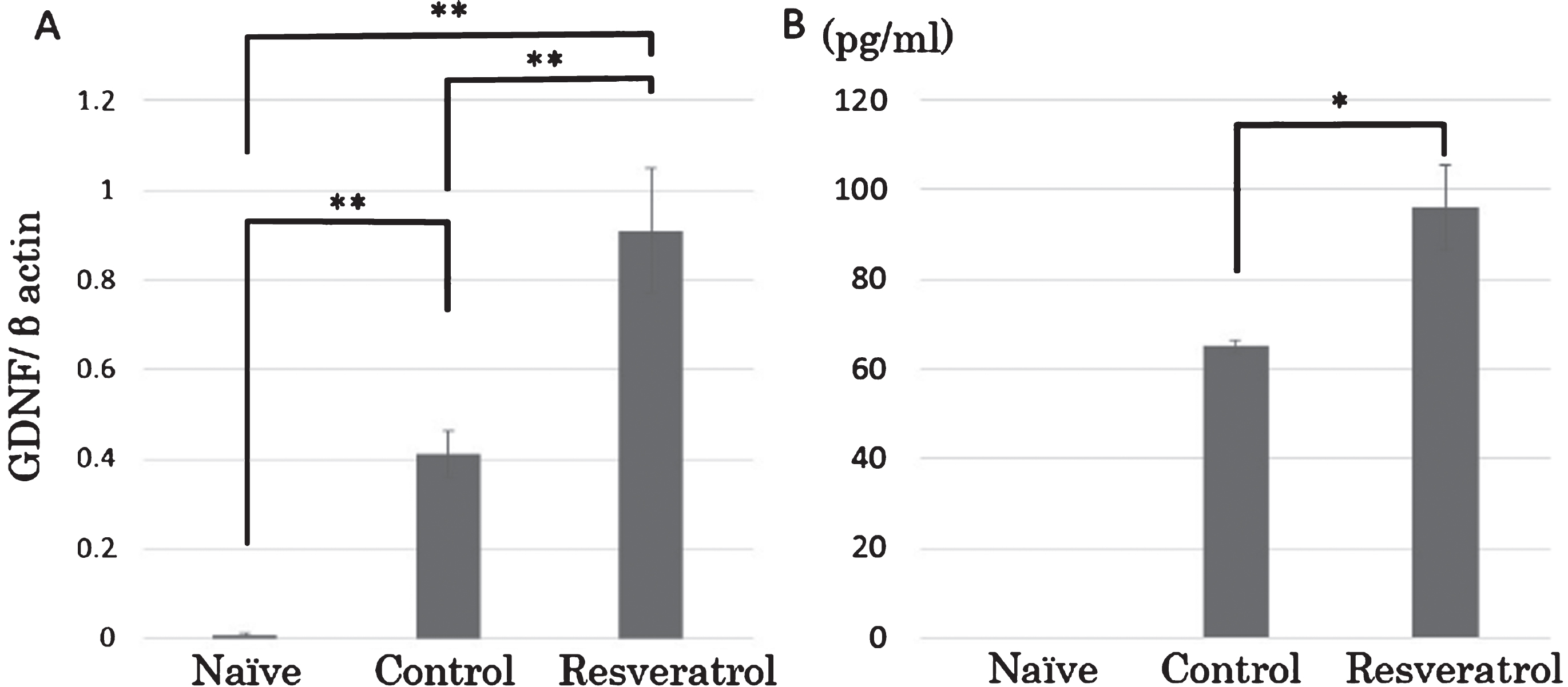
Glial cell line-derived neurotrophic factor (GDNF) concentration and mRNA expression in the nerve grafts of each group. GDNF mRNA expression was significantly higher in the control group than in the naïve group. Moreover, GDNF mRNA expression was significantly higher in the resveratrol group than in either the naïve group or the control group (A). GDNF was not detected in any sample of the naïve group. GDNF concentration was significantly higher in the resveratrol group than in the control group (B). *P < 0.05; **P < 0.01.
No GDNF expression was detected in any samples of the naïve group. In the control and resveratrol groups, GDNF expression was detected in four of 10 and nine of 10 samples, respectively. The mean concentration of GDNF was significantly higher in the resveratrol group than in the control group (control group: 65.09±1.45 pg/mL; 96.02±9.47 pg/mL; P = 0.0120) (Fig. 5B).
Discussion
The present study demonstrated that pretreatment of nerve autografts with resveratrol improves axonal regeneration in spinal motor neurons following replantation surgery in a rat model of nerve root avulsion. The nerve autografts were cultured in Schwann cell medium and pretreated with resveratrol for 1 week prior to replantation surgery. Our results indicated that the addition of resveratrol to the explant culture medium induced a significant increase in the number of dedifferentiated Schwann cells and increased GDNF expression in the explant-cultured nerves.
Pretreatment of nerve grafts promotes axonal regeneration
Following nerve transection, intraneural cellular changes known as Wallerian degeneration begin to occur distal to the lesion site within 2 days after injury. Wallerian degeneration includes axonal degeneration, degradation of the myelin sheath, and removal of myelin debris by the resident Schwann cells and recruited macrophages (Rotshenker, 2011). These degeneration steps also occur in the nerves harvested for autografting and may be accelerated by pretreatment of the nerve grafts.
Pre-degeneration is a pretreatment procedure employed to condition the nerve prior to grafting (Tomita et al., 2009). In in vivo pre-degeneration procedures, the proximal end of the grafted nerve is crushed or transected in advance. Several days later, the nerve graft is then harvested from a point distal to the site of injury. Previous studies have indicated that in vivo pre-degeneration improves axonal regeneration relative to that observed for fresh nerve grafts by decreasing the initial delay period without affecting the rate of regeneration (Danielsen et al., 1994). These effects have been attributed to increases in the number and migration of Schwann cells and macrophages (Danielsen et al., 1994; Keilhoff, Fansa, Schneider, & Wolf, 1999), and to increases in the release of neurotrophic factors (Carbonetto, 1991).
In the present study, nerve grafts were subjected to explant culturing for 1 week. As observed for in vivo pre-degeneration procedures, Wallerian degeneration begins within the explant-cultured nerve immediately after harvesting. Because macrophages from the circulation cannot invade the explant-cultured nerve, only resident cells such as Schwann cells and macrophages contribute to the early degeneration process under such conditions. In the normal Wallerian degeneration process, myelin clearance within the first 5 to 7 days after nerve injury is considered to result from autophagy of the dedifferentiated Schwann cells (Gaudet, Popovich, & Ramer, 2011; Thumm & Simons, 2015). Following Schwann cell autophagy, macrophages recruited from the blood circulation are considered to play a major role in myelin breakdown. Therefore, the lack of extrinsic macrophages invading from the blood circulation would have little effect on the early degeneration process in the harvested nerve graft. Our immunohistological findings revealed that addition of resveratrol to the explant culture medium induced a significant increase in the number of dedifferentiated Schwann cells in the explant-cultured nerve. In accordance with previous findings, these results suggest that resveratrol promotes Schwann cell autophagy, enabling the clearance of myelin debris that contains axonal regeneration-inhibiting molecules (Mueller et al., 2003). Although pretreatment with resveratrol did not significantly increase the number of resident macrophages, such treatment tended to increase the number of residual macrophages. Therefore, proliferation of macrophages in nerve autografts may also contribute to myelin clearance.
Pretreatment with resveratrol increases in GDNF expression in nerve grafts
Our previous study demonstrated that the number of motor neurons in the affected spinal cord segment decreased rapidly and was reduced by more than half at 4 weeks after nerve root avulsion injury (Noguchi et al., 2013). However, in the present study, the number of surviving motor neurons on the lesioned side was 67–69% of that on the contralateral unlesioned side at 8 weeks after replantation surgery. These findings suggest that the replanted nerve graft contained critical factors that exerted neuroprotective effects on spinal motor neurons. Following nerve injury, GDNF is rapidly produced by Schwann cells in the distal nerve segments (Hoke, Gordon, Zochodne, & Sulaiman, 2002) and is considered a potent survival factor for axotomized motor neurons (Chen, Chai, Cao, Lu, & He, 2001; Henderson et al., 1994). In our study, we observed no differences in the number of surviving motor neurons between resveratrol-treated (resveratrol group) and control groups, although the resveratrol-treated nerves produced a significantly larger amount of GDNF than the control nerves. However, our retrograde labeling investigation revealed that axonal regeneration was significantly greater for motor neurons in resveratrol-treated nerve grafts. It may exert limited neuroprotective effects on the survival of axotomized motor neurons; however, these findings indicate that resveratrol may exert its effects by increasing the expression of GDNF, which has been shown to promote axonal regeneration.
Use of the C5 spinal nerve as the donor nerve
In clinical cases of nerve injury, the sural nerve in the calf is commonly used as the donor nerve. In Experiment 1, we first selected the common peroneal nerve from the posterior region of the leg as the donor nerve. However, because this procedure was too complex and invasive for use in rats, the C5 spinal nerves were used as donor nerves for Experiment 2. Because the C5 spinal nerve is adjacent to the C6 spinal nerve, sharply harvesting the C5 spinal nerve root during C6 nerve root avulsion injury is not complicated, and the degree of subsequent paralysis does not differ greatly from that observed following avulsion injury alone. Indeed, our previous study revealed that precise excision of the nerve root outside of the vertebra results in less motor neuron death in the spinal cord segment than avulsion of the nerve root (Noguchi et al., 2013), as well as less damage to the nerve graft. Therefore, we utilized precise excision of C5 spinal nerves in the present study. The number of regenerated axons in the control group of Experiment 2, in which C5 spinal nerve grafts were used, was lower than that in the control group of Experiment 1, in which peroneal nerve grafts were used. Sensory nerve grafts have been associated with relatively poorer motor nerve regeneration than motor or mixed nerve grafts (Nichols et al., 2004). In the C5 spinal nerve, the epineurium allows for complete separation of the motor and sensory areas of the nerve fiber. In contrast, the peroneal nerve is a mixed sensory and motor nerve. Therefore, we speculated that these structural differences influenced the number of regenerated axons.
Total or subtotal brachial plexus injuries (BPIs) are often accompanied by other critical forms of trauma. Detailed diagnosis and treatments for upper limb paralysis are usually delayed until patients have recovered from critical trauma to some extent. Thus, at least several weeks may be required before initiating BPI treatment. Moreover, BPIs are usually associated with additional injury to the nerve roots, as well as rupture or traction injuries of the nerves outside of the vertebra. Several months of follow-up are often required to determine which surgery is indicated. Therefore, pretreatment of the nerve autografts may be feasible in clinical settings due to the length of the interval between BPI and replantation surgery. However, in such cases, some treatment would be necessary to prevent motoneuron death following nerve root avulsion injury (Noguchi et al., 2015).
In conclusion, the present findings demonstrated that axonal regeneration into the replanted nerve roots can be improved by pretreatment of the nerve grafts with resveratrol. However, further study is required before resveratrol pretreatment can be considered a viable clinical option in patients undergoing replantation surgery for nerve root avulsion injuries.
Footnotes
Acknowledgments
This study was supported by KAKENHI Grant Numbers JP24592232 and JP16K10857 from the Japan Society for the Promotion of Science. We would like to acknowledge Mr. Haruyasu Kohda and Ms. Keiko Furuta (Division of Electron Microscopic Study, Center for Anatomical Studies, Graduate School of Medicine, Kyoto University) for their technical assistance in histological studies.