






Abstract
BACKGROUND:
Several studies showed encouraging results after total disc replacement (TDR) in patients with cervical-brachial syndrome (CBS).
OBJECTIVE:
The aim of this study was to supplement the existing documentation of results after total disc replacement and to underline the importance of the correct indication.
METHODS:
The clinical and radiological outcome of 34 patients was evaluated in a 2-year follow-up by several parameters as the Visual Analogue Scale (VAS) for pain, the Neck Disability Index (NDI) and the Kellgren and Lawrence Score.
RESULTS:
The median values for NDI changed from 65% (20–90) before surgery to 20% (0–86) 2 years after surgery (
A median of 1 (0–3) was calculated for the Kellgren and Lawrence Score in the affected segment preoperatively. Due to loosening in five cases the TDR was removed and changed into anterior cervical decompression and fusion (ACDF). In all of these five cases a preoperative Kellgren and Lawrence Score of 2 or 3 was calculated and five of five patients (100%) were smokers.
CONCLUSION:
The use of TDR in nonsmoking patients with a low preoperative Kellgren and Lawrence Score of 0–1 lead to a clinically and radiologically successful outcome.
Introduction
More than 50% of adults suffer from a cervical-brachial syndrome (CBS) at least once in their lifetime [1]. The CBS includes neck-shoulder-arm pain, possibly accompanied by paralysis and/or numbness of the upper extremities [2]. An overload of the cervical discs in certain heavy physical activities or in case of a permanently abnormal posture will ultimately lead to an imbalance of their physiological functions. The aging process of intervertebral discs is associated with a loss of water and proteoglycans which has a considerable effect on the osmotic conditions of the tissue [3]. Moreover, early-onset degenerative disc disease (DDD) may result in adaptation processes of adjacent vertebral bodies with an increase of bony structures. Degenerative changes of the cervical intervertebral discs, facet joints and vertebrae are radiologically detected during routine plain radiographs with up to 85% of all people aged over 70 years [4]. They however do not automatically result in clinical symptoms.
Radiological degenerative changes that are found in patients with a cervical-brachial syndrome are uncovertebral arthrosis, spondylarthrosis, changes of the cervical disc, a disc protrusion or prolapse [5]. All these diseases can cause a stenosis of the neuroforamen and may thus lead to chemical and/or mechanical irritation of the nerve roots with emanations along their corresponding dermatomes [6]. Pain and sensory disturbances as well as paralysis are possible consequences. This plurality of different processes on the vertebrae and also on the ligaments causes a biomechanical change of the motion segment and may thus impair spinal statics [7]. Therapeutic measures depend on the cause of the symptoms and on their intensity. A surgical procedure should be taken into consideration by an intensive interdisciplinary discussion and approval of the previously informed patient only after a series of insufficient conservative treatments – such as pain medication, physiotherapy, massage, etc. [8]. To opt in for surgery, a good correlation of individual history, neurological findings and neuroradiological diagnosis is required. Magnetic resonance imaging (MRI) is considered the method of choice to detect stenosis and damage of the spinal cord and to rule out other processes like tumors or infections. Computed tomography (CT) is a diagnostic supplement to identify bony changes and to plan surgical procedures in detail.
The best surgical results are achieved if the interventions are performed within 6–12 months after the onset of mild symptoms [9]. The anterior cervical discectomy and fusion (ACDF) is a well-documented method that can be performed on several segments of the cervical spine [10]. A potential problem of this procedure and the concomitant spondylodesis is the increased biomechanical load transmitted to the adjacent segments which often requires revision surgery due to adjacent segment degeneration [11]. Alternatively, the implantation of an artificial disc prosthesis is a way of achieving a certain restoration of intervertebral height. It has been shown that this procedure enables recovering biomechanics of the cervical spine, thus resulting in less damage to the adjacent segments [12]. A systematic review on this topic mainly analyzed data of Evidence-Level I [13]. Two of the cited studies showed that disc replacement led to significantly better results than intersegmental fusion. It is emphasized that through prosthesis implantation the height and level of the foramina could be recovered, thereby reducing nerve compression. Another advantage of the prostheses was that, compared to fusion, less postoperative dysphagia and dysphonia had been recorded [14]. In a further review, beneficial clinical and radiological outcomes after artificial disc replacement of the cervical spine are also confirmed [15]. It is noted, however, that more clinical studies are necessary to prove a real superiority of disc replacement over spinal fusion regarding safety and efficacy.
The aim of the present observational study was to supplement the existing documentation of results after artificial disc replacement with a representative number of cases and to discuss the possibilities and limitations of this method with the help of 2 exemplary cases described in detail. This study is intended to additionally underline the importance of the right indication for the implantation of a cervical disc prosthesis. Clinical and radiological parameters should be recorded statistically.
Material and methods
For this prospective study, data of 34 patients with CBS who received an implantation of a cervical disc prosthesis were compiled. The decision to perform the operation was based on the duration of cervical-brachial-symptoms (
All patients were operated using a standard anterolateral cervical approach. Here, the patient is transferred supine on the radiolucent portion of an operating table. Antero-posterior (AP) and lateral x-rays using C-arm fluoroscopy have to be performed during the implantation. The standard left-sided approach is used for all patients included in this study. The skin is incised transversal via common landmarks, fat and platysma are cut with electrocautery. The spine will be palpated through the prevertebral fascia which has to be cleared with a dissector. An AP fluoroscopy is performed in order to identify the precise midline of the spine and to place the retainer screws, one in the superior vertebral body and one in the inferior vertebral body, both in the midline. A retainer is then placed over the two screws and locked into place, followed by pretensioning the disc space without any distraction. Disc material must then be removed. Afterwards, the distractor is inserted to the back of the vertebral bodies resulting in a fully completed discectomy and decompression. Implant samples are inserted and checked with an AP and lateral fluoroscopy. Keel cuts are created in the vertebral bodies by the use of powered mill and the appropriately sized implant is opened and inserted, making sure to line up the keels on the implant with the keel cuts in the bone. The retainer screws are then removed and the holes filled up with bone wax. Finally, after suturing the platysma and subcutaneous tissue, the wound is closed with intracutaneous suture [16].
Social and medical histories of all patients were documented preoperatively. Further, the clinical and radiological findings were recorded prior to inclusion, directly postoperatively and at a 24 months follow-up.
The Kellgren and Lawrence Score [17] of the affected segment was determined preoperatively by a lateral plain radiograph of the cervical spine and used for the classification of the severity of degenerative changes in the spine. Grade 0 means an absence of degenerative changes in the disc. Grade 1 describes only minimal anterior osteophytes and a low level of degenerative changes, whereas grade 4 stands for a severe narrowing of the disc space with sclerosis of vertebral plates and multiple large osteophytes (Table 1). In our study, the Kellgren and Lawrence Score in adjacent segments before surgery was compared with the score at the 24 months follow up. Furthermore the Neck Disability Index (NDI %), which depicts the cervical-spine-related complaints and problems regarding everyday activities, was calculated [18]. The following interpretation has been used: 0–4 points (0–9%): no disability, 5–14 points (10–29%): mild disability, 15–24 points (30–49%): moderate disability, 25–34 points (50–69%): severe disability, 35–50 points (70–100%): complete disability [19]. Additionally, the following parameters were documented: Pain intensity of the patients using the visual analogue scale (VAS: 0–10 cm), analgesic consumption according to the WHO scheme, assessment of patients with regard to overall satisfaction with the treatment outcome, as well as adverse events (AE) that might be attributable to the operation.
Radiographic grading of cervical disc degeneration on lateral views described by Kellgren [11]
Radiographic grading of cervical disc degeneration on lateral views described by Kellgren [11]
The resulting data were transferred to the BIAS program for statistical evaluations (H. Ackermann, Frankfurt). The level of statistical significance was set as
Thirty-four (
Demographic data
Demographic data
The following diagnoses of patients suffering from a CBS were documented: 74% of the patients (
A median of 1 (0–3) was calculated for the Kellgren and Lawrence Score in the affected segment during the initial examination. Here, the following distribution of the score was obtained: 0:
The median value of the Kellgren and Lawrence Score in the caudal segment (
By analyzing the cranial segment, the scores (median value) were 1 (0–3) before the operation and 1 (0–4) at the follow-up. In five patients (15%) an increase was registered; in 29 cases (85%) the score remained unchanged (
Regarding the pharmacological pain management, 31 patients were pretreated with analgesics (WHO I:
The median values for Neck Disability Index (NDI) changed from 65% (20–90) before surgery to 20% (0–86) two years after surgery. This corresponds to a decrease of 69% and a Rosenthal effect size of 0.6 (strong effect;
The intensity of the pain evaluated by the VAS had an average rate of reduction from 8.4
Five patients (15%) had revision surgery. In all cases, the TDR was loosened. One of these 5 patients had an additional adjacent segment degeneration with cervicobrachial pain. One patient had a malpositioning of the TDR according to the loosening. In 4 out of 5 cases a preoperative Kellgren and Lawrence Score was 2, in one the Kellgren and Lawrence Score was 3 (Table 3). Revision surgery with anterior cervical fusion (ACDF) was performed in all five affected patients.
Kellgren Score and ratio of patients with revision surgery
Correlation between Kellgren Score and clinical outcome. A. Improvement of NDI at follow up in correlation to the preoperative Kellgren Score. B. Improvement of VAS follow up in correlation to the preoperative Kellgren Score. C. Revision rate in correlation to the preoperative Kellgren Score. D. Patient satisfaction with the clinical outcome in correlation to the preoperative Kellgren Score (1 
Percentage of smokers in patient population with Kellgren Score 0–1 and 2–3 and revision rate in subpopulations.
The fact that no patients with a preoperative Kellgren and Lawrence Score of 0–1 had a revision surgery is presented in Fig. 1C, whereas all patients who had to undergo revision surgery had a preoperative Kellgren and Lawrence Score of 2–3 (
Five out of the 5 patients (100%) with loosening of the TDR were smokers (Fig. 2).
Patients’ satisfaction after the surgical procedure showed: very satisfied:
During the whole study the following AEs were recorded: Foreign body sensation in the throat and difficulties of swallowing:
As typical for this study, 2 individual cases are presented separately.
Case 1, example of a cervical disc prosthesis with a positive outcome (Fig. 3):
36-year-old female patient with massive CBS corresponding to the dermatome C6 left, with a muscle weakness 3/5 of the arm flexion. The VAS was 8 in spite of analgesics WHO II. Conservative treatments did not improve pain and neurological symptoms. The lateral X-ray showed no degeneration at the affected segment C5/6, corresponding to Kellgren and Lawrence Score 0. The MRI showed a herniated disc at C5/6 left.
Disc prosthesis implantation C5/6 (Aesculap, Activ C) was carried out. Postoperatively the patient was almost painless (VAS: 1 cm; Ibuprofen (400 mg) if necessary but very rarely). In the follow up, no further neurological deficits were observed. Postoperative X-ray showed good implant position. No subsequent degeneration at follow-up.
Radiological images of case 1. A. Preoperative X-ray. Segment C5/6 shows no degeneration accordingly a Kellgren Score 0. B. Preoperative MRI. Segment C5/6 shows a herniated disc on the left side. C. Postoperative X-ray. Correct implant position in C5/6 (Aesculap
Radiological images of case 2. A. Preoperative X-ray. Segment C5/6 shows a severe degeneration accordingly a Kellgren Score 3. B. Postoperative X-ray. Correct implant position in C5/6 (Aesculap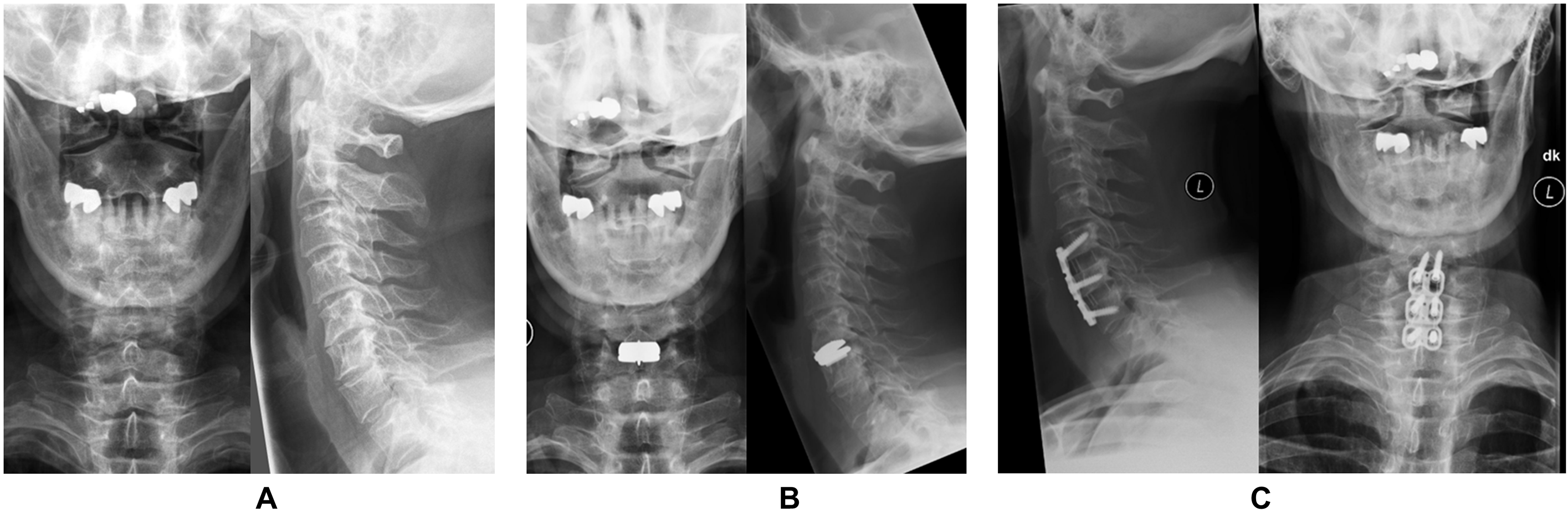
Case 2, example of a disc prosthesis with a negative outcome because of a necessary operative revision (Fig. 4):
54-year-old male patient with osteochondrosis C5/6 and CBS without neurological deficit. The preoperative VAS was 8 and the patient was refractory to conservative treatment for more than 6 weeks. Degenerative changes, corresponding to Kellgren and Lawrence Score 3 were found in the x-ray. In the first surgery the implantation of a disc prosthesis C5/6 (Aesculap, Active C) was performed.
Postoperatively the pain improved to VAS 3, but only one year later VAS reached 9 again. Due to painful nociceptive disorder, and degenerative changes, also affecting the adjacent segment, an anterior cervical discectomy and fusion was performed. During surgery a not bony integrated prosthesis was detected. Postoperatively after the second operation, the patient’s pain decreased (VAS 2), also X-ray showed good implant placement and no increased degeneration or loosening.
The diagnosis CBS stands for a wide spectrum of disorders with many different causes. All the patients included in this study had two things in common. First, they all suffered from a refractory CBS because of a degenerative disc disease and second, all 34 patients received a total disc replacement. By including patients with a wide range of age (24–66 years) and different grades of degeneration of the affected segment (Kellgren and Lawrence Score 0–3), this observational study is appropriate to examine the right indication for total disc replacement at the cervical spine. Furthermore, it was intended to present clinical and radiological results after TDR and to discuss the possibilities and limitations of this method.
The mean age of the patients included in our study (47.2
The surgical procedures of the implantation of disc prosthesis in general led to good clinically and radiologically-documented success in the majority of the patients. Pain intensity and functional impairment were reduced by an average of two thirds, with both variables correlating significantly with each other. Patients significantly reduced the consumption of analgesics objectified by a lower level of the WHO scheme. This corresponds to the results of other studies [22, 23, 24].
Recently published studies compared TDR with ACDF of the cervical spine. All patients included in these studies suffered from a cervical brachial syndrome because of a DDD of the cervical spine. None of these studies featured a differentiation of the varying types of DDD or a classification of the severity of DDD [20, 22, 23, 24].
Compared to others, this study had a high rate of revision surgery. Five out of 34 patients (15%) needed a change from TDR to ACDF within 24 months after surgery. All of these patients had a Kellgren and Lawrence Score of 2 or 3. Therefore, 5 of 9 patients (56%) with severe degeneration were in need of revision surgery after the implantation of a cervical disc replacement. In all cases the TDR was loosened. A possible reason for the lack of integration of the TDR could be the higher degree of sclerosis of the endplates. Additionally, all 5 patients were smokers. A recent study showed that smokers presented a significant lower rate in fusion after lumbar spondylodesis [25]. This fact indicates that smoking can lead to a lower rate of bony integration of the TDR and consequently to neck pain, which in turn requires revision surgery. These aspects signify that a high preoperative Kellgren and Lawrence Score, combined with smoking, can lead to a higher risk for secondary surgery.
By using the Kellgren and Lawrence Score, this study shows that grading of degeneration can be used as a good tool to evaluate the severity of the DDD and consequently to take the decision for either a TDR or ACDF.
As mentioned above, the results in general and especially the two case studies support this statement. The first case study shows a good indication for a disc prosthesis in a patient with symptoms of a cervical-brachial syndrome and a low level of degenerative disc disease (Kellgren and Lawrence Score 0). After implanting the disc prosthesis the patient’s symptoms improved, objectified by the improvement of the NDI and VAS scores and a good radiological postoperative success.
The second example presents a patient with a preoperatively higher level of degenerative disc disease (Kellgren and Lawrence Score 3). The patient had been operated a second time with a removal of the disc prosthesis and implantation of a spondylodesis of the affected segment because of a progressive DDD, also affecting the adjacent segments.
74% of the patients in this study had either no degeneration (Kellgren and Lawrence Score 0) or a low grade of degeneration (Kellgren and Lawrence Score 1) in the affected segment. Investigations on patients with a higher degree of degenerative disc disease could be interesting. However, the evaluation of the observational study also shows that this operation method is limited when there are additional pathological changes in the operating area, as for example a higher level of DDD. Thus, a decision for this surgical method should be made by mainly considering individual aspects. The use of TDR can be advisable as a protective measure e.g. in younger patients with little or no osteochondrosis and a lower preoperative Kellgren and Lawrence Score.
Conclusion
The results of this study show that the use of a TDR in patients with CBS with a low Kellgren and Lawrence Score of 0–1 can lead to clinically and radiologically successful outcomes on the long-term. Smokers with a Kellgren and Lawrence Score of 2–3 have a high risk for revision surgery after TDR.
Footnotes
Conflict of interest
None to report.