






Abstract
BACKGROUND:
Recurrent acute pancreatitis is a common acute abdominal disease in surgery.
OBJECTIVE:
To evaluate the radiographic features of pancreatic computed tomography (CT) imaging in the diagnosis of acute and chronic pancreatitis.
METHODS:
48 pancreatitis patients who met the criteria were selected in this retrospective study from 2010 to 2019. Each diagnosis was evaluated as functional abdominal pain, recurrent acute pancreatitis, or chronic pancreatitis. All clinical data were collected from the patient’s medical records. 54 radiological features were extracted from each region of interest in outline the pancreas and divided into five categories: first order statistics, the gray level co-occurrence matrix (GLCM), the gray level run-length matrix (GLRLM), the neighborhood gray level difference matrix (NGTDM), and morphological features by the MATLAB program.
RESULTS:
Of the 48 patients, 16 had functional abdominal pain (33.3%), 18 had recurrent acute pancreatitis (37.5%), and 14 had chronic pancreatitis (29.2%). In the univariate analysis, nine radiological features, eight GLCM features and one NGTDM feature were significantly different between groups. Nine radiological characteristics had important reference values with AUC values ranging from 0.73–0.91.
CONCLUSION:
Nine radiographic features of CT imaging demonstrate good evaluation efficiency in the diagnosis of pancreatitis and can distinguish patients with functional abdominal pain, recurrent acute pancreatitis, and chronic pancreatitis.
Introduction
Recurrent acute pancreatitis (RAP) is a common acute abdominal disease in surgery and is characterized by acute onset, rapid progression, and high mortality. The RAP diagnostic criteria are as follows: Two or more episodes of docunted acute pancreatitis, separated by at least three months, and complete remission was obtained between two episodes. The causes of this disease include gallstones or cholestasis, alcohol consumption, sphincter of Oddi dysfunction, gene mutations (such as PRSS1 and SPINK1), and pancreatic duct interference with the normal flow of pancreatic secretions [1, 2, 3, 4]. The recurrence rate in patients diagnosed with acute pancreatitis is 17–22%, and up to 36% of patients with acute pancreatitis will eventually develop chronic pancreatitis [5, 6]. Imaging examination plays an important role in the diagnosis of acute pancreatitis, of which superconducting magnetic resonance imaging (MRI) and multislice spiral computed tomography (CT) are most commonly used, with CT having the advantage of fast image acquisition speed, high spatial resolution, strong reproducibility, and ease of operation, which is more valuable for its promotion. In addition, Martin et al. shows that a dual-energy computed tomography (DECT) using iodine quantification allows for diagnosis of early acute pancreatitis with higher sensitivity compared to standard image evaluation [7]. However, the pancreas usually appears normal in CT in patients with RAP after the resolution of acute pancreatitis attacks and during recurrent attacks, which makes the diagnosis of RAP challenging [8, 9, 10]. Therefore, diagnostic tools are needed to further refine the clinical diagnosis of RAP and distinguish it from other abdominal pains. Radiomics is the application of computer-image processing to transform the image data of the region of interest (ROI) into extractable high-dimensional feature data, which uses texture analysis to study the quantitative characteristics of radiography and has shown potential value in breast and lung cancer [11, 12]. Since neither abdominal pain nor the elevation of pancreatic enzymes is a specific feature of acute pancreatitis, its diagnosis is challenging in patients with acute pancreatitis whose pancreas appears normal on CT after remission. Radiological features analyzed from CT imaging films are important in diagnosing lesion characteristics in patients with pancreatic disease [13]. This paper studies the pancreatic CT radiological features of patients with pancreatitis, analyzes whether the pancreatic radiation characteristics are able to distinguish patients with functional abdominal pain, acute pancreatitis, and chronic pancreatitis, and provides a theoretical basis for the application of the radiological features of CT imaging in the accurate diagnosis of pancreatitis.
Materials and methods
Research design and data collection
A total of 168 patients of the Department of Imaging of Taihe people’s hospital from 2019 to 2020 were enrolled in this retrospective single-center cohort study. All medical records and imaging studies were obtained and reviewed from medical institutions. Of the 168 patients, 120 were excluded based on the exclusion criteria, and 48 met the criteria. Based on clinical assessment, these 48 patients were subsequently divided into three groups: functional abdominal pain, acute pancreatitis, and chronic pancreatitis. All relevant clinical data, including age, gender, and body mass index, were collected from the patient’s medical records. The study protocol was approved by the medical ethics committee of our hospital, and the patients and their families signed the informed consent form for this study.
Patient selection
In the diagnosis of acute pancreatitis, the following three criteria need to be met: ⟀ abdominal pain, ⟁ abdominal imaging findings consistent with acute pancreatitis, ⟂ the elevation of serum amylase or lipase
Exclusion criteria: absence of abdominal enhanced CT; presence of foreign bodies near the pancreas (e.g., stents or surgical clips), producing bundle sclerosing pseudopacities on the pancreas; imaging features of pancreatitis (including peripancreatic fat retention, edema, and fluid in the CT); previous pancreatic surgery.
Image acquisition and region of interest division
Acute pancreatitis patients in this study obtained a standard arterial phase and venous phase CT of the abdomen 60 seconds after a contrast injection, and other pathients were obtained a standard venous phase CT. Images were scanned on a 256iCT scanner. Patients were injected with 80–100 mL of iohexol at an injection rate of 4–5 mL/s, and the scanning protocol was customized for each patient to minimize the dose (approximately 120 kVp, 300 mAseff, and 0.6–0.8 pitch). The 3-mm slices were used for radiomics analysis because these slices were uniformly available for intuitive images. Although the patient selection criteria refer to a previous study by Mashayekhi et al. [14]. However, our study only analyzed venous phase images, which were more standard in contrast timing and able to avoid differences in the phase acquisition times of pre-contrast and arterial phase images, with a more standard imaging protocol and image resolution. The DICOM files of the CT images were downloaded into an in-house MATLAB program to outline the pancreas segmentation on each slice using the ROI tool [15]. Segmentation of the pancreas was carefully checked by an experienced abdominal radiologist to ensure accuracy and modified, if required, to ensure correct anatomical contours.
Extraction of radiological features
The steps for feature extraction from radiographic images include the acquisition of radiographic images, the identification of ROI on the acquired images, and the extraction of radiographic features from the ROI. Fifty-four radiographic features were extracted by the MATLAB program and divided into five categories: first order statistics, the gray level co-occurrence matrix (GLCM), the gray level run-length matrix (GLRLM), the neighborhood gray level difference matrix (NGTDM), and morphological features. Each GLCM and GLRLM feature was generated at four different angles (0, 45, 90, 135), and the average was used for these features. We used 64 equally sized gradations and gray levels in our analysis. For the internal reader variability measurements, a total of 15 cases (five in each group) were segmented, and radiological features were extracted at two different times three months apart (see Fig. 1).
Statistical analysis
The Wilcoxon rank sum test was used to compare the radiographic features of patients with RAP, non-specific abdominal pain, and chronic pancreatitis. Receiver operating characteristic (ROC) curves with area under the curve (AUC) were used to assess the predictive performance of individual radiographic features. All data analyses were performed using SPSS 19.0 and MATLAB software programs, and
Results
According to the above clinical criteria, of the 48 patients included in the study cohort, 16 (33.3%) had functional abdominal pain, 18 (37.5%) had recurrent acute pancreatitis, and 14 (29.2%) had chronic pancreatitis. Compared with patients with acute pancreatitis and functional abdominal pain, patients with chronic pancreatitis were on average about nine years older, had a higher incidence of diabetes, and were more likely to be alcoholics and current smokers (see Table 1).
Baseline characteristics of patients according to clinical diagnosis (i.
,
%)
Baseline characteristics of patients according to clinical diagnosis (i.
CT scan and radiographic feature image analysis (The steps of feature extraction from radiographic images include: ⟀ acquisition of radiographic images; ⟁ identification of regions of interest on the acquired images; ⟂ extraction of radiographic features from regions of interest.).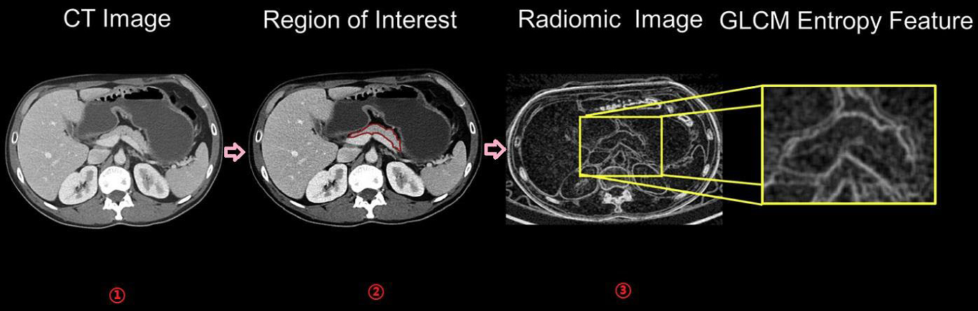
Important radiographic features based on clinical diagnosis (
Comparison of AUC and p-values for individual radiographic features in different groups
Note:
In univariate analysis, nine radiographic characteristics were significantly different in all three patient groups (Table 2). Of these nine features, eight were GLCM features and one was an NGTDM feature. For these nine radiographic features, we constructed separate ROC curves to measure AUC. When comparing patients with acute pancreatitis to those with functional abdominal pain and chronic pancreatitis, AUC values ranged from 0.76 to 0.93 and from 0.73 to 0.91, respectively (Table 3). It shows that some of the radiographic features of CT imaging demonstrate good evaluation efficiency in the diagnosis of pancreatitis and can distinguish patients with functional abdominal pain, recurrent acute pancreatitis, and chronic pancreatitis.
Imaging examination is an important part of the diagnosis of acute pancreatitis and can help to confirm the diagnosis and detection of complications. However, patients with RAP cannot be directly diagnosed based only on abdominal pain symptoms, laboratory values, or imaging studies obtained during an episode of acute pancreatitis [16]. Moreover, low temporal resolution, imperfect image contrast, ionizing radiation, high cost, and extended time requirements are the main limitations of conventional imaging techniques. Radiomics may therefore be a useful diagnostic aid [17]. Once the initial diagnosis is established, a careful diagnostic algorithm can be used to determine the cause of the disease and perform appropriate treatment, and the use of non-invasive methods to distinguish acute pancreatitis from other causes of abdominal pain (including functional pain) can reduce the overtreatment of invasive measures (such as endoscopic ultrasound and endoscopic retrograde cholangiopancreatography) [18].
Artificial intelligence (AI) is, at its core, a branch of computer science that attempts to both understand and build intelligent entities, often instantiated as software programs [19]. The use of AI in diagnostic medical imaging is undergoing extensive evaluation, and deep learning has shown substantial improvements in image classification tasks since 2012. AI has shown impressive accuracy and sensitivity in identifying imaging abnormalities and promises to enhance tissue-based detection and characterization [20]. After being trained and learned, AI algorithms are able to distinguish benign abnormalities from clinically significant lesions, which is beneficial for clinical diagnosis. With the help of modern deep learning methods, many radiological applications of AI, such as the detection of pulmonary nodules using computed tomography images, the diagnosis of tuberculosis and common lung diseases using chest radiography, and the identification of breast masses using mammography scans, have reached expert level diagnostic accuracy [21, 22, 23]. Lee et al. developed a deep learning-based computer-aided diagnostic (CAD) system for cervical lymph node metastasis of thyroid cancer diagnosed by CT scan [24]. Van Hamersvelt et al. used a variety of artificial intelligence techniques to detect early macroscopic cardiac ischemia and arterial stenosis [25]. However, at present, AI algorithms still have a long way to go in the application to the diagnosis of pancreatitis. Because the clinical symptoms are more intense and characteristic, the current diagnosis of acute and chronic pancreatitis is still mainly based on the clinician’s experience and serological diagnosis, which has largely limited the development of AI and deep learning in this field.
Our preliminary study suggests that GLCM features showed advantages in distinguishing these three disease characteristics. Of the individual radiographic features, eight GLCM features and one NGTDM feature were significant. AUC analysis of individual radiographic features in the comparison of different groups demonstrated that these nine characteristic parameters had significant diagnostic significance. GLCM features were generated by analyzing the distance between pairs of voxels and the intensity of angle-dependent pairs of voxels. These significantly different characteristics were related to the measurement of pancreatic homogeneity. Both homogeneity and correlation information measures are features that evaluate the homogeneity of ROI. The homogeneity and correlation information measures showed that the non-specific abdominal pain group always had the most homogeneous pancreas, while the pancreatic homogeneity in patients with acute and chronic pancreatitis was reduced and the pancreatic homogeneity in patients with chronic pancreatitis was the lowest. In addition, the entropy value is a measure of heterogeneity, and the analysis results showed that patients with chronic pancreatitis had the most heterogeneous pancreas, while patients with acute pancreatitis had the least heterogeneous pancreas [26]. Patients with functional abdominal pain had no change in pancreatic structure because the pain is not caused by pancreatic processes and pancreatic histology is normal, resulting in a more homogeneous image; however, patients with chronic pancreatitis usually had symptomatic changes in ductal dilatation and calcification with histological manifestations of chronic inflammation and fibrosis, resulting in a more heterogeneous image [27]. Patients with acute pancreatitis had experienced an episode of acute pancreatitis but had not developed chronic pancreatitis, so the intensity was intermediate between the other two groups; after research and analysis, it was found that this was consistent with the natural characteristics [28]. In analyzing NGTDM features that differ between one voxel and its neighbors, contrast was the only feature that was important, and contrast quantified the intensity difference between neighboring voxels. Patients with functional abdominal pain had the lowest contrast, while patients with chronic pancreatitis had the highest contrast due to the presence of calcified areas in their pancreas.
It has been shown that radiomics can reveal signals of tumor predictability, is able to capture tumor-intrinsic heterogeneity, and correlates with underlying gene expression types. A study of CT images from 53 patients found 14 GLCM features with individual AUCs ranging from 0.64 to 0.82 to differentiate high-grade from low-grade intraductal papillary mucinous neoplasms of the pancreas [29]. A study of the slice tumor using CT found that the entropy and correlation of GLCM were significant and could discriminate between pancreatic ductal adenocarcinoma and healthy pancreatic tissue [30]. In addition, another study examining the entire pancreas using CT found that correlation in GLCM is one of the five most relevant features [31]. The salient features we found were generally consistent with the results of radiomics characterization studies reported in the literature, which found that GLCM features were important in distinguishing pancreatic pathology.
However, there are still some limitations in the current study: the study sample is relatively small, with each group composed of 14–18 patients, and a larger sample is needed to evaluate the differences between the populations; this study is a retrospective study, the occurrence of initial symptoms was not used as a factor to assess the duration of the disease, and the total number of acute pancreatitis attacks in the patient population was not analyzed (a prospective study can be conducted in the future to track the progress of patients from the onset of disease symptoms); the CT images of patients were performed in medical institutions, and the use of different scanners could lead to variability in the extracted radiomics data; the pancreas segmentation was performed manually, and in order to ensure the accuracy of the results, a more effective classification algorithm is needed for evaluation validation; different classification algorithms are also needed for evaluation validation.
Conclusion
Extracting more information from CT images, such as radiographic features, as useful tools in medical devices, can be used to distinguish patients with functional abdominal pain, acute pancreatitis, and chronic pancreatitis using venous phase CT imaging, and the application of radiographic features in accurate diagnosis is important.
Ethics statement
This study was conducted in accordance with the Declaration of Helsinki and approved by the ethics committee of Taihe people’s Hospital. Informed consent was obtained from all patients.
Availability of data and materials
All data generated or analyzed during this study are included in this published article.
Competing interests
None of the authors have any personal, financial, commercial, or academic conflicts of interest to report.
Funding
Not applicable.
Author contributions
GX conceived the study, ZX and WD participated in its design and coordination, GX, ZX and WD helped draft the manuscript. All authors read and approved the final manuscript.
Footnotes
Acknowledgments
Not applicable.