
Abstract
BACKGROUND:
The minimally invasive endodontics could retain more peri-cervical dentin (PCD) and other important dental structures, thus realizing the minimal loss of teeth structures and preserving the strength and function of the endodontically treated tooth (ETT). The search for abnormal root canals or calcified root canals could be quite time-consuming and increase the risk of perforation.
OBJECTIVE:
This study introduced a novel multifunctional 3D printing guided splint inspired by the dice, which can achieve the minimally invasive access cavity preparation and canal orifice identification.
METHOD:
Data were collected from an outpatient with dens invaginatus. Cone-beam Computed Tomography (CBCT) revealed a type III invagination. The CBCT data of the patient were imported into a computer-aided design (CAD) software (Exocad 3.0; Exocad GmbH) for the 3D reconstruction of jaw bones and teeth. The dice-inspired 3D printing guided splint consists of the sleeve and guided splint. The sleeve with minimal invasive opening channel and orifice locating channel were designed with a reverse-engineering software (Geomagic Wrap 2021). The reconstructed models in the Standard Template Library (STL) format were imported into a CAD software. The design of the template was aided by the dental CAD software in Splint Design Mode. The sleeve and splint were exported into the STL files separately. A 3D printer (ProJet
RESULTS:
The novel multifunctional 3D printing guided splint could be set in position. The opening side in the sleeve was selected and the sleeve was inserted in place. The minimal invasive opening was made in the crown of the tooth to access the pulp. The sleeve was draw out and turned to the orifice location side, and then inserted in place. The target orifice was located rapidly.
CONCLUSION:
This novel dice-inspired multifunctional 3D printing guided splint allow dental practitioners to gain accurate, conservative, and safe cavity access from teeth with anatomical malformations. Complex operations might be carried out with less reliance on the operator’s experience than with conventional access preparations. This novel dice-inspired multifunctional 3D printing guided splint would have a broad application in the dental field.
Introduction
Digital dentistry and additive manufacturing (AM) have advanced rapidly with astounding achievements. Additive manufacturing, also known as 3D printing, is capable of producing highly complex and refined structures. Additive manufacturing techniques such as Selective laser melting (SLM) and Stereolithography (SLA) have been used in dentistry for nearly 20 years. Endodontic applications for guided endodontics, guided removal of fiber post, autotransplantation and dental education are reported in the literatures [1, 2, 3, 4, 5, 6, 7].
Root canal treatment of teeth with canal malformations and obliteration is a challenging task for general dentists and endodontic specialists. The identification of abnormal root canals or calcified root canals could not only be quite time-consuming but also increase the risk of perforation. As a result, inspired by implantology protocols, some researchers have applied digital planning in endodontics [8]. However, due to functional limitations, the traditional endodontic template cannot meet the various demands of clinical practice and is difficult to extend. The multifunctional guided splint with extensibility features for endodontics has not been reported so far.
The minimally invasive endodontics involves the treatment and prevention of pulpal lesion and apical periodontitis, aiming to cause the minimal loss of teeth structures and retain more peri-cervical dentin (PCD) and other important structures. The endodontically treated tooth (ETT) will be strong and functional for the rest of the patient’s life [9, 10, 11]. PCD has been defined as the dentin extending 4 mm above and below the level of alveolar bone [11]. Some researchers believed that the peri-cervical dentin was a key structure responsible for the retention rate of the teeth [10, 12, 13]. Infection control is the foundation for the treatment of irreversible pulpitis and apical periodontitis, which should be regarded as a higher pursuit of endodontic specialists and general practitioners to preserve healthier tooth structures and increase the long-term retention rate of the endodontically treated teeth.
The dice is a small cube with six equal square sides with a different number or note on each side. The novel multifunctional 3D printing guided splint described in this study was inspired by the dice, and it can be used for both the minimally invasive access cavity preparation and canal orifice identification. Access cavity preparation and canal orifice identification could be integrated in a multi-functional guided splint because the directions would change with rotation of the detachable part. The novel detachable guided splint consisting of the sleeve and guided splint could simultaneously realize minimally invasive access to cavity and orifice location.
Materials and methods
Design of the dice-inspired multifunctional 3D printing guided splint
Data were collected from an outpatient with dens invaginatus. The patient is a 32-year-old Asian female who came to the clinic with a chief complaint of right maxillary canine root discoloration. During clinical examination, gingival recession was observed on the buccal tooth surface of right maxillary canine and obvious discoloration on the exposed root. The right maxillary canine showed no response to both cold and electrical pulp sensitivity testing. On the periapical film, radiographic appearance showed a large area of destructed periapical tissues (Fig. 1). Possible external root resorption was observed. Diagnosis tests revealed an apical chronic periodontitis on tooth 13 and a root canal treatment was recommended.
Intraoral periapical radiographical examination of dens invaginatus on tooth 13.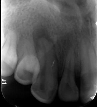
Cone-beam computed tomographic (CBCT) radiographical examination of dens invaginatus on tooth 13. CBCT horizontal cross-section shows a periapical lesion and the presence of the dens evaginatus.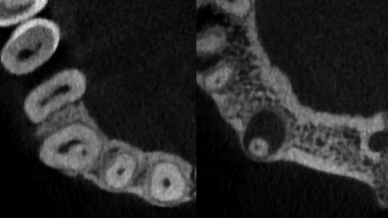
With the goal of explaining the internal anatomy of the affected tooth, a complementary radiographic examination was performed with CBCT (Newtom, Italy). CBCT revealed a type III invagination (Fig. 2). The Cone-beam Computed Tomography (CBCT) data of the patient were imported into a computer-aided design (CAD) software program (Exocad 3.0; Exocad GmbH) for the 3D reconstruction of jaw bones and teeth. The dice-inspired multifunctional 3D printing guided splint consists of the sleeve and guided splint. The sleeve with minimal invasive opening channel and orifice locating channel were designed with a reverse-engineering software (Geomagic Wrap 2021) (Fig. 3). The reconstructed models in the Standard Template Library (STL) format were imported into a computer-aided design (CAD) software program (Exocad 3.0; Exocad GmbH). The design of the template was aided by the dental CAD software in Splint Design Mode. The sleeve and splint were exported into the STL files separately (Fig. 4).
Design of minimally invasive access cavity preparation and canal orifice identification.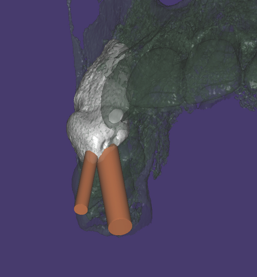
Design of the novel dice-inspired multifunctional 3D printing guided splint.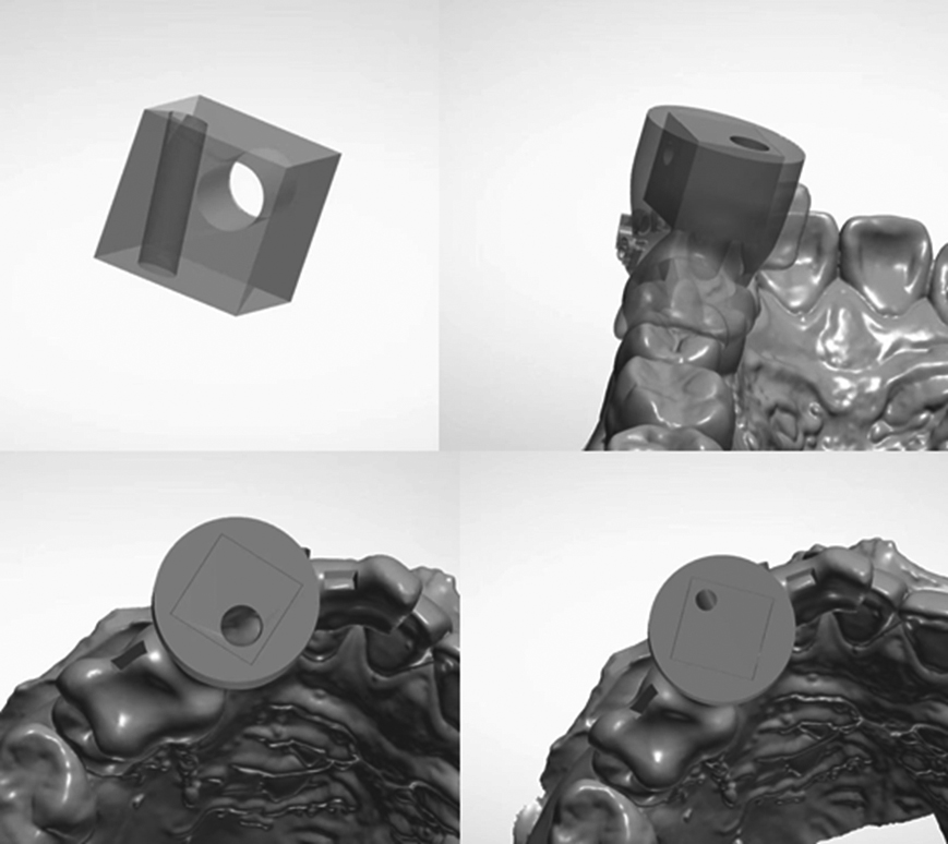
The novel dice-inspired multifunctional 3D printing guided splint was printed and placed stably.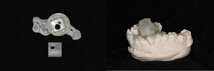
A 3D printer (ProJet
Usage of the dice-inspired multifunctional 3D printing guided splint
The rubber dam was placed around the affected tooth to isolates 3
Discussion
3D printing in dentistry could be classified into the following major categories: stereolithography apparatus (SLA), fused deposition modeling (FDM), MultiJet printing (MJP), PolyJet printing, ColorJet printing (CJP), digital light processing (DLP) and selective laser sintering (SLS) [14]. The SLA technology uses an ultraviolet (UV) laser to turn photosensitive resin into solid 3D objects, layer by layer. SLA was the earliest and the most commonly used technology employed in dentistry [15]. With the wide application of CBCT in the dental field, CBCT imaging become a more accurate data source for 3D printing applications and is of great significance for treatment of root canal calcification and malformation. CBCT has been applied in conjunction with professional software to evaluate the complex three-dimensional structure of root canals and confirm location and number of canals, providing more useful information for the predictable treatment plans [16, 17, 18, 19].
This research introduced a novel 3D printing guided splint inspired by the dice, which could achieve the minimally invasive access cavity preparation and canal orifice identification. Combined with CBCT and 3D printing, the method has solved clinical problems. The inspiration was from the dice, and a multifunctional splint consisting of the special sleeve and guided splint was created. A dice owns six surfaces and controls three directions. Each direction is endowed with different function including minimal invasive opening and orifice location. The dice would be easily turned into next side after one side functioned. This novel dice-inspired multifunctional 3D printing guided splint had high inter-appointment repeatability and was less affected by the experience of the operators, which would boost the efficiency of RCT significantly.
Root canal treatment of teeth with canal malformations and obliteration remains a major challenge for both general dentist and endodontic specialists. The search for abnormal root canals or calcified root canals could both be quite time-consuming and increase the risk of perforation. In recent years, some clinicians reported that splint-guided or dynamic-navigation aided endodontic access showed promise as an option for treating calcified and malformed canals [20, 21, 22, 23].
In dental practice, access to a complex root canal system is of great significance. It could be a time-consuming process even for an experienced endodontic specialist, let alone the general dentists. Once the treatment is attempted, misdirected instruments could result in root canal perforation, increasing iatrogenic complications. To this end, it is frequently at the cost of substantial tooth loss even if the canal is found [24]. Connert et al. reported that the guided endodontics access led to a more accurate and expeditious location of calcified root canals with significantly less dental hard tissue loss compared with the conventional approach [25, 26]. Therefore, splint-guided or dynamic-navigation aided endodontic access can be better options in the clinical practice.
However, there are some limitations to this technique. It might be difficult to place the splint and bur into the guide simultaneously in the posterior region, especially for Asian patients. Guided endodontic treatment could be restricted to the interocclusal space. The sleeve could control three directions, so the target root canal cannot exceed the number of three. In addition, planning and manufacturing the template is time-consuming.
Conclusion
This novel dice-inspired multifunctional 3D printing guided splint allow dental practitioners to gain accurate, conservative, and safe cavity access from teeth with anatomical malformations. Complex operations might be carried out with less reliance on the operator’s experience than with conventional access preparations.
Footnotes
Acknowledgments
This work was supported by the Research Foundation of the School and Hospital of Stomatology, Peking University (PKUSS20200111), the Clinical Research Foundation of Peking University School and Hospital of Stomatology (PKUSS-2023CRF101), and the Beijing Natural Science Foundation (7214274).
Conflict of interest
The authors have no conflicts of interest relevant to this article.