






Abstract
BACKGROUND:
Cervical malignancy is considered among the most perilous cancers affecting women in numerous East African and South Asian nations, both in terms of its prevalence and fatality rates.
OBJECTIVE:
This research aims to propose an efficient automated system for the segmentation of cancerous regions in cervical images.
METHODS:
The proposed techniques encompass preprocessing, feature extraction with an optimized feature set, classification, and segmentation. The original cervical image undergoes smoothing using the Gaussian Filter technique, followed by the extraction of Local Binary Pattern (LBP) and Grey Level Co-occurrence Matrix (GLCM) features from the enhanced cervical images. LBP features capture pixel relationships within a mask window, while GLCM features quantify energy metrics across all pixels in the images. These features serve to distinguish normal cervical images from abnormal ones. The extracted features are optimized using Genetic Algorithm (GA) as an optimization method, and the optimized sets of features are classified using the Co-Active Adaptive Neuro-Fuzzy Inference System (CANFIS) classification method. Subsequently, a morphological segmentation technique is employed to categorize irregular cervical images, identifying and segmenting malignant regions within them.
RESULTS:
The proposed approach achieved a sensitivity of 99.09%, specificity of 99.39%, and accuracy of 99.36%.
CONCLUSION:
The proposed approach demonstrated superior performance compared to state-of-the-art techniques, and the results have been validated by expert radiologists.
Introduction
Cervical malignancy stands out as one of the most perilous cancers affecting women in various East African and South Asian nations, both in terms of its prevalence and fatality rates. In 2008 alone, around 530,000 women globally received diagnoses of invasive cervical cancer, with 275,000 succumbing to this disease. Breast and cervical cancers disproportionately impact women [1]. The onset of cervical cancer, characterized by malignant cells in the cervix region, often leads to bleeding in later stages, resulting in sudden fatalities [2].
Figure 1a depicts a typical cervical section in females without malignancy, while Fig. 1b illustrates the abnormal malignancy region in a woman afflicted by cervical cancer. The primary culprit for cervical malignancy in females is the presence of the Human Papilloma Virus (HPV) in the cervical section. According to the World Health Organization (WHO), over eighty percent of cervical malignancy cases are on the rise in emerging nations, primarily due to a lack of information and awareness about the illness [3]. The shortage of clinician or surgeon resources in developing nations emphasizes the critical need for examining cervical malignancy using computerized machine learning methods [4]. The ongoing research for malignancy section identification in cervical images serves various clinical and health settings, aiding physicians in the automated detection of malignancy sections [5].
In light of these challenges, this research proposes a machine learning-based intuitive detection and categorization method for cervical malignancy recognition and fragmentation. While numerous machine learning algorithms have been employed for cancer image classifications in the past, the CANFIS method emerges as particularly effective for cervical cancer detection systems, owing to its proficient supervised internal layers. The CANFIS classifier, a modification of the ANFIS classifier, enhances fuzzy rules to yield superior classification results. Additionally, the Genetic Algorithm (GA) optimization algorithm operates on external feature metrics to refine computed feature values in cervical images. In this paper, the GA method optimizes the computed feature set, which is subsequently classified using the CANFIS classifier.
Cervical images (a) Normal case (b) Abnormal case.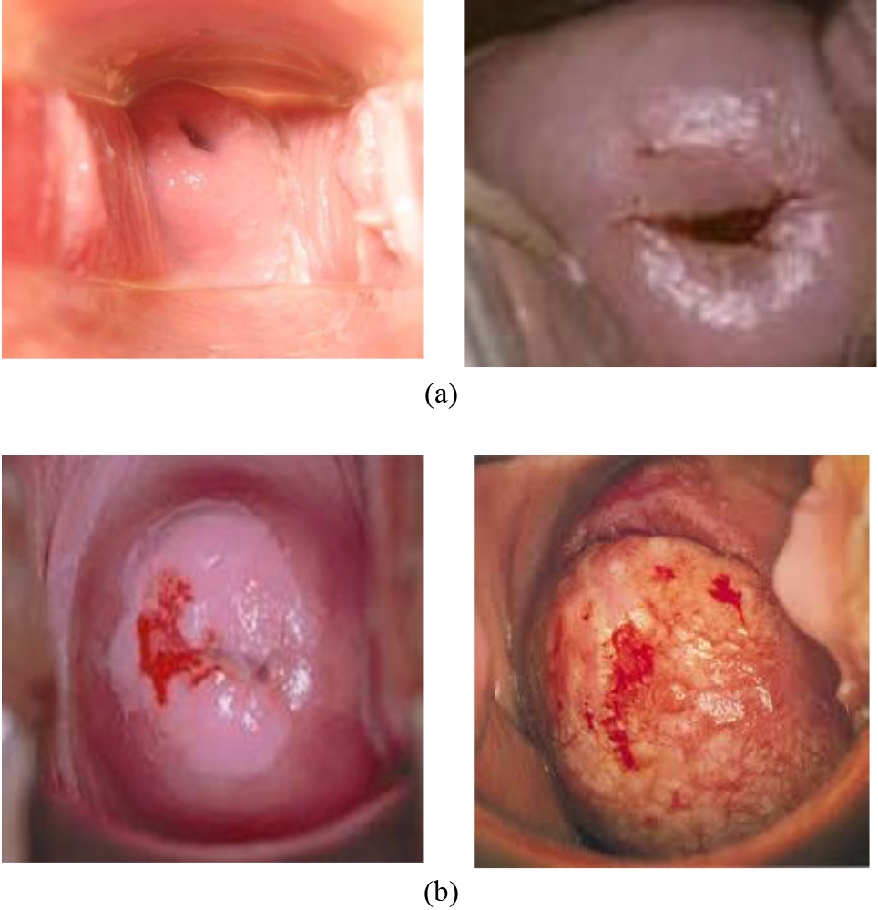
Section 2 of this research work describes the present approaches for cervical malignance finding and fragmentation. Sections 3 and 4 create a novel strategy for detecting and classifying cervical malignance. Section 5 discusses simulation outcomes. Section 6 designates this research work conclusion, and Section 7 discusses future improvements.
Pan et al. [6] employed a multimodal system or methodology for screening cervical cancer using an MRI staging model. The authors discussed various non-linear factors to distinguish between normal and abnormal images. Ghaffar et al. [7] utilized various optimization techniques and explored their implications with elderly disabled systems. Lin et al. proposed a cervical healthy computing system for analyzing various healthcare parameters to predict or screen cervical abnormalities. Umesh Kumar Lilhore et al. [8] implemented machine modeling algorithms for the detection of cervical cancer using cervigram images in their work. The authors used the Boruta analysis algorithm to predict features from cervical images. These features were combined and classified through a hybrid machine model that employed the combination behavior of the Support Vector Machine (SVM) and the Random Forest (RF) to improve the classification rate of the cervical cancer detection system. Chitra et al. [9] utilized the fuzzy local information c-means clustering algorithm to cluster abnormal pixels in cervical images. The authors applied the Sooty Tern Optimization framework model to optimize the region of pixels belonging to the cancer region in their work. Furthermore, a CNN-Long Short Term Memory (LSTM) model was employed to produce classification results. Pallavi et al. [10] analyzed Pap smear cell images to predict cervical cancer using various supervised learning techniques. The hyperbolic tangent functional logic was employed in the supervised classifier. This work utilized an SVM classifier to predict non-linear features and produce results. Vijayanand Sellamuthu Palanisamy et al. [11] designed a cervical cancer detection algorithm using residual neural networks. The activation function was optimized using the Adam optimization algorithm in their work. Ontor et al. [12] proposed a YOLO deep learning model for the detection of cervical cancer. This method used a minimized set of layers to optimize the computational complexity of the cancer detection algorithm. The authors achieved a 96.1% classification rate using the proposed model. Wang et al. [13] proposed a feature computational modeling method for detecting abnormalities in medical images. Kamal et al. [14] developed a system between the human brain and a computer system to acquire brain activity signals, which were then used by soft computing algorithms to predict disease status. Sahli et al. [15] computed a geometrical feature set from medical images, and these computed geometrical features were used by various classification algorithms.
Alyafeai Zaid et al. [16] employed a deep learning-based classification to detect and segment malignancy sections in cervical images. The authors utilized the Guanacase Dataset 2005 and achieved a standard sensitivity (SE) of 97.2%, standard specificity (SP) of 98.3%, and standard accuracy (AC) of 98.8% compared to ground truth images. Thiyaneswaran et al. [17] utilized the k-means clustering method to detect and segment malignancy sections in surface images. The authors achieved a standard accuracy (AC) of 90.0% when using an unfolded admittance database. Wasswa William et al. [18] proposed a pap smear investigation mechanism for cervical malignancy detection from pap smear images. They achieved a standard sensitivity (SE) of 99.28%, standard specificity (SP) of 97.4%, and standard accuracy (AC) of 98.8%. Sachan et al. utilized the random forest classification strategy on cervical images. They achieved 90.5% standard sensitivity (SE), 93.2% standard specificity (SP), and 92.9% standard accuracy (AC) from the unfolded admittance database. Karthiga Jaya et al. [19] detected and segmented malignancy sections in cervical images using the ANFIS categorization strategy. The authors utilized the Guanacase Database 2005 and achieved a standard sensitivity (SE) of 97.42%, standard specificity (SP) of 98.3%, and standard accuracy (AC) of 99.12% compared to ground truth images.
Kumarganesh et al. [20] proposed an ANFIS classifier strategy for tumor categorization from original images, achieving a categorization standard accuracy (AC) of 96.6%. Kumarganesh et al. [21] proposed using an ANFIS categorizer strategy to detect tumors in original images. They achieved a standard sensitivity (SE) of 93.07%, standard specificity (SP) of 98.79%, and standard accuracy (AC) of 97.63% for cancer categorization. Teck et al. [22] employed the contourlet transformation strategy in conjunction with the Gabor orientation strategy to transform the original cervical images. The converted coefficients were combined by the second order arithmetical component specifications, and the resulting component set was well qualified and classified using the SVM categorizer. The authors achieved a standard categorization accuracy (AC) of 81% in detecting malignancy portions in original cervical images.
Yuexiang et al. [23] introduced a direct strategy for identifying malignancy portions in cervical images used in the input test. The original cervical images were employed to determine first and second-order arithmetical components, which were then fine-tuned and classified using the linear categorization strategy. Finally, to detect cancer regions in the categorized outcome images, a morphological fragmentation strategy was applied. Fei et al. [24] proposed a procedure to distinguish abnormal regions in the cervix areas of female patients. The authors used biopsy images to scan the cervical portions of the patients using a Computer-Aided Detection (CAD) strategy. This computer-aided strategy, employing various soft computing techniques, enhanced the categorization precision of the cervical malignancy exploration scheme. In comparison to ground truth images, the authors achieved a standard sensitivity (SE) of 86% and a standard tumor sectionalization accuracy (AC) of 91%.
Slowik et al. [25] designed an optimal strategy for deciding and separating the Pap smear cells of patients. Based on their morphological attributes, the authors compared and evaluated the fragmented nucleus section of cervical images using the physician fragmented nucleus portions, achieving a standard categorization accuracy (AC) of 93%. For fragmenting the cancer portion in cervical images, Elayaraja et al. [26] developed a GA-based CANFIS categorization mechanism that achieved 99.37% standard sensitivity (SE), 98.9% standard specificity (SP), and 99.21% standard accuracy (AC). Senthil Kumar et al. [27] improved accuracy by extracting performance assessment measures using an image thickening and background thinning technique. Thiyaneswaran et al. [28] calculated that an AlexNet with an ADAM solver achieved a system accuracy of 98.21%. Prakash et al. proposed a hybrid CNN method and RIDGELET transform for the detection and classification of Meningioma brain tumors, attaining a classification accuracy of 99.7% in the BRATS 2019 dataset and 99.36% in the Nanfang dataset [29].
Materials and methods
Materials
The Guanacaste database was utilized to acquire the original cervical images for this study. This dataset includes a substantial number of original cervical images paired with corresponding ground truth images that have been validated by a specialist physician. In this research work, the images from this database are categorically assigned into various severity levels.
Methodology
This research introduces an effective approach to identifying and isolating malignant regions in cervical images at their source. The method utilizes a Genetic Algorithm (GA)-based optimization strategy. Initially, a Gaussian filter is applied to enhance the pixels in the cancer-affected areas of cervical assessment images, ensuring accurate classification. Subsequently, specific attributes are removed from the filtered images. These discarded parameters are then refined using GA. The improved attributes are employed for classification through the CANFIS categorization method. Finally, morphological operations are applied to segment the malignant regions in abnormal cervical images [30]. The proposed strategy for detecting and categorizing these malignant areas in cervical images is illustrated in Fig. 2.
Proposed cervical cancer detection and segmentation method.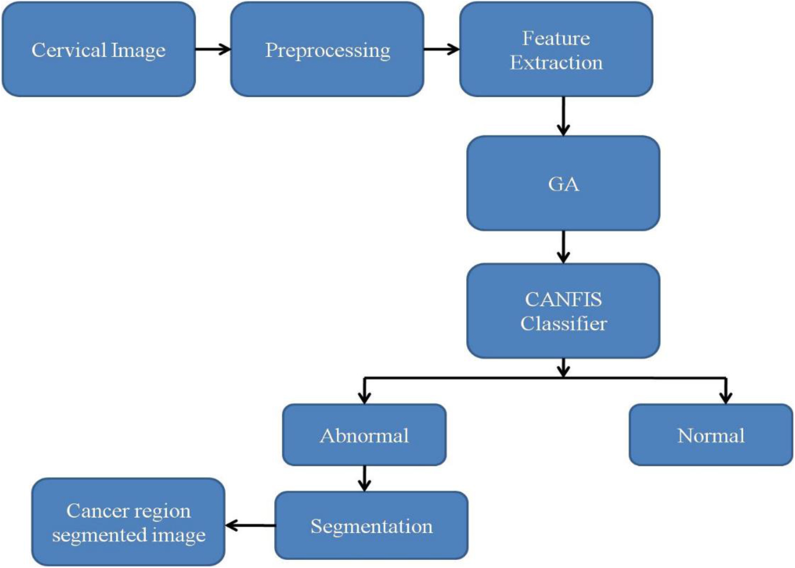
Image smoothing is one of the preprocessing techniques employed in this research, achieved by utilizing the Gaussian filter [31] on the cervical images. The Gaussian kernel is defined as:
Where ‘
The process of capturing or eliminating attributes from the original cervical images is referred to as feature extraction. This operation is conducted to facilitate the classification of the original cervical images. By extracting features from various regions of the image, the original cervical image is categorized. The resulting cervical images are classified into one of two categories: normal or abnormal. The following features are extracted from the cervical images that have undergone a Gaussian filter in order to categorize them.
3.2.2.1 LBP
This non-parametric descriptor generates a unique pattern based on the values of its neighboring pixels. It encodes all the pixels in a Gabor-transformed image into a distinct attribute parameter, highly useful for pattern recognition in various computer vision applications. In the process of computing the Local Binary Pattern (LBP), as described by Prasad et al. in 2023 [32], a 3x3 overlapping window is applied over the Gabor-transformed image. For each center pixel within this window, the value is computed by subtracting it from the surrounding pixels within the window. A binary value of ‘0’ is assigned if the subtraction result is negative, and ‘1’ is assigned if the subtraction result is positive or equal to ‘0’. The LBP feature of the center pixel in the 3x3 window is calculated using the following equation.
Whereas
The LBP features are primarily used to detect variations in non-smoothed regions in cervical images. The LBP-extracted image of the Gabor-transformed cervical picture is depicted in Fig. 3.
LBP extracted cervical picture.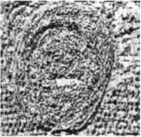
3.2.2.2 GLCM features
GLCM (Gray-Level Co-occurrence Matrix) attributes, as described by Lin et al. in 2022 [33], are texture-related features that depend on the number of pixel pairs with specific concentration values. These attributes are established in a specific spatial arrangement and are used to analyze the relationships between the distributions of pixel values in a cervical image. For the computation of GLCM attributes, one of the directional distributions at angles 0∘, 45∘, 90∘, and 180∘ can be utilized. In this context, the rows and columns of the GLCM matrix are denoted by ‘i’ and ‘j,’ respectively. ‘I(i, j)’ represents a cervical image that has undergone Gaussian filtering. The GLCM feature set includes the following characteristics: homogeneity, contrast, energy, correlation coefficient, variance, maximum probability, and inverse dissimilarity. These GLCM attributes are employed to distinguish between abnormal and normal regions in the cervical image and are extracted from the Gabor-transformed cervical image to aid in this differentiation process.
The standards for GLCM attributes for the regular cervical picture, which is free of lesions, and the aberrant cervical picture, which has cancer patterns, are listed in Table 1. The equations depicted in Table 1 are used to compute the GLCM features, and these values vary with respect to normal and abnormal cervical images.
GLCM features for normal and abnormal cervical images
The non-linearity performances of the retrieved attributes are developed using GA. In this proposed study, the dimensions of the attributes are reduced using GA, and the optimized set of attributes is then fed into the categorization procedure to classify images of the cervical region.
The feature set extracted from the original cervical image needs to be organized into a suitable matrix-vector format. To streamline and reduce the length of this feature set, it is crucial to apply an optimized algorithm. With the assistance of a Genetic Algorithm (GA), the nonlinearity characteristics of the extracted feature set are fine-tuned. This optimization process aims to enhance the classification performance of the proposed system by focusing on the most relevant and optimized features. In this paper, the application of GA allows for the reduction of the dimensions of the extracted attributes. These optimized attribute sets are subsequently utilized as inputs for the cervical image classification method.
The GA (Genetic Algorithm) algorithm is outlined in the following steps:
Step 1: Initiate the process with a randomly selected population, where each chromosome comprises a variety of genes. To form the initial population, replicate it 70 times. Step 2: Initialize the weights for all genes on the chromosomes with randomly assigned starting values. Step 3: Apply the crossover function to the genes within the chromosome along with their associated weight values. The crossover probability is set to 0.5 to achieve the best optimization in this context. Step 4: After the crossover process, apply the mutation function to the resulting chromosomes. A mutation probability of 0.2 is chosen to achieve the best optimization. Step 5: Once the mutation process is complete, compute the Euclidean distance between each gene and its corresponding chromosome. Select the chromosome with the smallest Euclidean distance. Step 6: Repeat steps 2 through 6 for each feature in the attribute set matrix until all features have been processed and optimized.
This algorithm outlines the steps involved in optimizing a set of features using a genetic algorithm for a specific task or application.
The possibility cost of spilling and merge processes stabilizes at the conclusion of the 1st iteration, and the resulting mathematical formulas can be used to calculate them.
The formula for calculating the possibility of a split process is
Whereas, the populace volume N in this work is initially fixed to 70.
The formula for calculating the possibility of a merging process is
ANFIS (Adaptive Neuro-Fuzzy Inference System), as extended by Senthil Kumar et al. in 2020 [34], has been expanded to coactive ANFIS, designed to handle multiple outputs and can be considered as a generalization of ANFIS. A modified classification architecture derived from ANFIS is known as CANFIS (Coactive Neuro-Fuzzy Inference System).
The fundamental component of CANFIS is the fuzzy neuron, responsible for assigning membership values to the input. Two commonly used types of membership functions are the universal Bell and Gaussian membership functions. The membership function for a fuzzy set over the input universe X is defined as
The core element of CANFIS is the fuzzy neuron, which assigns membership values to the input. Two commonly employed types of membership functions are the universal Bell and Gaussian membership functions. The membership function for a fuzzy set over the input universe X is defined as
CANFIS categorization structure of the proposed method.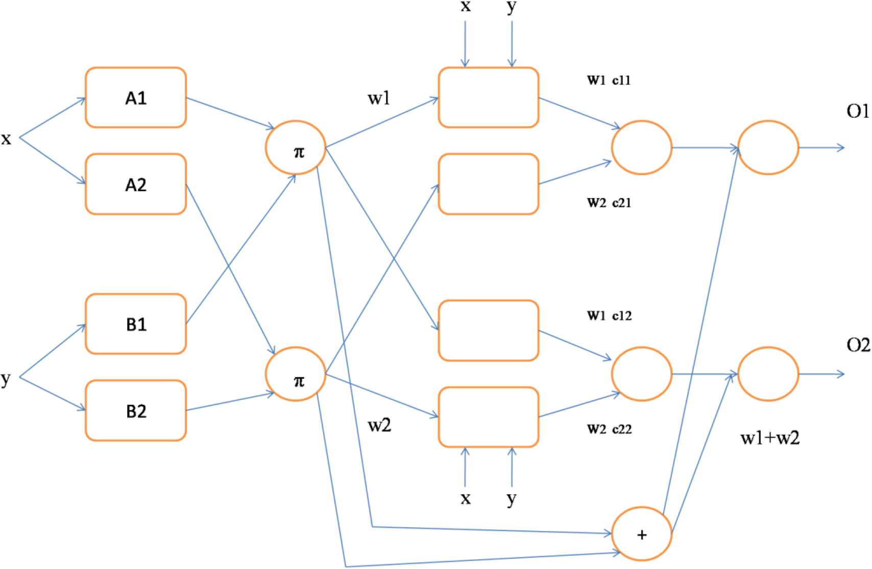
In Fig. 5, each layer plays a specific role in predicting cancer, described as follows:
Layer 1: Each node in Layer 1 assesses the extent to which the given input belongs to one of the fuzzy sets (A, B, C, or D) by computing the membership grade for each fuzzy set. Three membership functions define these fuzzy sets. Layer 2: The combination of all the outputs from Layer 1 serves as the input to Layer 2. Layer 2 consists of two components. The lower component represents the modular network, calculating the total firing strength for each output. The higher component utilizes the membership functions assigned to each of the inputs. This architecture integrates fuzzy logic principles with neural network elements to predict cancer based on input data. The membership grades from Layer 1 and the modular network’s computations in Layer 3 contribute to making accurate predictions regarding cancer-related outcomes.
The final output of the network is computed in the 4th layer, which calculates the weight regularization of the results from the two components of the 3rd layer. In this design, there is no need for a separate Layer 5, as the functions it would typically perform are handled within Layer 4 itself. This streamlined architecture simplifies the network while achieving the desired outcome.
CANFIS categorization architecture for predicting cancer.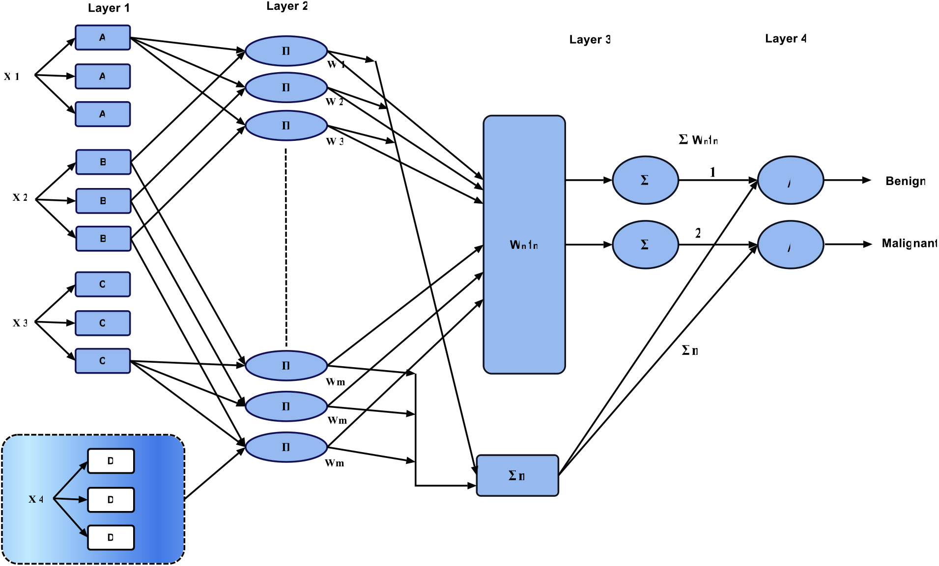
In the CANFIS categorization structure, the X node receives input features from the original cervical image, while the Y node receives the trained patterns, encompassing both normal and anomalous examples. Based on the learned patterns, this structure generates an output of either 0 or 1, where binary values 0 and 1 represent the categories of normal and abnormal, respectively.
The inputs for this categorizer are denoted as X and Y, and the output can be either a training pattern or a categorized pattern, depending on the mode of this classifier. The CANFIS categorization process for each layer is described by the following equations, detailing how the network processes input data and generates the final categorization result.
The symbols for Layer 1 are,
Where
Layer 2 is represented by,
Where,
Layer 3 is represented by,
where,
The symbols for Layer 4 are,
Where as,
Layer 5 is represented by,
Whereas,
In the CANFIS categorization process, the cervical image is classified as “normal” (benign) when the response from Layer 5 is low, and it is classified as “abnormal” (malignant) when the response from Layer 5 is high. Using CANFIS categorization, the features are effectively categorized into normal or abnormal classes. Following the categorization, further morphological operations are applied to the classified abnormal cervical image to segment the malignancy section. This segmentation involves the use of two fundamental morphological operations:
Erosion Operation: This operation is applied to the irregular cervical image to obtain an eroded image. Dilation Operation: The dilation operation is applied to the irregular cervical image to obtain an expanded or dilated image.
To identify and isolate the cancerous region within the cervical image, the eroded image is subtracted from the dilated image. This process helps to highlight and separate the cancerous area from the surrounding tissues, aiding in the accurate localization and analysis of malignant regions in the cervical image.
The cervical images utilized in this research were obtained from the Guanacaste database [35]. This dataset contains cervical images with manually segmented cancer regions, marked by experienced radiologists, providing ground truth annotations. These ground truth cervical images play a crucial role in assessing the performance of the proposed system using various performance metrics. The dataset comprises a total of 295 cervical images, further divided into training and testing sets. The dataset includes 120 cervical images with normal cases and 175 cervical images with abnormal cases. The proposed cervical malignancy detection and segmentation approach were tested on these images. The experimental results obtained on the 295 cervical images from this database demonstrate the effectiveness of the proposed method. Additionally, the same experimental results can be extended to other sets of cervical images obtained from real-time clinical datasets or similar cervical image databases, indicating the generalizability of the approach to various datasets and clinical scenarios. The projected strategy is simulated in this work using MATLAB R2015 and incorporates the GA strategy along with the CANFIS categorization approach.
The categorization rate, which is described in the following mathematical formula for both normal and abnormal cases, is used to analyze the effectiveness of the suggested cervical malignance finding method.
Table 2 presents an evaluation of the anticipated strategy’s performance based on the categorization rate for the average case. The projected technique GA
Table 2 clearly demonstrates that the proposed strategy attains a significantly higher classification rate for normal cases compared to other conventional systems. The performance of the proposed approach is further illustrated in Fig. 6, where it is graphically compared based on the classification rate for the average case.
Performance comparisons of projected strategy in terms of categorization rate for normal case
Table 3 provides a comprehensive analysis of the proposed technique’s performance in terms of classifying abnormal cases. The GA
The proposed strategy, as demonstrated in Table 3, clearly outperforms other conventional methods, achieving a significantly higher classification rate for the abnormal cases.
Performance comparisons of projected strategy in terms of classification rate for abnormal case
Graphical comparisons of proposed methods in terms of categorization rate for standard case.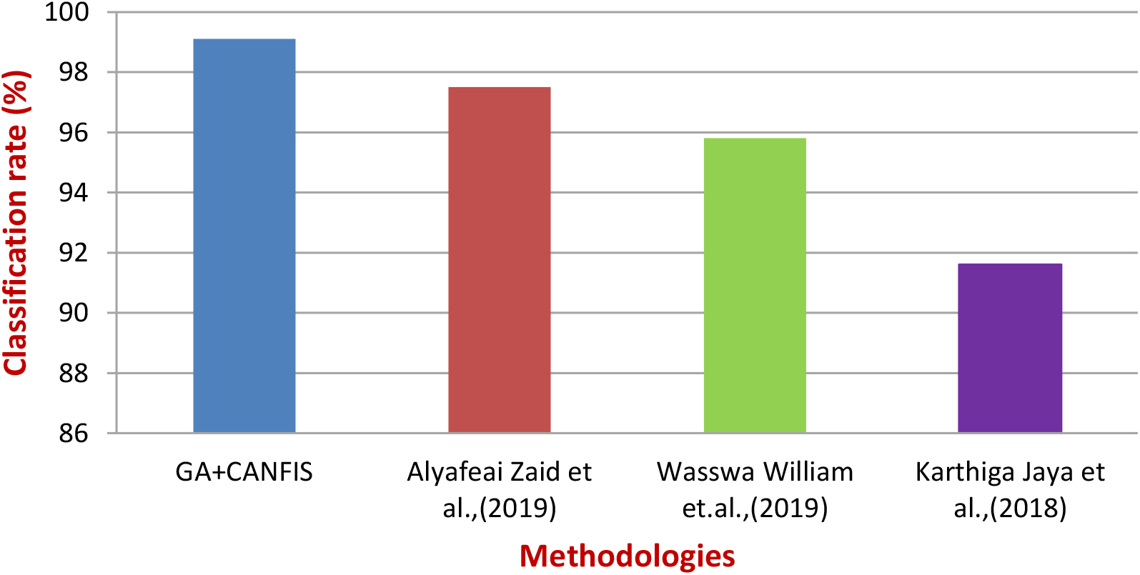
Figure 7 shows the graphical comparisons of the projected strategy in terms of the categorization rate for the abnormal case.
With the assistance of performance assessment specifications, the effectiveness of the suggested cervical malignancy classification and separation approach is examined. The metrics include sensitivity (SE), specificity (SP), accuracy (AC), Positive Predictive Value (PPV), and Negative Predictive Value (NPV), as defined in the previous chapter.
The proposed GA with CANFIS categorization method for the detection and categorization of cervical malignancy, as stated in this research study, achieves 99.09% standard sensitivity, 99.39% standard specificity, and 99.36% standard accuracy, 99.04% standard PPV, and 90.96% standard NPV, as represented in Table 4.
Performance estimation of projected cervical malignance finding scheme
Graphical comparisons of proposed strategy in terms of categorization rate for abnormal case.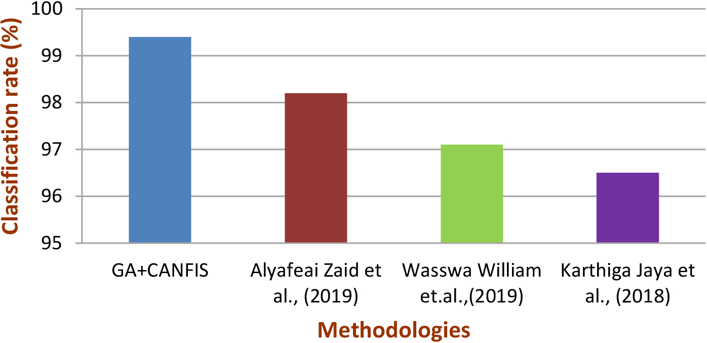
Figure 8 demonstrates the graphical analysis of performance evaluation for the planned cervical cancer detection system. Table 5 provides performance assessments of the projected strategy compared to predictable cervical malignancy detection technologies.
The GA with CANFIS categorization technology specified in this work achieved 99.09% standard SE (sensitivity), 99.39% standard SP (specificity), and 99.36% standard AC (accuracy). In comparison, the conservative process specified by Alyafeai Zaid et al. [16] accomplished 97.2% standard SE (sensitivity), 98.3% standard SP (specificity), and 98.8% standard AC (accuracy) in relation to images that represent the real world.
The conventional method Wasswa William et al. [18] achieved 99.28% standard SE (sensitivity), 97.4% standard SP (specificity), and 98.8% standard AC (accuracy). Karthiga Jaya et al. [19] accomplished 97.42% standard SE (sensitivity), 98.3% standard SP (specificity), and 99.12% standard AC (accuracy) in relation to images that represent the real world.
Performance comparisons of projected strategy with conservative cervical malignance finding technologies
Graphical analysis of Performance assessment for projected cervical malignance finding scheme.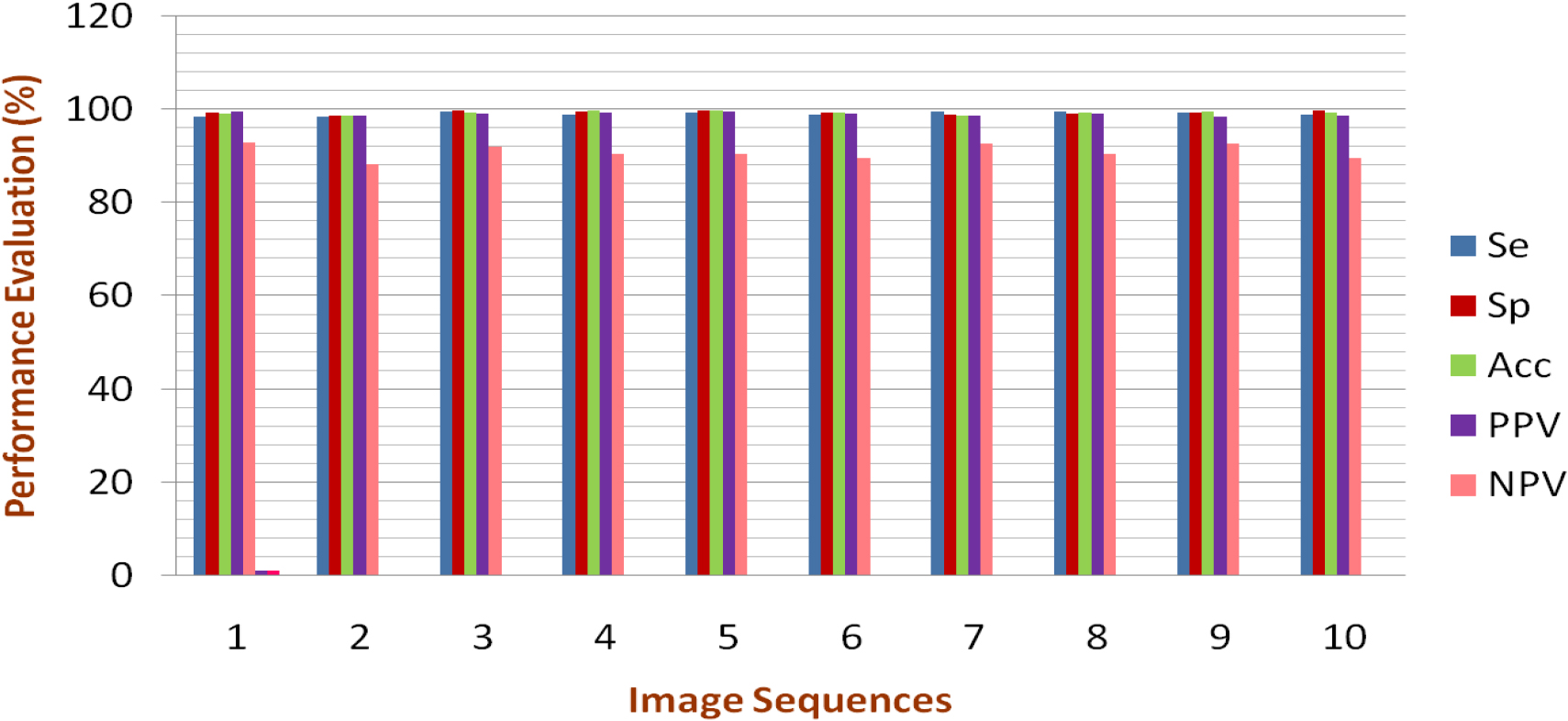
Figure 9 displays the graphic illustrations of performance assessments of proposed the process with conservative cervical malignance finding technologies.
Graphical illustrations of performance comparisons of proposed method with conventional cervical cancer detection methodologies.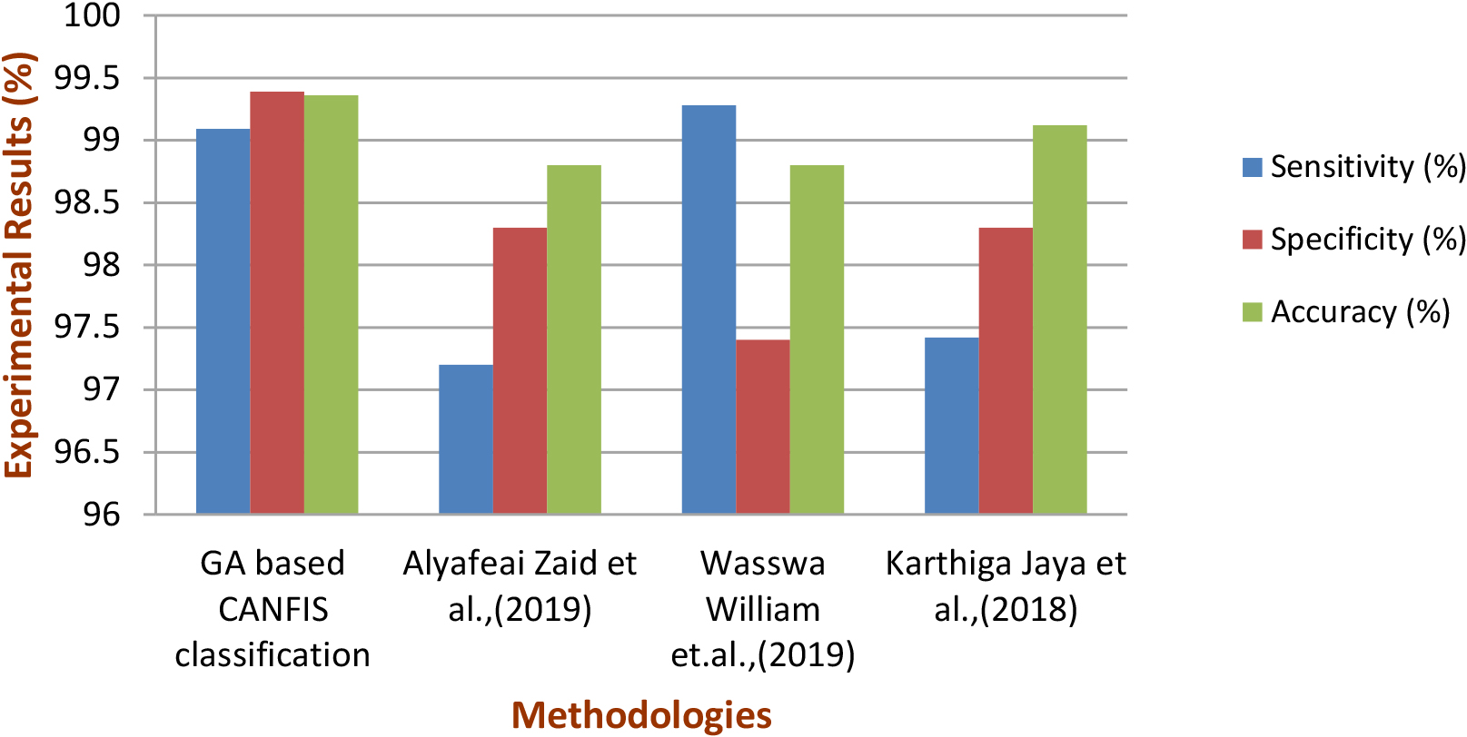
This research introduces an enhanced categorization process utilizing the CANFIS categorization procedure to detect and classify malignancies in cervical images. The methodology involves initial image enhancement through the Gaussian Filter technique, followed by attribute extraction using LBP and GLCM from the enhanced cervical images. These extracted attributes undergo optimization using the Genetic Algorithm (GA), and the optimized attribute sets are then classified using the CANFIS categorization method. Subsequently, a morphological segmentation technique is applied to the categorized abnormal cervical images for the detection and segmentation of cancerous regions. The GA-based CANFIS categorization methodology achieves impressive standard sensitivity (SE) of 99.09%, specificity (SP) of 99.39%, and accuracy (AC) of 99.36%. The GA
Looking ahead, future approaches will likely revolutionize cervical imaging detection by classifying cervical images into severity categories, such as “Early,” “Advanced,” or “Surgery,” enhancing medical decision-making. Streamlining pre-processing, segmentation, and categorization will significantly reduce the time required for each stage, offering faster results for healthcare providers. The final step involves developing a cervical image analysis system utilizing emerging hybrid technologies like Field Programmable Gate Array (FPGA), Internet of Things (IoT), and Very Large-Scale Integration (VLSI). This innovative approach will empower medical practitioners to deliver treatment recommendations more efficiently. The completion and evaluation of an FPGA-based hardware implementation, alongside an IoT-based real-time patient monitoring system, mark the next steps in advancing cervical cancer detection technology.
Data availability statement
The data used for this research is a publicly archived dataset available from: http://www.cse.lehigh.edu/idealab/cervitor/downloads.html.
Funding
No funding was received for this research.
Footnotes
Conflict of interest
The authors declare that there is no conflict of interest.