






Abstract
OBJECTIVE:
This study aimed to investigate the underlying pathogenesis of acute unilateral vestibulopathy (AUV) using gene expression profiling combined with bioinformatics analysis.
METHODS:
Total RNA was extracted from the peripheral blood mononuclear cells of ten AUV patients in the acute phase and from ten controls. The differentially expressed genes (DEGs) between these two groups were screened using microarray analysis with the cut-off criteria (|fold changes| > 1.5 and p-value < 0.05). Functional enrichment analysis of DEGs was performed using Gene Ontology and Kyoto Encyclopedia of Genes and Genomes pathway analysis, and the protein-protein interaction (PPI) network was constructed using the STRING (Search Tool for the Retrieval of Interacting Genes) database.
RESULTS:
There were 57 DEGs (50 up-regulated and 7 down-regulated) identified in the AUV group. Functional enrichment analysis showed that most of the up-regulated DEGs were significantly enriched in terms related to the neutrophil-mediated immune pathway. From the PPI network, the top ten hub genes were extracted by calculating four topological properties, and most of them were related to the innate immune system, inflammatory processes and vascular disorders. The complete blood count tests showed that the neutrophil-to-lymphocyte ratio was significantly higher in the 72 AUV patients than in the age-matched controls (2.93±2.25 vs 1.54±0.61, p < 0.001).
CONCLUSIONS:
This study showed that the neutrophil-mediated immune pathway may contribute to the development of AUV by mediating inflammatory and thrombotic changes in the vestibular organ.
Introduction
Acute unilateral vestibulopathy (AUV) is the sixth most common cause of vertigo/dizziness and the third most common peripheral vestibular disorder after benign paroxysmal positional vertigo and Meniere’s disease [1, 2]. The term “vestibular neuritis” has been widely used although it is unclear whether it is always caused by inflammation [1]. Several hypotheses have been proposed for the etiology of AUV, but the exact pathogenesis remains unknown [3]. The most probable cause is reactivation of herpes simplex virus type 1 (HSV-1), which resides in a latent state in the vestibular ganglia [1–3]. This hypothesis is supported by HSV-1 DNA being detected in the human vestibular ganglia [4], an animal model by inoculating HSV-1 into the auricle of mice [5], and the histopathological similarity between AUV and other viral disorders [6]. Despite these findings, a viral etiology has not yet been conclusively demonstrated due to the lack of compelling structural evidence for a consistent inflammatory component [2, 3]. Besides a viral etiology, other hypotheses such as vascular occlusion and autoimmune disease have been suggested [7–11].
Recently, the bioinformatics analysis of gene expression profiling has been widely used to identify the molecular biomarkers and pathogenesis in various malignancies and autoimmune diseases [12, 13]. However, there are no reports of a bioinformatics analysis being applied to AUV. Thus, the aim of this study was to identify the underlying pathogenesis of AUV using gene expression profiling combined with bioinformatics analysis.
Materials and methods
Patients
The flow diagram of this study is shown in Fig. 1. We recruited ten patients in the acute phase of AUV (≤3 days after the onset), comprising six males and four females aged from 40 to 84 years (mean age 60.2±11.2 years). The diagnosis of AUV was made based on the patient’s history and the findings of clinical and laboratory examinations [2]. The inclusion criteria were as follows: (1) key symptoms of the acute onset of sustained rotatory vertigo and unsteadiness with a tendency to fall toward the side of the affected ear, with all symptoms lasting for several days and following a monophasic illness pattern; and (2) key signs of spontaneous horizontal-torsional nystagmus beating toward the nonaffected side and pathological vestibulo-ocular reflex (VOR) function of the affected horizontal canal (HC) documented in a caloric test (canal paresis >25%) or a video head impulse test (gain of the HC VOR <0.83 obtained from 31 normal controls). None of the included patients had clinical evidence of other vestibular disorders such as Meniere’s disease, vestibular migraine, bilateral vestibulopathy, or central vestibular disorders.
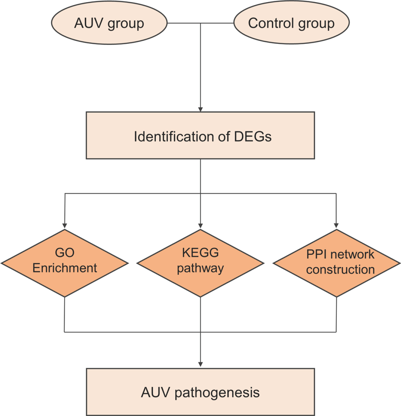
Flow diagram of this study. AUV=Acute Unilateral Vestibulopathy; DEG=Differentially Expressed Gene; GO=Gene Ontology; KEGG=Kyoto Encyclopedia of Genes and Genomes; PPI=Protein-Protein Interaction.
All experiments followed the tenets of the Declaration of Helsinki, and informed consents were obtained after the nature and possible consequences of this study had been explained to the participants. This study was approved by the Institutional Review Boards of Pusan National University Yangsan Hospital.
Peripheral whole blood (2.5 mL) was collected from the patients and ten healthy normal controls. The samples were placed immediately into PAXgene Blood RNA tubes (QIAGEN), and then were stored under refrigerated conditions (2–8°C) before RNA extraction. Total RNA was extracted from whole blood using the PAXgene Blood RNA kit within 3 days after sample collection according to the manufacturer’s protocol. Briefly, the samples were incubated for a minimum of 2 hours at room temperature (15–25°C) to achieve complete lysis of blood cells, and then, centrifuged for 10 minutes at 5000×g using a swing-out rotor. The supernatant was decanted, and 4 mL of RNase-free water was added to the pellet. The samples were vortexed until the pellet was visibly dissolved, and then centrifugation was applied for another 10 minutes at 5000×g. After the entire supernatant was removed, the remaining pellet was resuspended in 350μL of BR1 buffer and transferred to a microcentrifuge tube, followed by automated RNA purification using the QIAcube system. The purity and quantity of RNA were measured using a NanoDrop ND-1000 device (Thermo Fisher Scientific, Wilmington, DE, USA), and the quality of RNA was checked using an Agilent 2100 bioanalyzer (Agilent Technologies, Santa Clara, CA, USA) with an RNA Integrity Number.
Microarray analysis of RNA expression profiles
Isolated total RNA was amplified, labeled, and hybridized using the Affymetrix Human Clariom S assay according to the manufacturer’s protocol. Briefly, the purified RNA sample was transcribed to double-strand cDNA using the GeneChip Whole Transcript (WT) amplification kit. The sense cDNA was then fragmented and biotin-labeled with TdT (terminal deoxynucleotidyl transferase) using the GeneChip WT terminal labeling kit. Approximately 5.5μg of the labeled cDNA was hybridized onto the Affymetrix GeneChip array at 45°C for 16 hours. After washing, the arrays were stained on the GeneChip Fluidics Station 450 and scanned using the GeneChip Scanner 3000. Raw data were analyzed using Affymetrix GeneChip Command Console software.
Identification of differentially expressed genes
The raw data were summarized and normalized with the Robust Multi-array Average module implemented in Affymetrix Power Tools. The differentially expressed genes (DEGs) between AUV and normal control samples were screened based on fold-change filtering combined with Student’s t-test. The false discovery rate (FDR) was controlled by adjusted p-value using the Benjamini-Hochberg algorithm. Genes were considered to be differentially expressed using criteria of |fold changes| > 1.5 and adjusted p-value < 0.05.
Functional enrichment analysis
To explain functional annotation for DEGs, Gene Ontology (GO) and Kyoto Encyclopedia of Genes and Genomes (KEGG) pathway enrichment analyses were performed using g:Profiler (https://biit.cs.ut.ee/gprofiler/). GO covers three biological terms: biological process (BP), cellular component (CC), and molecular function (MF). The KEGG pathway provides a molecular interaction/reaction network pathway regarding metabolism, genetic information processing, environmental information processing, cellular processes, organism systems, human diseases and drug development. An adjusted p-value (FDR) <0.05 was used as the criterion for statistical significance.
Construction and analysis of the protein-protein interaction network
To construct the protein-protein interaction (PPI) network, DEGs were imported into the Search Tool for the Retrieval of Interacting Genes (STRING) online database (http://string-db.org). Using the Python NetworkX package, the hub genes were extracted from the PPI network by calculating the following four topological properties: the degree (number of interactions per node), the closeness (reciprocal of the sum of the geodesic distances from one node to all other nodes), the betweenness (number of shortest paths that pass through each node), and the subgraph centrality (weighted sum of all closed walks originating from each node). For each hub gene, the SuperPath, the unified pathway extracted from the different sources including KEGG, Cell Signaling Technology, R&D Systems, GeneGo, Reactome, BioSystems, Tocris Bioscience, and QIAGEN, was analyzed from GeneCards (http://genecards.org).
Comparison of complete blood cell count parameters
To investigate the alterations in inflammatory markers, we compared the complete blood cell count (CBC) parameters obtained from the peripheral venous blood samples of 72 AUV patients and age-matched controls. The parameters included the absolute counts of neutrophils, lymphocytes, monocytes, and platelets. The neutrophil-to-lymphocyte ratio (NLR) and the platelet-to-lymphocyte ratio (PLR) were also compared since these parameters have previously been suggested to be useful inflammatory biomarkers [8, 14]. The CBC parameters displayed as mean±standard deviation (SD). Statistical analyses were conducted using SPSS software (version 22, SPSS, Chicago, IL, USA). Student’s t-test was used to compare the CBC parameters between the two study groups, and Pearson’s correlation test was performed to assess the association between NLR and PLR. P-value < 0.05 was used as the criterion for significance.
Results
Identification of DEGs
We detected 21,448 DEGs (10,663 up-regulated and 10,785 down-regulated) between AUV and control samples. Among them, 50 up-regulated DEGs and 7 down-regulated DEGs were identified in the AUV group based on the cut-off criteria (|fold changes| > 1.5 and p-value < 0.05) (Table 1). The DEGs between the two groups are depicted on a scatter plot and a volcano plot in Fig. 2A and 2B. Hierarchical clustering analysis of up-regulated and down-regulated DEGs revealed distinctive expression patterns between the two groups (Fig. 2C and 2D).
All differentially expressed genes
All differentially expressed genes
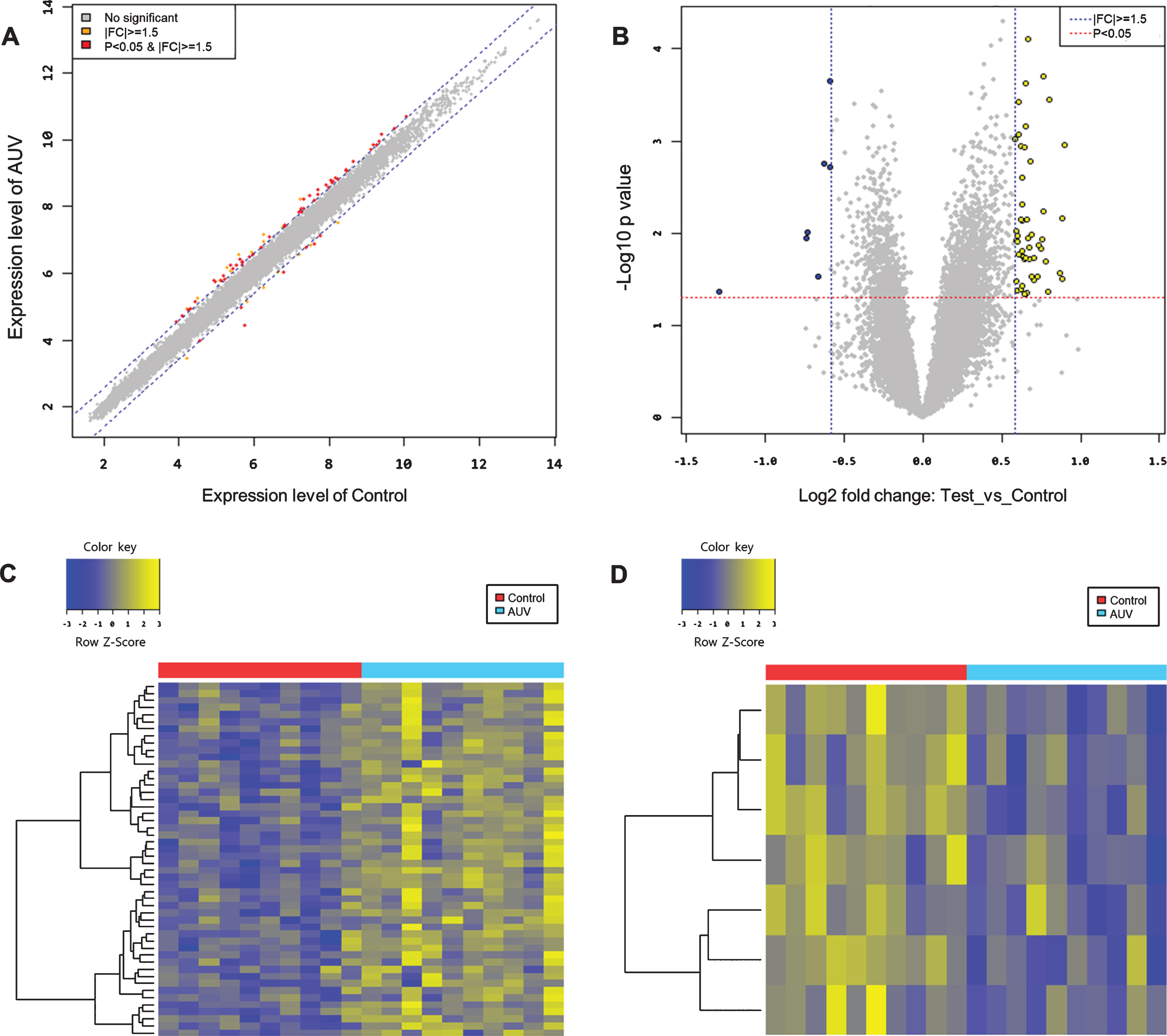
Scatter plot (A) and volcano plot (B) of differentially expressed genes. (A) A total of 57 DEGs (50 up-regulated and 7 down-regulated, red points) are identified in the AUV group based on the cut-off criteria (|fold changes| > 1.5 and p-value < 0.05) (B) The x-axis shows the fold changes in gene expression between two groups, and the y-axis shows the statistical significance of the differences. Yellow and blue points represent up-regulated and down-regulated DEGs, respectively. Results of a hierarchial clustering analysis of up-regulated (C) and down-regulated (D) differentially expressed genes between AUV and control groups. Each group includes ten individuals and each column represents one individual’s sample. Yellow and blue colors indicate high and low relative expression, respectively. FC=Fold Change.
GO enrichment analysis was performed for all DEGs to obtain their underlying functions, which revealed that the up-regulated DEGs were enriched in 32 GO terms: 15 BP, 8 CC, and 9 MF (Fig. 3). For BP terms, the majority of the up-regulated enrichments were related to the terms for immune system processes including neutrophil activation (GO:0042119, GO:0002283), neutrophil degranulation (GO:0043312), neutrophil mediated immunity (GO:0002446), cytokine production involved in immune responses (GO:0002367), and adaptive immune responses (GO:0002369, GO:0042088, GO:0002827) (Fig. 3A). For CC terms, the up-regulated DEGs were mostly enriched in terms for secretory granules that were found primarily in mature neutrophils, including tertiary granules (GO:0070820, GO:0070821), ficolin-1-rich granule membrane (GO:0101003), and specific granules (GO:0035579, GO:0042581) (Fig. 3B). For MF terms, the up-regulated DEGs were significantly enriched in terms associated with carbohydrate binding (GO:0030246, GO:0005536), transporter activity (GO:0015145, GO:0005355, GO:0051119, GO:0015149), protein kinase activity (GO:0097472, GO:0004693), and immune receptor activity (GO:0004896) (Fig. 3C). The down-regulated DEGs were enriched in only two BP terms: epithelial cell proliferation (GO:0050680) and DNA biosynthetic process (GO:0071897) (Fig. 3D).
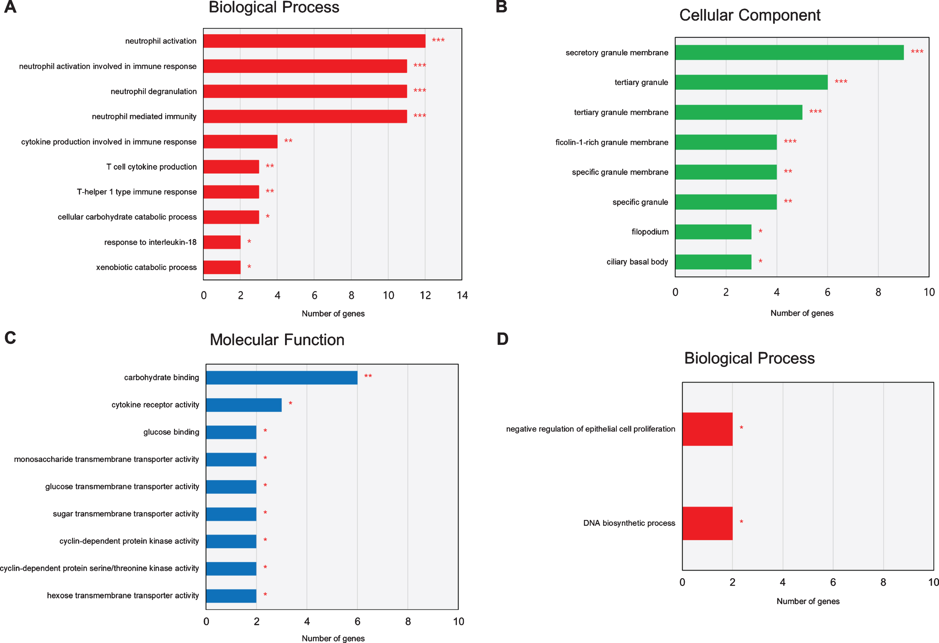
Results of a GO enrichment analysis of up-regulated (A, B, C) and down-regulated (D) DEGs in the AUV group. The x-axis represents the number of genes enriched in each GO term. The y-axis represents GO terms with adjusted p-values of < 0.05 (*), <0.01 (**), and < 0.001 (***).
KEGG pathway analysis showed that up-regulated DEGs were predicted to participate in the following six pathways: metabolic pathways (hsa:01100), cytokine-cytokine receptor interaction (hsa:04060), glutathione metabolism (hsa:00480), viral protein interaction with cytokine and cytokine receptor (hsa:04061), insulin signaling pathway (hsa:04910), and fluid shear stress and atherosclerosis (hsa:05418) (Fig. 4A). In contrast, the down-regulated DEGs had no significant enrichment in any pathway.
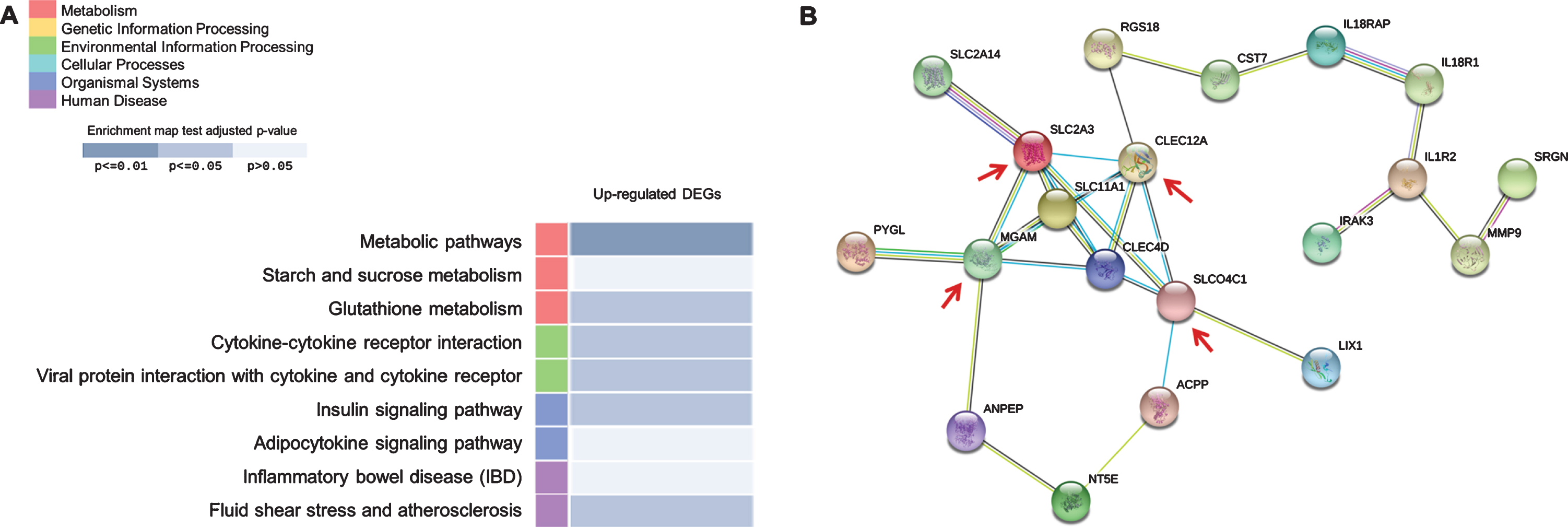
(A) Results of a KEGG pathway analysis of up-regulated DEGs in the AUV group. The adjusted p-values indicated that up-regulated DEGs are enriched in six pathways. (B) PPI among the 20 DEGs. The important hub genes that are included in all four topological properties, are indicated by the red arrows.
The PPI network of all DEGs was constructed using the STRING database. The PPI network complex comprised 20 proteins encoded by DEGs (Fig. 4B). CLEC12A, MGAM, SLC2A3, SLCO4C1, CLEC4D, SLC11A1, IL1R2, IL18RAP, RGS18, IL18R1, ACPP, CST7, ANPEP, and MMP9 were included in the top ten hub genes of each topological property, and CLEC12A was the top hub gene for all topological properties (Table 2). The innate immune system pathway was one of the SuperPaths that they had in common (Table 3).
Top ten hub genes presented by four topological properties extracted from the protein-protein interaction network
Top ten hub genes presented by four topological properties extracted from the protein-protein interaction network
SuperPaths for hub genes identified by protein-protein network analysis
Bioinformatics analysis revealed that the up-regulated DEGs were mainly enriched in immune system pathways associated with neutrophil activation. We compared the CBC parameters between 72 AUV patients and age-matched controls (Table 4), which revealed that the NLR value was significantly higher in the AUV group than in the controls (2.93±2.25 vs 1.54±0.61, p < 0.001) (Fig. 5A). The mean PLR value did not differ significantly between the two groups, but the 37 patients with an NLR that was higher than the mean+SD in the control group tended to have a higher PLR value than the controls (148.11±66.47 vs 125.55±33.78, p = 0.058). Furthermore, there was a strong positive correlation between NLR and PLR values in the AUV group (Pearson correlation coefficient r = 0.759, p < 0.001) (Fig. 5B).
Comparison of complete blood count parameters between the two study groups
Comparison of complete blood count parameters between the two study groups
Data are n or mean±standard deviation values. AUV=Acute Unilateral Vestibulopathy; NLR=Neutrophil-to-Lymphocyte Ratio; PLR=Platelet-to-Lymphocyte Ratio; WBC=white blood cell.
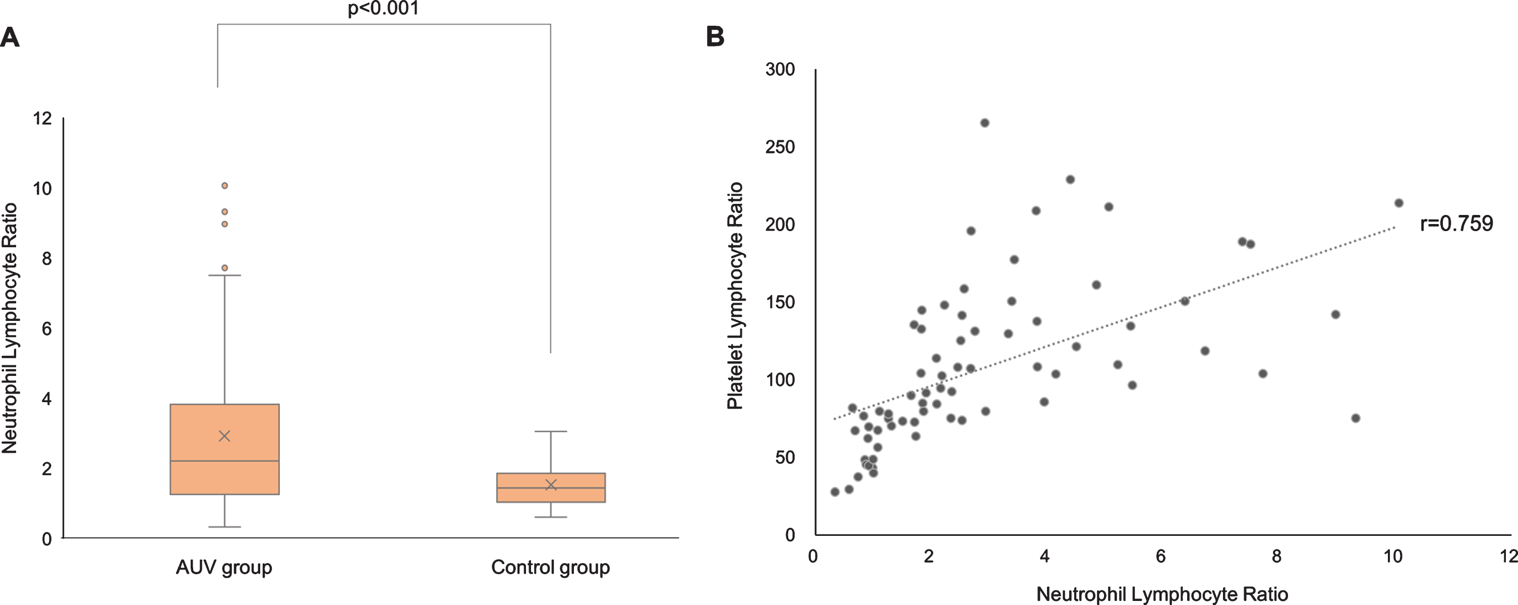
(A) Comparison of the neutrophil-to-lymphocyte ratio (NLR) between the AUV and control groups. The mean NLR value was significantly higher in the AUV group than in the controls. (B) Correlation between NLR and platelet-to-lymphocyte ratio (PLR) values in the AUV group. There was a strong positive correlation between the NLR and PLR values (Pearson correlation coefficient r = 0.759, p < 0.001).
To our knowledge, this is the first study to have evaluated the pathogenesis of AUV using gene expression profiling combined with bioinformatics analysis. We profiled numerous DEGs associated with the neutrophil-mediated immune pathway in the AUV group. These bioinformatics results were supported by the high value of NLR for the CBC parameters, and suggest that neutrophil-mediated immune mechanisms play a more critical role in the development of AUV, with direct viral infection associated with lymphocyte activation being less likely to contribute to AUV.
The viral-infection hypothesis has long been suggested for the etiology of AUV based on the presence of concurrent infectious illness [1–3, 15], the detection of HSV-1 DNA in the human vestibular ganglia [4], an animal model by inoculating HSV-1 into the auricle of mice [5], and the temporal bone histopathology findings in AUV [6]. In the present study, bioinformatics analysis showed that numerous up-regulated DEGs in the AUV group were mainly enriched in the immune system process. However, most of them were related to the terms for neutrophil-mediated immunity, such as neutrophil activation, neutrophil degranulation, specific granules, tertiary granules, and ficolin-1-rich granule membrane. Neutrophils are important components of immunity associated with inflammatory responses against a broad spectrum of pathogens [16]. In particular, specific and tertiary granules are later-developing granules found in mature neutrophils, and play a role in phagocytosis and exocytosis, respectively [16]. This neutrophil-mediated immunity was confirmed by CBC parameters showing a higher value of NLR in the AUV patients than in the controls, which is similar to the findings of two previous studies [8, 9]. On the other hand, viral infection usually induces an increase in the lymphocyte count and a decrease in the neutrophil counts, and hence leads to lower NLR values [17]. A previous study found that approximately 50% of AUV patients showed decreases in the total and CD8 T-lymphocytes, leading to an immunological imbalance of the increased T-helper (CD4)/ T-suppressor (CD8) lymphocyte ratio [11]. Based on all of these findings and the characteristic time interval between infectious illness and the onset of AUV, this disease may be caused by neutrophil-mediated immune complications following the infection rather than direct viral infection along the vestibular nerve.
The present study constructed a PPI network to predict the associations of protein functions of the identified 57 DEGs. By calculating four topological properties, the hub genes identified in PPI network included CLEC12A, SLC2A3, MGAM, SLCO4C1, CLEC4D, SLC11A1, IL1R2, IL18RAP, RGS18, IL18R1, ACPP, CST7, ANPEP, and MMP9. Most of these genes share an innate immune system pathway and are known to be associated with various autoimmune diseases such as rheumatoid arthritis (RA) [18, 19], ankylosing spondylitis [20], Behcet’s disease [21–23], inflammatory bowel disease [13, 24], multiple sclerosis [25], and celiac disease [24]. Notably, CLEC12A was the top hub gene for all topological properties, suggesting that it plays a critical role in the PPI network. CLEC12A encodes a myeloid inhibitory C-type lectin-like receptor that is expressed primarily on granulocytes and monocytes [26]. It has diverse functions, such as cell adhesion, cell-cell signaling, glycoprotein turnover, and roles in inflammation and immune responses. With CLEC4D, one of up-regulated DEGs, CLEC12A has been reported to be associated with the development of RA [18, 19]. A recent study of experimental autoimmune encephalomyelitis mice found that CLEC12A was involved in the binding and transmigration of dendritic cells across the blood-brain barrier, and administering a blocking antibody against the CLEC12A receptor prevented significant disease progression [27]. Besides, SLC2A3 encodes GLUT3, which is a glucose transporter that provides glucose to activated immune cells as their primary energy source [28]. IL1R2, IL18RAP, and IL18R1 encode cytokine receptors involved in inducing cell-mediated immunity [29]. Thus, the hub genes identified in the PPI network may be closely related to the development of AUV by altering the immune system.
It is obvious that the inflammatory response plays a crucial role in the pathogenesis of AUV, but it remains unclear how this process leads to damage to the vestibular nerve. Several authors have proposed the vascular theory based on the presence of a proinflammatory state in AUV [7]. By analyzing peripheral blood mononuclear cells, it was found that AUV patients had a significantly increased percentage of CD40 + monocytes and macrophages, which may cause the formation of platelet-monocyte aggregates and subsequently lead to microvascular occlusion in the vestibular organ. Other studies have also supported the vascular theory based on the high values of PLR and the mean platelet volume, and a higher prevalence of cardiovascular risk factors in AUV patients [8–10].
The present study found neutrophil degranulation to be one of the immune system pathways that the hub genes had in common. This is an exocytosis process in which a neutrophil releases proteases, lipases, and inflammatory mediators in secretory granules. This process is obviously important in the immune response, but can also result in harmful effects including tissue damage, thrombosis, and exposure of autoantigens [16]. It was reported that neutrophil degranulation might mediate the vascular damage by promoting the detachment of endothelial cells and the adherence of platelets [30]. Indeed, many studies have found that an immune response induced by neutrophil activation was associated with cardiovascular disorders [31–34]. Gene expression profiling for acute ischemic stroke has identified several immune-related genes including ARG1, LY96, and MMP9, which were positively correlated with NLR, and stroke severity [35]. Furthermore, some hub genes identified in the present study, including MGAM, SLC11A1, MMP9, IL1R2, IL18RAP, and IL18R1 have been reported to be associated with coronary artery disease, acute ischemic stroke and vasculitis [35–41]. One of the enriched pathways in the KEGG analysis was the fluid shear stress and atherosclerosis pathway. RGS18 and SRGN, which were found to be up-regulated DEGs in AUV, have a role in platelet formation, activation, and degranulation [42, 43]. In terms of CBC parameters, there was a strong positive correlation between the NLR and PLR values in the present AUV group. All of these results may represent plausible evidence for the vascular theory of AUV, but it remains unclear why the inflammatory and thrombotic response only insult unilateral and peripheral end organ of vestibule without any systemic signs. Further investigations are needed to confirm the validity of the vascular theory.
Gene expression profiling has been reported in other vestibular disorders such as Meniere’s disease and vestibular migraine [44–46]. RNA sequencing study found DEGs associated with oxidative stress or ion homeostasis in sporadic Meniere’s disease, suggesting that immune factors might be involved in the pathogenesis [44]. Another study applied gene expression profiling for the differential diagnosis of Meniere’s disease and vestibular migraine, which found that two disorders have a different pro-inflammatory signature [46]. All of these including the present study showed that gene expression profiling can be an excellent method to identify the underlying pathogenesis of various vestibular disorders.
This study was subject to some potential limitations. First, our results from the microarray analysis were based on a small sample, and so the expression levels of the hub genes should be verified using qRT-PCR for a large number of patients. Second, we performed gene expression profiling during the acute period of AUV. To have more confidence in the role of a neutrophil-mediated immune response in AUV, the changes in the expression levels of mRNA should be examined in the acute and chronic periods of AUV as well as in control groups.
In conclusion, we have identified the DEGs associated with the immune system pathway in AUV. The findings of bioinformatics analysis indicate that they are likely to contribute to the development of AUV via the neutrophil-mediated immune pathway that leads to inflammatory and thrombotic changes in the vestibular organ.
Footnotes
Acknowledgments
This research was supported by Basic Science Research Program through the National Research Foundation of Korea funded by the Ministry of Education (NRF-2018R1D1A1A09081786).