
Abstract
Objective of this study is to present and test a new method for metal artifact reduction (MAR) by segmenting raw CT data (sinogram). The artifact suppression technique incorporates two steps namely, metal projection segmentation in the sinogram and replacement of segmented regions by new values using an interpolation method. The proposed segmentation algorithm uses the sinogram instead of reconstructed CT slices. First, one of the best and newest region-based geometric active contour models is used to detect projection data affected by metal objects (missing projections). Then, the Hough-transform method is applied to detect all sinusoidal-like curves belonging to metal objects. Finally, a post image processing technique is used aiming to increase accuracy of the segmentation process. To provide a proof of performance, CT data of two patients with metallic teeth filling and pelvis prosthesis were included in the study as well as CT data of a phantom with metallic teeth inserts. Accuracy was determined by comparing mean, variance, mean squared error (MSE) and, peak signal to noise ratio (PSNR) as evaluation measurements of distortion in phantom images with respect to metallic teeth (original and suppressed) and without metallic teeth inserts. Quantitative results showed an average improvement of 12 dB in terms of PSNR and 517 in terms of MSE when the new MAR method was applied to remove metal artifacts. Qualitative improvement was also assessed by comparing uncorrected clinical images with artifact suppressed images. Moreover, qualitative comparison of the results of the proposed new method with the existing methods of MAR showed the superiority of the new method tested in this study.
Introduction
A common problem in computed tomography (CT) is streak artifacts caused by the presence of high-density objects in the field of view of scanner device. Metal implants such as prosthetic devices, dental fillings, surgical clips and electrodes produce this kind of artifacts. The results of scanning a metal object are bright sinusoidal curves in the raw CT data (sinogram). The reconstruction of this sinogram during the process of filtered back projection (FBP), which is the common image reconstruction technique in CT, causes dark and bright streaks in the reconstructed image. This problem arises due to four reasons: 1) significant beam hardening, 2) poor signal to noise ratio from photon starvation in the metal reconstruction, 3) scattering and 4) edge-gradient effects [1].
Thus, the metal artifacts create serious limits to the clinical value of the CT scanning since they affect diagnosis and radiotherapy treatment planning. The need for more improved metal artifact reduction methods in CT grows with the increasing importance of CT imaging [2, 3]. In a routine practice, a slight reduction in metal artifacts can be achieved by selecting appropriate acquisition parameters such as high tube voltage and high tube current, but it augments the risk of absorbing more radiation by patients [4]. Moreover, we can use iterative reconstruction methods [5, 6] which are less sensitive to metal artifacts but they arise another problem, i.e. long reconstruction time [7]. Several image processing solutions have been provided to remove artifacts caused by metal implants. The first publications on metal artefact reduction come back to the works done by Glover et al. [8] and by Kalender et al. [9]. The image-based method used by Kalender et al., for instance, comprised segmentation of the metal implant in reconstructed images. An advantage is that segmentation in reconstructed CT images is relatively easy since these images do not substantially suffer from overprojection. Moreover, pixel values do not vary substantially within materials, facilitating segmentation in this kind of images.
In addition to the segmentation of the metal prosthesis, projection data of metal objects could also be exploited using the Radon transformation. Then a linear interpolation could be performed in the projected metal regions in original raw data to substitute the metal traces by lower intensities. A filtered backprojection was finally used to create a new image with improved image quality. It should be noted that reprojecting regions to Radon space in the same geometry as the original raw data may be quite complicated, especially in the case of modern multirow detector spiral data [10]. As an alternative, artificial raw data, created from reconstructed slices using a simpler geometry, can be used instead of original raw data [4]. A disadvantage of this latter approach is that generation of artificial raw data from a reconstructed image as a base for the correction is suboptimal in terms of spatial resolution. The most publications on MAR methods are based on segmentation of metallic objects in the CT images followed by forward projection of the metal-only image, interpolation in raw data, and image reconstruction by backprojection of the modified raw data [11–13]. A comparison study of MAR techniques based on segmentation of metal affected regions in original CT images has been done by Rinkel et al. [10].
Segmentation of metal traces directly in the sinogram makes it efficient and different from current methods that perform segmentation in reconstructed images. Implementing segmentation and correction directly in original sinograms would avoid the need for a computationally complex forward projection (or the disadvantage associated with using artificial raw data). Moreover, such approach can correct metal artifacts that originate from prosthesis positioned out of the field of view. In [14], metal sinogram segmentation was achieved by using a Markov random field model. The method proposed in [15] was based on a region-based active contour model which can be applied to a sinogram of industrial dual energy CT image that is treated as an image formed by two regions of approximately piecewise-constant intensities, thus it is a suitable case for the active contour model to be applied [16]. However, this kind of active contour models cannot be applicable to medical images with intensity inhomogeneities because this method relies on intensity homogeneity.
By studying recent works on MAR [17, 18], we can conclude that the use of sinogram segmentation is a better technique to achieve a superior image quality in the case of CT scanning of patients with metal implants. However, the detection of metal traces in sinogram is still a challenging task. Moreover, in the most recent comparison of commercial techniques of MAR methods, Huang et al. [19] suggested that MAR methods in general can work well for the patients with metal implants, however, they should be used with caution in certain cases such as dental artifacts.
In this study, a new method of the segmentation of metal trace in spiral fan-beam CT sinogram is proposed which can be effectively applied in both cases of dental implants and pelvis prostheses. This method is based on the region-based geometric active contour model and 2D Hough transform. Recently, two newest region-based geometric active contour models [20, 21] have been proposed for images with intensity inhomogeneities and can be applied on the sinogram, however we selected the model in [20], because after our observation the undesired segmented regions in the result of using of the model in [20] were less compared with model in [21]. After applying the selected active contour model that originally termed as local binary fitting model (LBF) on the sinogram, the region of the missing projections can be obtained. Then, the Hough-transform method based on the parametric model of the fan-beam sinogram is applied to the regions between the detected boundaries for determining all the sinusoidal-like curves belonging to metal objects. Finally, in order to make segmented regions more accurate, a post-processing method is used followed by interpolation in the segmented regions. To evaluate our raw-data based method, we performed phantom tests for quantitative analysis. We additionally performed clinical tests based on scans of two patients with metallic teeth filling and pelvis prosthesis.
Methods and materials
Local binary fitting model
Li et al. [20] proposed an LBF model for applying active contours for image segmentation based on techniques of curve evolution, i.e. Mumford-Shah function [21, 22] and level sets. Consider a given vector valued image
This energy is then incorporated into a variation level set formulation [23] with a level set regularization term. By minimizing the energy function with respect to the level set function φ, and with C representing the zero level set, using the standard gradient descent method, the evolving equation, with respect to an artificial time t, can be driven as:
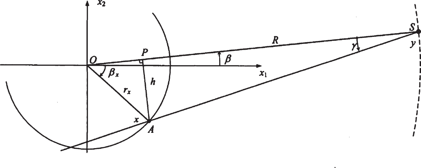
The most popular presentation of the projection data set is the so-called sinogram, i.e. the data collected over all of the projection angles form a 2D image with its intensities representing the magnitude of the projection samples. In this study, we are focusing on the helical CT single slice scanner and in order to use Hough-transform method, it is necessary to drive the equations of a fan-beam sinogram. These equations can be derived based on Fig. 1 [24].
Fan-beam geometry.
Consider a point of scanned object as x that can be represented by its polar coordinate (r
x
, β
x
). The circular trajectory of the X-ray source S that rotates around the object is given by the Eq. (5):
Equation (6) can be used to define a sinogram curve S (r
x
, β
x
) as Eq. (7):
Actually for each r x and β x there is a sinogram curve or S-like curve. Since any object can be approximated by a collection of points located in the space, it is expected its projection to be a set of overlapped S-like curves in the fan-beam sinogram space.
In this study, we focused our attention on the Siemens Somatom helical CT single-slice scanner. Figure 2(a) shows the phantom used in this study. The inserts were constructed from metallic pieces as dental filling showed in Fig. 2(b). Two spiral acquisitions of the phantom were performed: one with metallic teeth insert and one without teeth insert. Moreover, spiral CT scans of two patients were acquired: one patient has metallic teeth filling and another has pelvis prosthesis. Specifically, suppression of the metal artifacts by using raw data is based on two main steps; segmentation of the missing projections in the original sinogram and replacement of raw data pixels in metal region by more appropriate values.
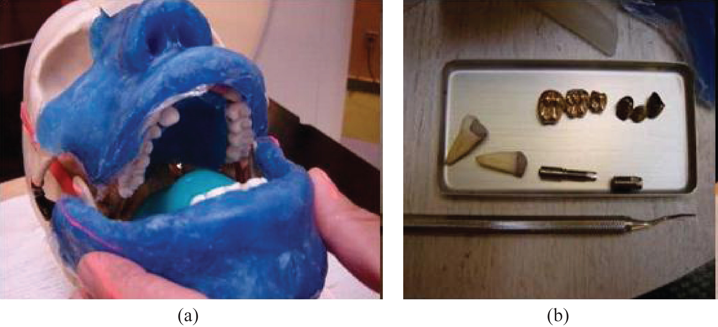
Constructed phantom for probing the effect of the MAR method. (a) The phantom without inserts. (b) Metallic teeth insert.
In the first step of segmentation, we used the active contour model in [20]. In fact, because the results of scanning a metal object are bright S-like curves, the sinogram can be treated as an image formed by two regions with different inhomogeneity intensities, thus it is a suitable case for segmentation. Compared to simple thresholding segmentation method which requires adjusting the threshold with different sizes of metal and non-metal objects, the active contour model based segmentation is self-adaptive. However, the segmentation only based on active contour model is not adequate for CT images with the small size and relatively large number of the metal objects. The traces of small metal objects in the sinogram are tiny and narrow curves, and also the distance between the traces is sometimes very small. Therefore, precise detection of the projected traces (i.e. the position and edges) is a major challenge.
In the proposed method, the active contour model is used only to determine approximately the boundaries of the missing projections region (i.e. the area containing all projections affected by metallic objects), as shown in the Figs. 3(b) and 4(b), and then in the second step, Hough-transform is used to correct these boundaries. The results are shown in Figs. 3(c) and 4(c).
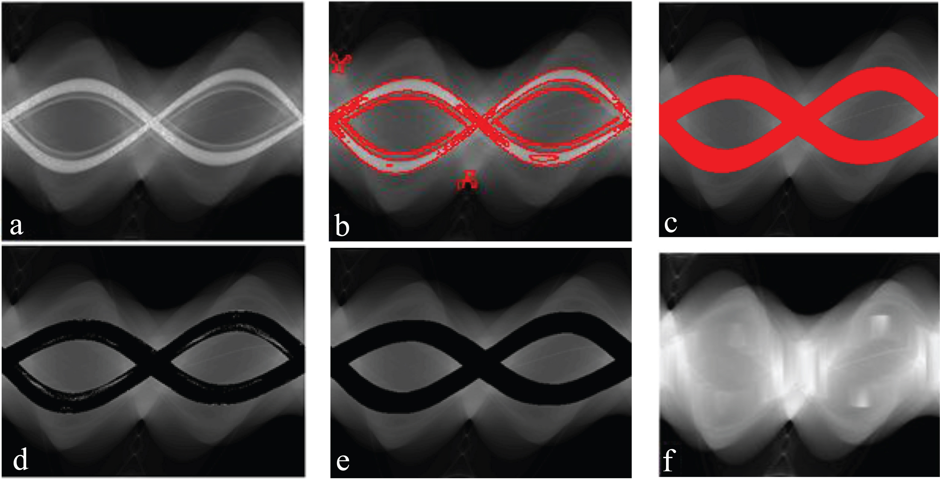
Example of a patient with pelvis prosthesis. (a) Original sinogram. (b) The preliminary segmentation using LBF model. (c) The result of applying the Hough-transform method to the regions detected in (b). (d) The result of removing the metal projections detected in (c). (e) The result of applying the post processing method to (d). (f) The result of replacement of the all removed projections in (e) with the appropriate values using the interpolation technique proposed in [25].
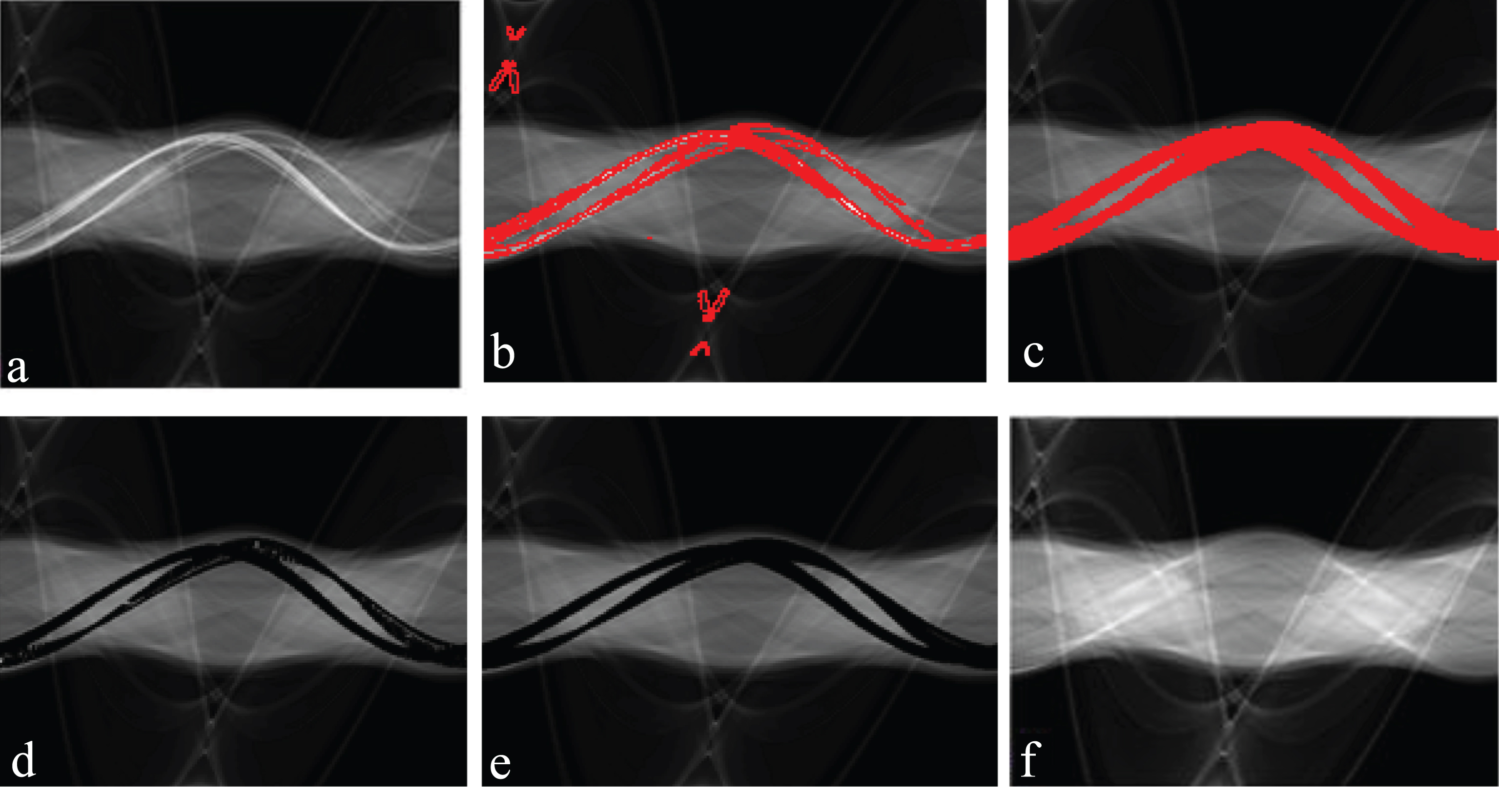
Example of a patient with teeth fillings. (a) Original sinogram. (b) The preliminary segmentation using LBF model. (c) The result of applying the Hough-transform method to the regions detected in (b). (d) The result of removing the metal projections detected in (c). (e) The result of applying the post processing method to (d). (f) The result of replacement of the all removed projections in (e) with the appropriate values using the replacing technique proposed in [13].
2.3.1.1. Hough-Transform parametric shape detection The Hough transform is a technique used in image analysis, computer vision, and digital image processing. In this paper, the purpose of the technique is to find all of the S-like curves belonging to the metal objects by a voting procedure. This voting procedure is carried out in a parametric space, from which missing projection candidates are obtained by using local maxima in a so-called accumulator space that is explicitly constructed by the fan-beam sinogram equation (Eq. (7)).
In the implementation of this Hough-transform method, the data of the regions between the detected boundaries by the first step are used. The Hough space is constructed based on the Eq. (7). In fact because the points achieved by the first step are (γ, β) and R is concerned to the property of the device, there are just two unknown parameters, i.e. (r
x
, β
x
). So we can use 2D Hough transform to obtain two parameters r
x
and β
x
. As can be seen in the Eq. (8), an arbitrary point (γ, β) in the regions between the detected boundaries is mapped to a curve in the parametric space. The ranges of r
x
andβ
x
are set to 0 ≤ r
x
≤ rx.max and 0 ≤ β
x
≤ 2π where rx.max is determined by the range of the field of view of scanner device in the practice.
The steps to parameterize an S-like curve are given as follows: Construct an 2D accumulator array for the parameter space (r
x
, β
x
) and initialize all elements of accumulator to zero; For each point in the regions between the detected boundaries, carry out the following steps; i.e. 3) through 8); For β
x
incremented from 0 to 2π, carry out the following steps; i.e. 4) through 5); Compute r
x
by Eq. (8); If r
x
is within the range (0, rx.max), increment the element in the accumulator array; Repeat until all points of the regions are computed; Scan the accumulator array to find the local maxima where each maximum corresponds to r
x
and β
x
of an S-like curve; End;
2.3.1.2. Post-Processing In this step, after removing the detected pixels corresponding to missing projections in the sinogram, as shown in Figs. 3(d) and 4(d), a mask of 3×3 pixels is applied to refine region segmentation such that if there are more than four black pixels around each removed pixel, all pixels in that mask are removed. We use the post-processing step due to the fact that some discontinuities may be found in the boundary of black region because the Hough transform can obtain the S-like curves inside and near the boundary of metallic projection region and cannot precisely form a continuous and smooth boundary for the detected region. Post-processing step tries to remove these discontinuities and produces a smoother boundary for the region. The results are shown in Figs. 3(e) and 4(e).
In our previous works [13, 25], we have proposed two techniques for replacing detected missing projections. In [25], an interpolation technique for MAR was introduced which is more efficient for the case of pelvis prosthesis. In this work, an optimization scheme was proposed by exploiting both the distance and the value of not affected projections to determine the interpolation values in order to preserve better the continuity of replaced values in the modified sinogram. This scheme computes more effectively the interpolation values based on the structure of nearest not affected projections and results in an excellent performance in the case of pelvis prosthesis as shown in Fig. 3(f). In [13], we proposed another replacing technique for MAR in the case of patients with dental fillings. In this technique, the missing projections are replaced by their corresponding unaffected projections in the opposite angular position in spiral scanning and the same angular position of the next slice in step scanning. The result of applying this method for the case of dental fillings is shown in Fig. 4(f).
Summary of the proposed method
After applying the LBF model on the sinogram, the region of the missing projections is obtained. Then, the Hough-transform method is applied to the regions between the detected boundaries for determining all the sinusoidal-like curves corresponding to metallic objects. Finally, a post-processing refinement is used to make the segmentation more accurate followed by a replacing technique to achieve a modified sinogram which will be used next to reconstruct non-artifact CT images.
Experimental results
The obtained results for the phantom experiment are shown in Fig. 5. Three different cases of the metallic inserts for the phantom were studied for this experiment and one slice is shown as a represent of that case. The first row (see Figs. 5(a),(b) and (c)) shows the original images for each case where different metal teeth have been inserted. The artifacts can be clearly observed in these images due to the metallic objects causing streak patterns and low quality contents. At the second row (see Figs. 5(d), (e) and (f)), in order to have ground truths for each case, we scanned the phantom without inserting any metallic tooth and extracted corresponding images related to each image in the first row. Third row (see Figs. 5(g), (h) and (i)) shows reconstructed images after applying the proposed MAR method. As can be seen, the proposed MAR method has successfully suppressed most artifacts and augmented the quality of CT images for all cases.
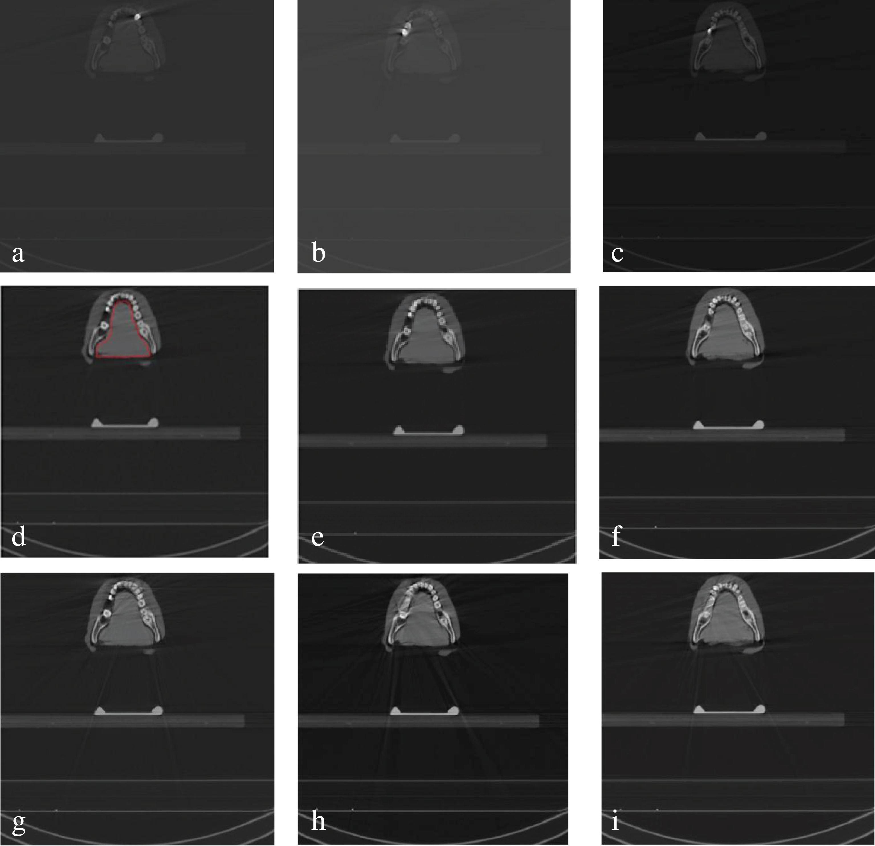
Results of applying proposed method on phantom case. a), b) and c) show three original images of scanned phantom with different metallic inserts. d), e) and f) show the original images of scanned phantom (corresponding to images a), b) and c) respectively) without inserting metallic teeth (served as ground truths). g), h), and i) show the reconstructed images (corresponding to images a), b) and c) respectively) after applying proposed MAR.
Where the original image I and its enhanced version
The same analysis on variances show that before applying MAR method the differences of variance for the uniform region of tongue are 167, 171 and 51 while after applying MAR method, they have been reduced to 19, 37 and 2 for cases 1, 2 and 3 respectively. It shows again that the uniformity of intensity values was more preserved by reducing metal artifacts. In terms of MSE and PSNR, we can see from Table 1 that after applying MAR method, MSE was reduced by 517 and PSNR was augmented by 12 dB in average for three cases which show clearly a reduction of artifacts in the region of tongue.
As we can conclude from Table 1, based on the mentioned metrics, that the proposed MAR method can successfully reduce metal artifacts and produce CT images in the presence of metallic objects as similar as those without metallic objects. In another experiment, we applied the proposed MAR method on two clinical cases. Figure 6 shows the obtained results on a patient with metallic teeth and Fig. 7 shows those on a patient with pelvis prosthesis. In each case, three CT images have been shown. These results show clearly that the streaking artifacts have been reduced and quality of the image has been improved.
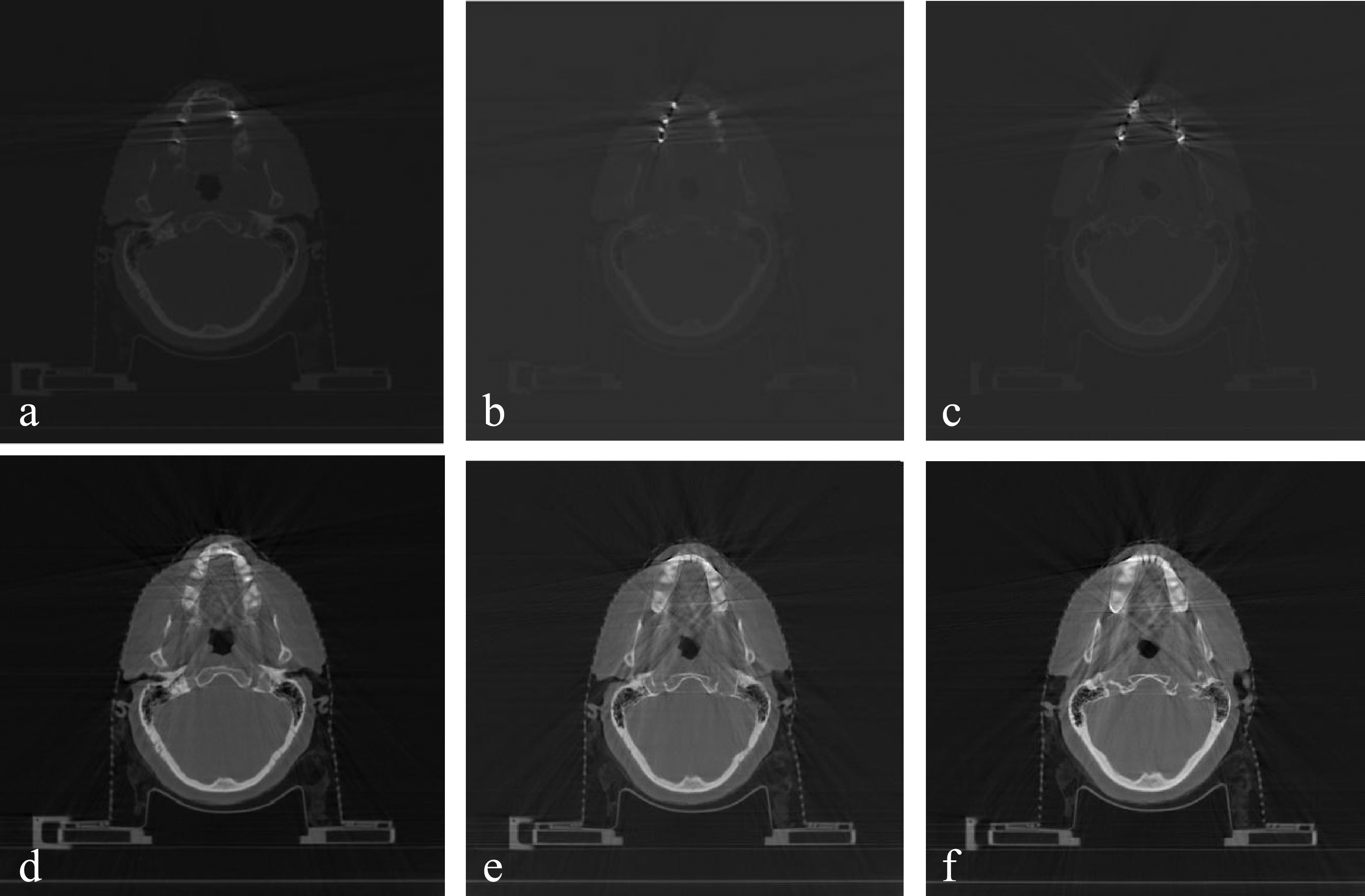
The results of applying our MAR method on a patient with metallic teeth. a), b), and c) show three examples of original CT images. d), e), and f) show their corresponding CT images after applying the proposed method.
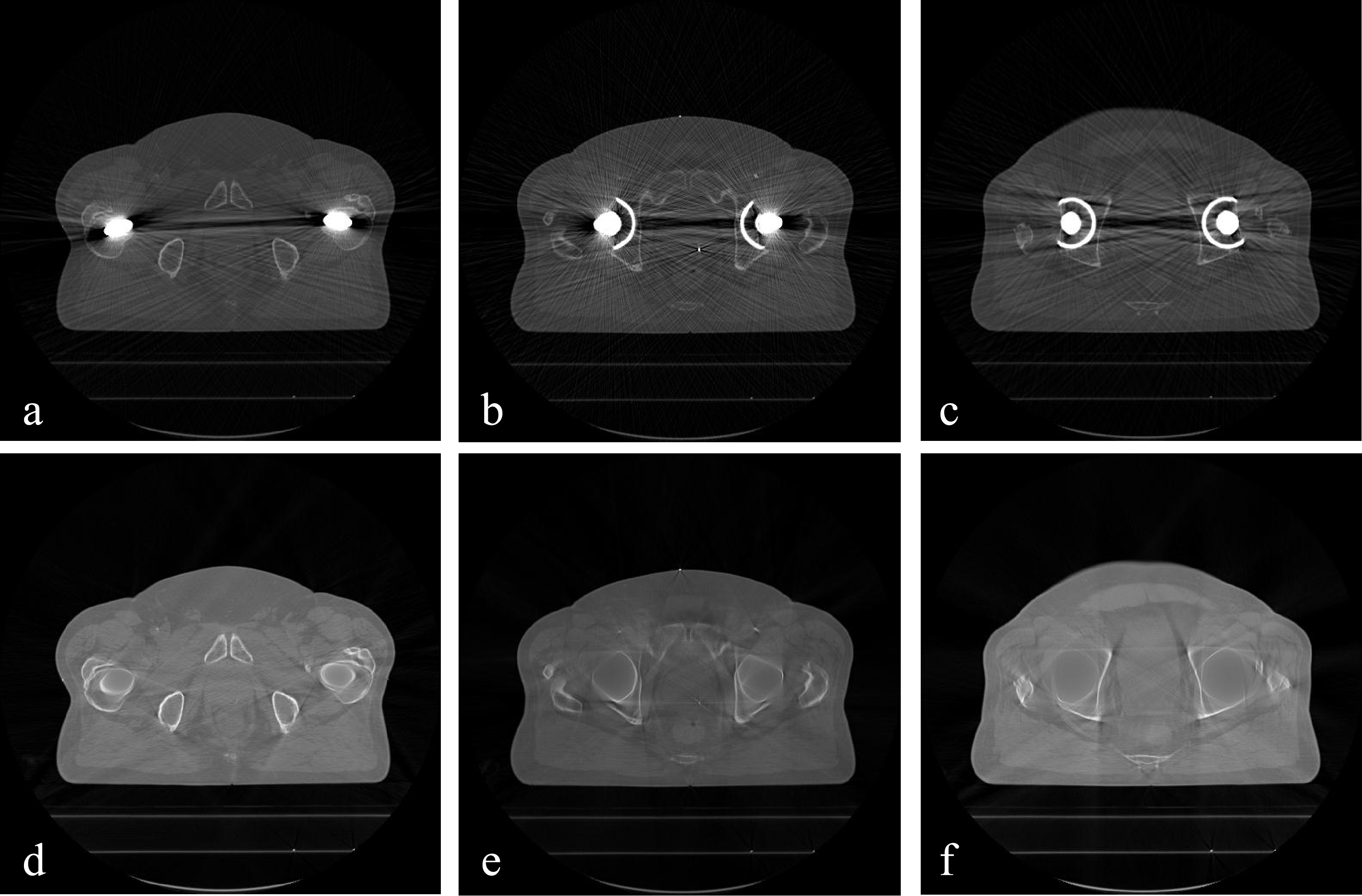
The results of applying our MAR method on a patient with two pelvis prostheses. a), b), and c) show three examples of original CT images. d), e), and f) show their corresponding CT images after applying the proposed method.
Finally, we compare our method with the recent work down in [15] which used an active contour model. Figure 8 shows the results which show the superiority of our method related to other as more streak artifacts are present in the resulted images of other method. The visual improvements for these clinical cases have been also confirmed by a specialist of radiology from Rajaii Hospital in Shiraz, Iran.
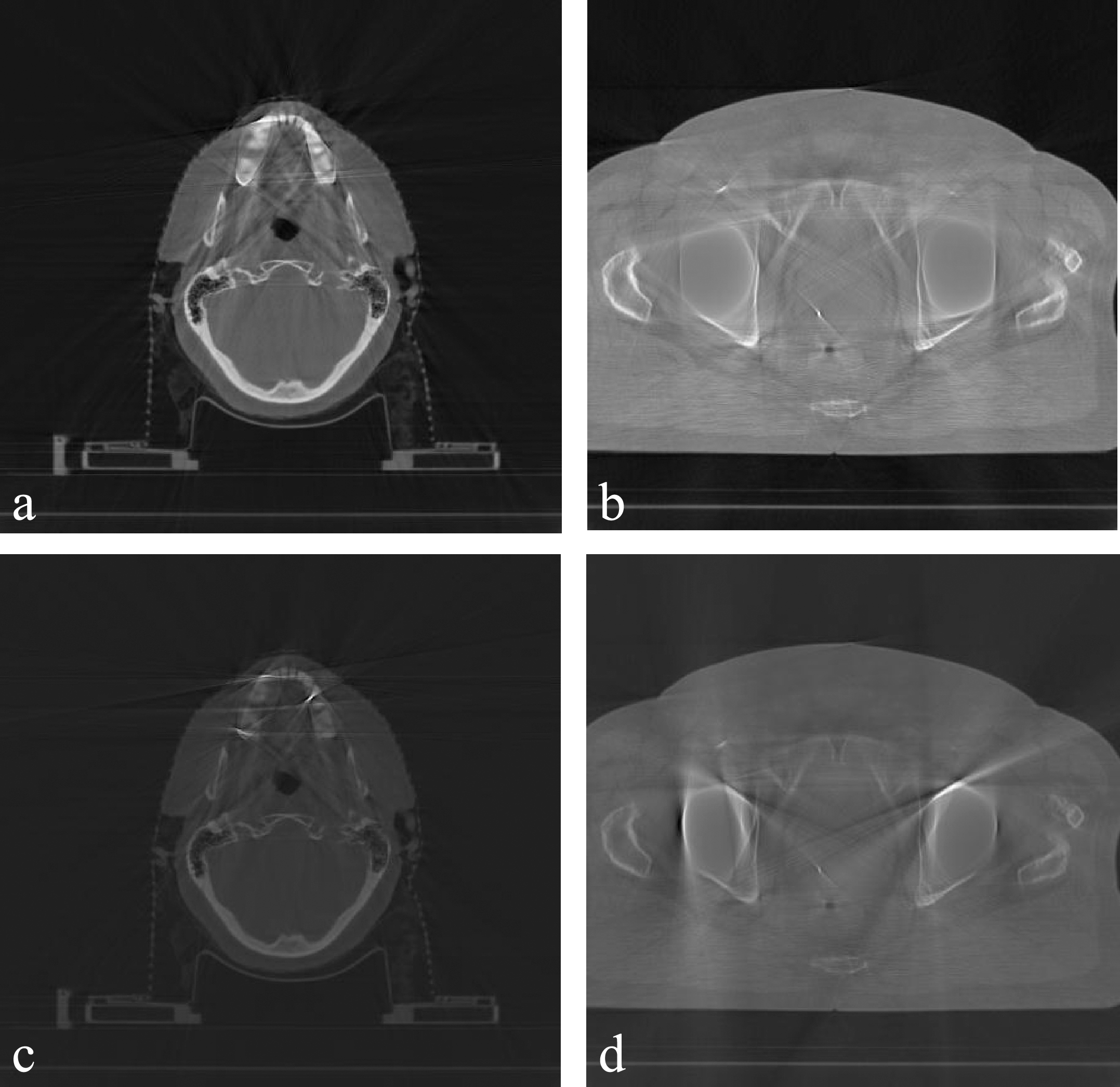
The results of comparing the proposed method with other active contour based method [15]; a) and b) show the results of our method, c) and d) show the results of other method.
An efficient and automated algorithm for metal artifact reduction based on missing projection segmentation was developed which can be applied on both cases of teeth filings and pelvis prosthesis. It is based on segmenting the sinogram, the raw data before CT image reconstruction, using an active contour algorithm to detect the missing projections corresponding to metallic objects. Then a replacing technique is used to assign appropriate values to missing projection. Finally, the modified sinogram is used to reconstruct CT images.
The unique aspect of our work is that we considered the sinogram as an image of intensity inhomogeneity and used an appropriate active contour algorithm (i.e. BFM) capable to handle this concept for efficiently determining the boundary of metallic project regions in the sinogram. Then, Hough transform was also used to accurately detect Sinusoidal-like curves belonging to metallic projections in these regions. The combination of these two strategies gives a more accurate segmentation of sinogram than the previous method based on active contour models tending to rely on intensity homogeneity. To prove the efficiency of the proposed method, in this work we use not only clinical evidences which visually illustrated the superior performance of the proposed method but also provide a phantom case to bring quantitative evaluations and comparisons. The phantom and real studies demonstrated that this algorithm provides a better quality of images by reducing metallic artifacts which is very promising to be used as a routine for such cases.
It should also be mentioned that in some cases (e.g. very close multiple metallic objects) some artifacts may still be present after reconstruction. It is due to the overlap of metallic projections in the sinogram which causes some difficulties to detect and separate these projections. Our future works concern to develop more sophisticated combination of active contour models and other transforms such as Curvelet transform to handle such challenges.
Footnotes
Acknowledgments
The authors would like to thank Prof. Chunming Li for providing the MATLAB codes on his website for implementation of the active contour models, Prof. Luc Beaulieu for his cooperation in carrying out all experiments in this paper and Prof. Ali Reza Shakibafar for his cooperation and judgment.