
Abstract

Encapsulating peritoneal sclerosis is a severe complication of peritoneal dialysis. Progressive sclerotic thickening of the peritoneum results in tethering and cocooning of the bowel, leading to chronic bowel obstruction, malabsorption, malnutrition, and high mortality. Conservative treatment is often unsuccessful and a surgical enterolysis is required for management. Pseudoachalasia is a rare condition that mimics the clinical and radiological features of achalasia of the cardia. Pseudoachalasia is most commonly caused by infiltrating or metastasizing cancers. In this report, we present a case of pseudoachalasia associated with encapsulating peritoneal sclerosis. The clinical symptoms settled after peritonectomy and enterolysis.
Encapsulating peritoneal sclerosis (EPS) is a severe life-threatening complication of peritoneal dialysis (PD). Progressive sclerotic thickening of the peritoneum causes intestinal adhesions, encapsulation, decreased peristaltic activity, and eventual small bowel obstruction, resulting in “chronic gut failure.” This in turn leads to malabsorption and malnutrition. Patients with EPS can present in a number of ways, including early satiety, abdominal bloating, ascites, significant weight loss, chronic sepsis, and subacute or acute bowel obstruction (1). Most patients with advanced EPS have limited oral intake and vomiting is an important feature. Diagnostic features on computed tomographic (CT) scanning include bowel encapsulation due to peritoneal fibrosis and peritoneal calcification (2). Due largely to difficulties with nutrition, established EPS is usually fatal without surgical intervention.
Achalasia of the cardia is a disease characterized by incomplete relaxation of the lower esophageal sphincter and absence of peristalsis of the esophagus. The dysfunction in motility of the esophagus results in dysphagia, recurrent vomiting, and weight loss. In the majority of cases, the etiology is unknown. Degenerative lesions are found in the vagus nerve as well as a decrease in ganglion cells in the myenteric nerve plexus of the esophageal wall (3).
Pseudoachalasia is a disorder indistinguishable from achalasia. The most common cause of pseudoachalasia is primary or metastatic neoplasm infiltrating the myenteric ganglions (4). To our knowledge, EPS has never been reported as presenting with or mimicking pseudoachalasia. We report a case where the primary pathology, EPS, was associated with a clinical and radiological picture of pseudoachalasia.
Patient and Method
A 38-year-old female patient presented with PD peritonitis requiring PD catheter removal after an uneventful PD period of 5 years. Culture of the PD fluid was positive for Stenotrophomonas maltophilia. Her history of kidney disease went back over 10 years, when she presented with nephrotic syndrome. An initial kidney biopsy demonstrated a mesangioproliferative glomerulonephritis. She was started on continuous ambulatory PD. Her medical history also included Crohn's disease, which was limited mainly to the perianal region, a microwave endometrial ablation for menorrhagia, and a parathyroidectomy for tertiary hyperparathyroidism.
Following removal of the PD catheter, she had a persistently raised C-reactive protein (CRP) level of 350 mg/L, despite not growing any organisms in culture of the dialysate. She then developed a couple of abdominal collections, one in the right flank, which was subhepatic and extended to the right iliac fossa, and another smaller one in the left flank. Both collections were initially percutaneously drained and cultured, and were negative for micro-organisms, including acid-fast bacilli. Over the next 3 months, she developed vomiting nearly every day soon after eating and, although she was still able to keep down small amounts of food and pass a small amount of feces every couple of days, she lost about 15 kg in body weight. A CT scan of the abdomen showed the two above-mentioned residual fluid collections but no obvious bowel obstruction. She then underwent a laparotomy during which features of EPS were found: thickened peritoneum and extensive encapsulation of the small bowel with retraction of the bowel loops to the center of her abdomen. No definitive procedure was performed at the time and her abdomen was closed. She was commenced on total parenteral nutrition. She developed signs of sepsis postoperatively. Peripheral blood cultures grew Klebsiella. A repeat abdominal CT showed the previous fluid collections, which had enlarged. Both collections were further percutaneously drained. The fluid grew enterococci in culture. She was then treated with antibiotics and became afebrile after a few days. Further blood cultures grew vancomycin-resistant enterococci. At this stage she was transferred to our center. CT scan of the chest and abdomen was performed. The chest CT showed a widely dilated esophagus, suggesting achalasia (Figures 1 and 2). The abdominal CT was consistent with features of EPS. She was prepared for a definitive procedure of enterolysis and peritonectomy with total parenteral nutrition and daily dialysis. After 1 week of preparation, she was operated on. A total peritonectomy and enterolysis was performed. In addition to excising the thickened parietal and visceral peritoneum in the main abdominal compartment and releasing the small bowel, the thickened peritoneum below the left hemidiaphragm, the anterior surface of the stomach, and the gastroesophageal junction was excised. She made a rapid and uneventful recovery postoperatively and began taking oral feeds 6 days after her surgery. She was also commenced on tapering doses of steroids. Her vomiting subsided after her operation and she started to gain weight. A gastroscopy done after definitive surgery did not show any evidence of achalasia or the esophageal dilatation seen on the CT scan. Subsequently she was transferred back to her base hospital. She has now made an uneventful recovery, came off total parenteral nutrition within 2 weeks of the operation, and regained all the weight she lost during her illness. She remains symptom free from the EPS 18 months after surgery and has had a successful deceased donor kidney transplantation.
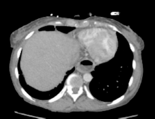
CT image of the dilated esophagus (horizontal view).
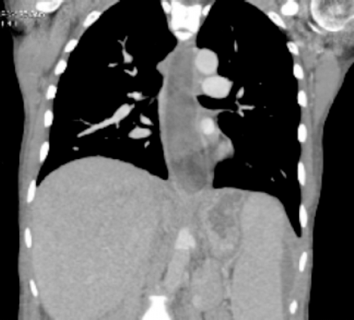
CT image of the dilated esophagus (frontal view).
Discussion
Encapsulating peritoneal sclerosis is a rare life-threatening complication of long-term PD and is characterized by progressive weight loss, abdominal symptoms of early satiety and vomiting associated with a very poor nutritional status, anemia, elevated CRP level, and low serum albumin level. Due to its rarity, there is no accurate estimate of the incidence of the disease. Experience from Japan, which has the largest numbers, suggests a prevalence between 0.54% and 7.3% (5,6). Usually requiring long-term hospital stay and treatment, advanced EPS is a very severe condition, with mortality rates of up to 56% (7). Chronic fibrosis and calcification of the parietal and visceral peritoneum result in severe adhesions, with cocooning and dysmotility of the small bowel. Esophageal dysmotility associated with radiological features have not been described to date in EPS. The optimum management of EPS is still debated and the rarity of the disease and non-standardized diagnosis and reporting makes controlled trials of conservative versus surgical management impossible. Conservative treatment with tamoxifen, steroids, immunosuppression, and parenteral nutrition has been described in very small numbers and with different degrees of success. The largest outcome series describing management of EPS are Japanese and show excellent outcomes with surgery (8). Surgical management aims to release the adhesions and remove the thickened peritoneum and cocoon, restoring the normal peristaltic activity of the small bowel and allowing fluid absorption from the fresh peritoneal surface. In about a third of patients, multiple interventions are necessary due to complications (enteric leaks, hemorrhage, fluid collections, and/or recurrence). Our experience shows that a planned multidisciplinary (surgical, renal, critical care, and dietetic) approach with peritonectomy and enterolysis can be successful.
Pseudoachalasia mimics the clinical and radiological picture of achalasia. About 3% – 4% of all patients diagnosed with achalasia do not suffer from primary motility dysfunction but have secondary motility disturbances caused by primary or metastatic neoplasias or benign tumors and postoperative complications at the level of the cardia. Liu et al. reported 13 cases, of which 8 cases were associated with carcinoma of the esophageal junction, 4 cases with different types of metastatic cancers, and 1 case with mediastinal fibrosis (9). Gockel et al. reviewed the literature and found 264 cases of pseudoachalasia in 122 publications from 1968 to 2002 (4). In 69% of all cases, the cause of achalasia was primary or secondary malignancy; in 13% the cause was benign lesions, in 12% postoperative complications, in 4% diseases of the central nervous system, and in 2% paraneoplastic syndromes. Four types of mechanisms of esophageal motor dysfunction were distinguished:
Circumferential obstruction of the distal esophagus (postoperative achalasia);
Infiltration of the myenteric plexus by tumor cells;
Neuronal degradation distant from the primary tumor site, with reduction in ganglion cells in the dorsal nucleus of the vagal nerve or in the vagal nerve itself;
Interaction of tumor factors with the myenteric plexus of the esophagogastric junction with no direct infiltration (paraneoplastic).
Patients with pseudoachalasia are older and have a shorter duration of dysphagia and more significant weight loss in comparison to patients with primary achalasia. Thus the clinical, radiological, and manometric picture of achalasia and pseudoachalasia are very similar and distinguishing between these two conditions is a challenging clinical problem.
To the best of our knowledge this clinical picture of esophageal dilatation mimicking pseudoachalasia with frequent vomiting has not been described in patients with EPS. The mechanism of presentation in this case remains unclear; however, we postulate that the thickened and sclerotic peritoneum on the abdominal surface of the diaphragm and the lower esophagus caused a mechanical constriction of the lower esophageal sphincter, presenting as pseudoachalasia. This was relieved by surgical excision of the thickened peritoneum leading to symptomatic relief confirmed by endoscopy. With an increasing number of patients with EPS being reported, and with surgical enterolysis, pseudoachalasia should be actively sought and excision of the thickened peritoneal membrane around the lower esophageal sphincter considered if present.
Footnotes
The authors declare that no financial conflict of interest exists.