





Abstract
Being able to drink milk from a bottle with sufficient sucking pressure is the first step in becoming an efficient feeder. Infants generate two types of sucking pressures while feeding from a bottle—compression and suction—by moving their tongue in a continuous peristaltic pattern. At first, the anterior portion of the tongue works together with the lower jaw and elevates to compress the nipple against the palate. The upward movement of the tongue and lower jaw produces a positive pressure called compression. Then suction, a negative intraoral pressure, is formed as the tongue and lower jaw move downward to enlarge the oral cavity. Suction is strengthened through inward movements of the cheeks and a tight lip seal on the nipple (Bosma, Hepburn, Josell, & Baker, 1990; Bu’Lock, Woolridge, & Baum, 1990; Hayashi, Hoashi, & Nara, 1997; Iwayama & Eishima, 1997; Smith, Erenberg, Nowak, & Franken, 1985; Wolf & Glass, 1992).
Preterm infants are often unable to produce sufficient compression and suction to feed efficiently from a bottle, partly because of low muscle tone or immature oral–motor control. Common deficits seen in the tongue movement of preterm infants during feeding include fragmentary peristaltic movements, purposeless nonperistaltic movements, twitching movements, retraction to the back of the mouth, and lack of the typical cupped-shaped tongue posture. Other problems likely to affect feeding efficiency, such as loose lip seal, wide jaw excursion, and inconsistent or arrhythmic jaw movements, are often noted when these infants are fed from a bottle (Bu’Lock et al., 1990; Case-Smith, Cooper, & Scala, 1989; Lau, Alagugurusamy, Schanler, Smith, & Shulman, 2000; Palmer & VandenBerg, 1998; Ross & Browne, 2002; Shaker, 1990).
Cheek and jaw support (oral support) have been widely used by therapists and nurses to enhance the feeding efficiency of preterm infants who are poor feeders. The theoretical basis for this technique derives from a neurodevelopmental approach. Neurodevelopmental theorists have proposed that the stability of a proximal body segment provides the foundation for skilled movement (or action) of a more distal part (Bobath, 1980). Therefore, from a neurodevelopmental perspective, it is believed that the infant is able to perform coordinated, refined tongue movement (skilled distal action) on the foundation of cheek and jaw stability (proximal body segment; Morris & Klein, 2000). On the basis of this theory, forward and inward support on the cheeks—cheek support—during feeding is hypothesized to improve suction by increasing the stability of the cheeks and promoting lip seal (Morris & Klein, 2000; Wolf & Glass, 1992).
Feeding efficiency also is influenced by the anatomical interconnections between tongue and jaw. Unstable or wide jaw movements may interfere with tongue movement, causing an inefficient sucking pattern (Case-Smith et al., 1989; Daniëls, Casaer, Devlieger, & Eggermont, 1986). In this case, provision of jaw support during feeding is believed to help stabilize the infant’s lower jaw, thereby promoting more coordinated tongue movement and better feeding efficiency. Moreover, from a biomechanical view, jaw support may reduce the effort required from the infant to move and sustain the lower jaw action as the infant compresses the nipple during feeding. Provision of jaw support would act as an affordance, that is, provide a stabilizing influence that would change degrees of freedom and therefore improve motor control (Lockman & Thelen, 1993). Therefore, speculation that jaw support may also help preterm infants maintain a more efficient rhythm during feeding exists among neonatal intensive care unit (NICU) practitioners (Hunter, 2005; Morris & Klein, 2000; Wolf & Glass, 1992). Moreover, the tactile stimulation resulting from the cheek support may help infants maintain optimal alertness for feeding (Einarsson-Backes, Dietz, Price, Glass, & Hays, 1994).
Two studies have provided empirical evidence on the immediate effectiveness of oral support on the feeding performance of preterm infants. Einarsson-Backes et al. (1994) investigated the effect of cheek and jaw support on the first 2 min of formula intake of 13 preterm infants between 34 and 40 wk postconceptional age (PCA) identified by nursing staff as poor feeders. The infants had significantly greater formula intake when they were given cheek and jaw support during feeding than when fed without support. A large effect size of .95 shows that the intervention is very effective in increasing the infant’s intake rate in the initial few minutes of feeding (Daley & Kennedy, 2000). Another study included 20 younger preterm infants (32–34 wk PCA) who were able to nipple feed less than half of the prescribed amount of formula in the first 5 min of feeding at the time of the study (Hill, Kurkowski, & Garcia, 2000). The study indicated that compared with the control condition (no support), the infants had less frequent and shorter rest periods during the initial and last 3 min of feeding when support was provided. However, data about feeding efficiency, such as intake rate, feeding duration, and the percentage of the prescribed volume ingested, were not reported in this study.
By contrast, coordinating swallowing and breathing may be more challenging if the volume flowing into the mouth has been increased by oral support (Ross & Browne, 2002; Shaker, 1999). Therefore, one concern is whether oral support provided during feeding may increase coughing and choking and other physiological alterations. The study by Hill et al. (2000) indicated that preterm infants had similar changes in oxygen saturation values, heart rate, and respiratory rate during the initial and last 3 min of feeding with and without oral support. Moreover, after completing the entire feeding, the infants’ oxygen saturation returned to the prefeeding levels sooner in the intervention feeding condition. Thus, the results to date do not support that the intervention leads to more physiological distress. However, more evidence is needed to confirm that oral support is a safe intervention for preterm infants.
A major limitation of the two studies has been the brief feeding period examined (2–3 min). The question of whether oral support provided throughout the entire feeding improves feeding performance remains unclear on the basis of available research. In the current study, we aimed to examine the effects of oral support more thoroughly and for the duration of the entire feeding. The following research questions were addressed: (1) Does oral support enhance feeding performance of preterm infants? (2) Do preterm infants exhibit a more mature sucking pattern, less leakage, longer alertness, and less feeding-related physiological alterations when oral support is provided during the feeding?
Method
Participants
Twenty preterm infants (7 boys, 13 girls) born between 25 and 36 wk of gestation were recruited from the NICU of a medical center in Tainan, Taiwan. Their postmenstrual ages (PMAs) were 32 to 41 wk at the time of the study. The infants weighed between 520 and 1,834 g at birth. Among the infants, 7 were considered small for their gestational age. All participants met the following criteria: (1) born at <37 wk gestation, (2) allowed to take ≥15 ml by mouth per feeding, and (3) inefficient feeders (i.e., unable to consume an average of ≥4 ml of feeding intake per min in a 5-min feeding assessment).1 Infants who had any of the following conditions were excluded: (1) congenital anomalies affecting feeding and digestive functions, (2) chromosomal or genetic problems, (3) medically unstable status (e.g., receiving mechanical ventilation for life support; frequent bradycardia, apnea, or sepsis), and (4) unresolved metabolic problems.
None of the enrolled infants required oxygen supplementation at rest; however, 5 infants still needed oxygen supplementation while feeding. The infants had a history of the following medical conditions: 15 (75%) had respiratory distress syndrome, 6 (30%) had chronic lung disease, 5 (25%) had brain insults (i.e., ultrasound documented Grade III intraventricular hemorrhage, hydrocephalus, periventricular leukomalacia, or meningitis), and 2 (10%) had clinically confirmed necrotizing enterocolitis. Other birth and medical conditions of the infants are described in Table 1. The study was approved by the research ethics committees of both the medical center and Sargent College, Boston University. Informed written consents were obtained from one parent of each infant before data collection.
Characteristics of Enrolled Infants
Note. N = 20 (7 boys, 13 girls). M = mean; SD = standard deviation.
Oral Support Intervention
Oral support was administered in the following manner. The first author (Hwang) held the infant’s cheeks inward and forward by placing her right ring finger (i.e., the hand holding the bottle) on the infant’s left cheek and the thumb of the other hand (i.e., the hand supporting the infant’s head) on the infant’s opposite cheek to assist the infant in sealing the lips around the nipple. Simultaneously, Hwang placed her right little finger under the infant’s chin to stabilize the lower jaw (see Figure 1).
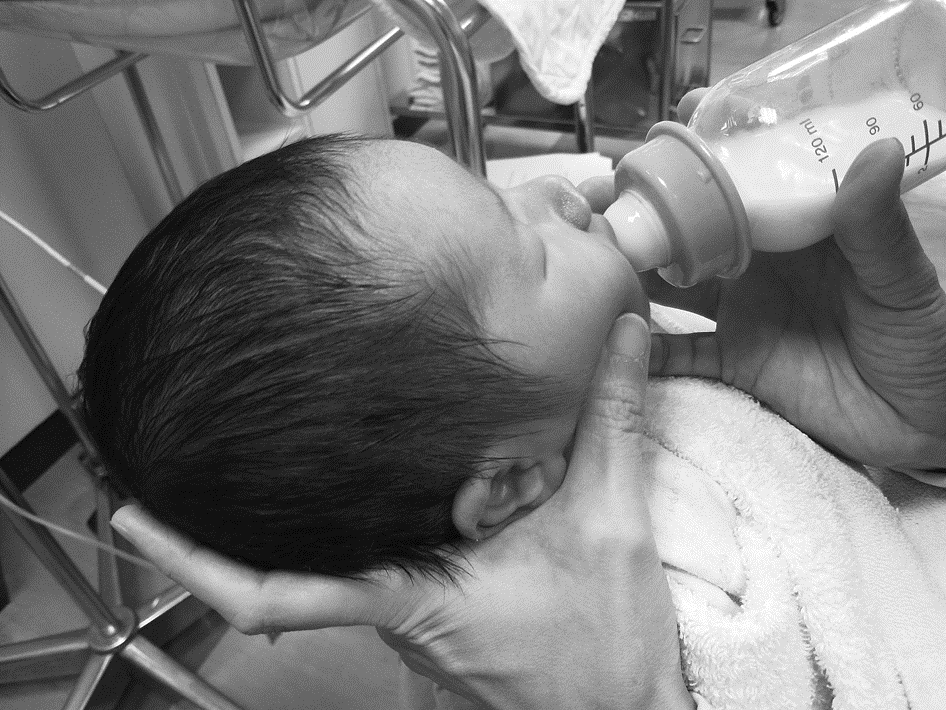
Oral support intervention.
Outcome Measures
Feeding Parameters.
The following indicators of feeding performance were measured for analysis: (1) feeding duration in minutes, (2) percentage of volume ingested, (3) percentage of leakage, (4) intake rate (ml/min), (5) sucking frequency (sucks/min), and (6) mean volume ingested per suck (ml/suck). A description of how these variables were measured and calculated for this study is included in Table 2 and in the Procedure section.
Definition and Measurement of Feeding Parameters
The measurement of sucking frequency was based on a modification of the method used in the study by Rybski, Almli, Gisel, Powers, and Maurer (1984). Two trained research assistants blinded to the hypotheses of the study coded the number of sucks from the initial 5-min clips of the videotaped feedings. Before beginning the coding work, a 1-min feeding session of each infant was randomly selected to examine the reliability of the two coders. Intercoder reliability was .98 (Coders A and B), whereas intracoder reliability was .99 (Coder A), and .96 (Coder B), using the intraclass correlation coefficient tests as the index of reliability.
Alertness Parameters.
The infants’ level of alertness was determined on the basis of a modification of the behavioral state definitions of the Neonatal Behavioral Assessment Scale (Brazelton & Nugent, 1995). Six states—deep sleep (State 1), light sleep (State 2), drowsy (State 3), quiet alert (State 4), active awake (State 5), and crying (State 6)—were used to determine the infants’ alertness. One trained research assistant blinded to the hypotheses of the study coded the highest level of alertness achieved by each infant during a 30-s block immediately before the feeding and at the end of the 3rd and 5th minute of the feeding. Intercoder agreement reliability with Hwang (r = .92) and intracoder reliability (r = .96) were established using the intraclass correlation coefficient test on 20 observations randomly selected from among enrolled infants.
Physiological Parameters.
Physiological changes in saturation of peripheral oxygen (SpO2) and pulse rate (PR) were measured with a MARS pulse oximeter (Novametrix Medical Systems, Wallingford, CT) designed to reduce the interference of motion or other artifacts on SpO2 levels and PR. Use of the pulse oximeter is based on the principle that oxygenated hemoglobin and reduced hemoglobin (i.e., oxygen-free hemoglobin) have different spectra of light absorption. Oxygenated hemoglobin absorbs more light in the infrared band (which ranges from 850 to 1,000 nm), whereas reduced hemoglobin absorbs more light in the red band (which ranges from 600 to 750 nm; Poets & Southall, 1994). A Y-sensor consisting of a light-emitting diode (LED) source and a photodiode was attached to one foot of each infant. The LED emits the light energy from red (660 nm) and infrared (940 nm) beams to the photodiode on the opposite side of the LED, passing through a pulsating arterial bed. The light energy not absorbed by the pulsating arterial bed is sent to the pulse oximeter by the photodiode, where it is digitized and processed into a functional saturation score2 (Levesque, Pollack, Griffin, & Nielsen, 2000; Thilo, Andersen, Wasserstein, Schmidt, & Luckey, 1993).
Procedure
A crossover design was used in which participants served as their own controls. Each infant was fed each day under two feeding conditions: (1) with oral support (intervention) and (2) without oral support (control). The procedure for the intervention feeding was the same as for the control feeding except that oral support was provided only during the intervention feeding. The order of the conditions was reversed for each infant between Day 1 and Day 2 to prevent possible confounding effects. The order of the conditions was also randomly assigned to every other infant by tossing a coin; that is, when an infant was randomly assigned to the intervention condition first, the next infant was first given the control condition. Conditions were randomized again for the subsequent infant, and so on. This strategy resulted in four feedings total for each infant, two conditions per day on two consecutive days.
Because routine nursing care was being administered by the infant’s assigned nurse before feeding, the research assistant weighed the bottle filled with the prescribed feeding volume and the tissue used to collect any leakage. After completion of routine care, the infant was held in a semiupright position with neck and head support provided, which is the typical holding pattern used for feeding infants at this institution. Physiological data recording (SpO2 and PR) began with a 2-min baseline period before feeding and continued throughout the entire feeding. Infants were videotaped simultaneously with the recording of physiological data. An audible sound was made by marking a relevant event on the oximeter to subsequently align videotaped feeding behavior and physiological data for analysis. No stimulation was applied to infants during the baseline period. A few strategies including hand swaddle or patting infant’s body were used to console the infants who cried.
At the end of the baseline period, Hwang placed the tissue underneath the infant’s chin and then initiated the feeding using the typical preterm infant bottle and nipple of the NICU. Eight infants were fed at their bedside, and the others were fed inside the isolette as per nursing indications. Oxygen supplementation was provided to infants who needed it during their feeding, according to the prescription of their assigned physician. If an infant coughed, choked, or had significant physiological changes (e.g., SpO2 < 80%, PR > 180 beats/min or < 100 beats/min, cyanosis) at any time during the feeding, the nipple was pulled out of the mouth immediately. The feeding was stopped momentarily until the infant recovered.
At the end of the initial 5-min feeding period, the feeding was stopped to weigh the bottle and tissue to measure oral intake and leakage for this period. The feeding was restarted immediately after taking the measurement. When the infant paused >1 min and presented no stress signs, a variety of common strategies, including burping, slowly pulling out and reinserting the nipple, or gently moving the infant, were used to encourage resumption of sucking as needed. A 30-min limit was imposed for the duration of each feeding session in compliance with common practice. Feeding was discontinued before the 30-min limit in the following two situations: (1) the infant finished the prescribed volume in <30 min and (2) the infant did not resume sucking after at least two sucking facilitation strategies were used. After the end of the feeding, the research assistant weighed the tissue and the bottle with any remaining volume to determine the amount of total leakage and the total feeding intake. Any unfinished volume remaining at the end of the feeding was given by means of a nasogastric or orogastric tube by the infant’s assigned nurse.
The actual amount ingested in the feeding was computed by subtracting the amount leaked from the amount extracted from the bottle for the same period of time. The weight was then converted to volume (ml; pilot testing revealed that 1 ml of formula or milk weighs approximately 1 g).
Data Analysis
The data from the same condition (intervention or control) on Day 1 and Day 2 were pooled for analysis because no significant differences were found in the data between the same two feeding conditions over the 2 days. Performance on the measured feeding parameters (i.e., feeding duration, percentage of volume ingested, percentage of leakage, intake rate, sucking frequency, mean volume ingested per suck) across the two feeding conditions was compared using paired t tests.
Previous studies have suggested that preterm infants demonstrate the strongest engagement in sucking and greater physiological alterations from baseline in the first few minutes of feeding (Mathew, 1991; Shivpuri, Martin, Carlo, & Fanaroff, 1983). Thus, statistical analyses on the variables were run only for the initial 5-min segment. Two-way repeated-measures analyses of variance (ANOVA; Condition × Time) were conducted to determine the influence of the condition on the changes in SpO2 and PR from baseline across time in the first 5 min of the feeding. ANOVA analyses were followed by Bonferroni tests to identify the paired comparisons that were significantly different. Sign tests were used to compare the level of alertness before and after feeding across the two feeding conditions on Day 1 and Day 2. The effectiveness of the intervention was further examined through post hoc effect size calculations for each feeding parameter, using Cohen’s d (Portney & Watkins, 2000).
Results
The feeding performance of the infants in the two feeding conditions is presented in Table 3. The prescribed volume ranged from 14 to 50 ml. In the initial 5-min feeding period, the intake rate was significantly higher during the intervention condition than during the control condition (t = 2.13, df = 19, p = .046, d = 0.68). The percentage of leakage for the initial period in the intervention condition was also lower than in the control condition (t = –2.2, df = 19, p = .040, d = 0.79). Over the entire feeding session, feeding duration was shorter for the intervention condition (t = –2.15, df = 19, p = .044, d = 0.68). Intake rate for the entire feeding session was also higher during the intervention condition (t = 2.47, df = 19, p = .023, d = 0.78), but no significant differences between the two conditions were found in the percentage of the prescribed volume consumed (t = 1.70, df = 19, p = .11, d = 0.55) or the percentage of leakage (t = 0.20, df = 19, p = .84, d = 0.06).
The quality of the videotape recordings, along with limited amplitude of the sucking movements, made it difficult to correctly code the sucking activity of 3 infants. Therefore, their sucking performance was excluded from the final sucking analyses. The 3 excluded infants were older at birth (28.0 ± 2.2 vs. 32.2 ± 4.9 wk of gestation; p = .02) and at the time of study (35.7 ± 2.0 vs. 38.3 ± 0.9 wk of gestation; p = .046) than the analyzed infants. An analysis of the sucking data for the remaining 17 infants indicated that differences in sucking frequency (t = –0.61, df = 16, p = .55, d = 0.20) and volume ingested per suck (t = 1.46, p = .17, df = 16, d = 0.50) across the two conditions did not reach statistical significance.
Feeding Performance of Infants in Both Feedings, With and Without Support (N = 20)
Note. M = mean; SD = standard deviation.
N = 17.
p < .05.
Because of technical problems, the physiological data for 1 infant were lost. The data analyses of the remaining 19 infants indicated that oral support did not have a significant impact on the infants’ SpO2 levels (p = .16) or PR (p = .58) in the initial 5 min of feeding. The only significant finding was that infants’ physiological status changed with feeding time (SpO2: p < .001; PR: p = .002). There was no interaction effect between oral support and feeding time on either of the physiological parameters (SpO2: p = .13; PR: p = .79). The change from baseline to the 1st min of feeding significantly differed from that to the 3rd min of feeding (SpO2 levels: p = .026; PR: p = .029). The result meant that there was a small initial decline in SpO2, which rapidly returned to baseline after the first few minutes of the feeding. By contrast, PR increased slightly from baseline to the 1st min but subsequently declined to baseline throughout the rest of the feeding (see Table 4).
Physiological Changes in the Initial 5 Min of Feeding
Note. N = 19. SpO2 = saturation of peripheral oxygen; bpm = beats per minute; M = mean; SD = standard deviation.
Most infants were in the drowsy or quiet alert states at each observed time point, on both Day 1 and Day 2. No significant effects of oral support on the infants’ level of alertness during feeding were found.
Discussion
The results of the current study indicated that oral support may improve the feeding performance of preterm infants by increasing intake rate and decreasing leakage during feeding. A post hoc effect-size calculation revealed that oral support had a moderate effect on intake rate during the initial 5 min as well as throughout the entire feeding. Consistent with the findings of an earlier study conducted by Einarsson-Backes and colleagues (1994), the current results provide empirical evidence to substantiate the effectiveness of oral support to improve the feeding performance of inefficient feeders born prematurely, at least for some of the variables studied. In addition, a recent study demonstrated that preterm infants who were fed with oral support twice a day during the transition to full oral feeding displayed superior sucking ability (e.g., higher nonnutritive sucking pressure and sucking activity) and better oral feeding progress (e.g., more daily bottle feedings, greater daily formula intake, and shorter transition time to full oral feeding) than the control infants who did not receive support during feeding (Boiron, Da Nobrega, Roux, Henrot, & Saliba, 2007).
Several neurodevelopmental and biomechanical mechanisms have been proposed to explain how oral support might benefit an infant’s feeding efficiency. Possible mechanisms include promoting lip seal on the nipple to decrease leakage, increasing cheek and jaw stability to strengthen suction and compression, and assisting the upward movement of the jaw to decrease the effort required by the infant to suck from a bottle. Under these assumptions, infants receiving oral support during feeding would have been expected to have less leakage from the sides of the mouth, greater intake per suck, and higher sucking frequency. Contrary to our expectations, oral support did not appear to improve the infants’ feeding performance in terms of increasing sucking frequency or volume ingested per suck, although it did decrease the percentage of leakage.
Because of methodological limitations, the sucking data of only 17 infants could be included in the final analyses. The lack of significant effects in sucking variables may have resulted from low power (i.e., a small sample size) rather than true absence of an effect. The moderate effect size (ES = 0.5) for the mean volume ingested per suck suggests that the intervention may in fact be improving this aspect of feeding efficiency but that the difference did not reach significance possibly because of the small sample size. Whether a significant difference in sucking efficiency could be obtained in a larger sample remains inconclusive. The 3 excluded infants were older than the rest of the infants either at birth or at the time of study. How the characteristics of infants affect the impact of oral support on sucking ability also needs further investigation. Another possibility is that the instruments used in this study were not sufficiently sensitive to adequately measure the intervention effect on sucking efficiency. The use of more sensitive instruments to measure the infant’s oral–motor function, such as real-time techniques to observe tongue movement (Miller & Kang, 2007) or more sophisticated sucking apparatus (e.g., a suckometer to measure sucking components such as negative and positive sucking pressure and the number of sucks per burst; Lau, Sheena, Shulman, & Schanler, 1997), would help clarify the mechanisms that explain how oral support enhances the feeding efficiency of preterm infants.
Consistent with the finding of Hill et al. (2000), our study indicated that physiological changes from baseline in SpO2 and PR are no different when infants are given oral support during the first 5 min of a feeding than when fed without support. These findings support the use of oral support as a safe feeding strategy to foster preterm infants’ feeding efficiency.
Previous research suggested that frequent sucking and swallowing during feeding could cause an obvious decrease in breathing rate among preterm infants, especially in the initial continuous sucking feeding phase, potentially compromising oxygenation (Shivpuri et al., 1983). A similar trend was shown in our study; however, the decrease in SpO2 levels at the initiation of the feeding observed in our study was smaller than in other research (Marinelli, Burke, & Dodd, 2001; Shiao, Youngblut, Anderson, DiFiore, & Martin, 1995). All infants included in our study were inefficient feeders, whereas the infants enrolled in other studies were typical preterm infants who may not have had feeding problems. A lower sucking frequency was found in the current study than in previous studies (Bromiker et al., 2005; Lau, Smith, & Schanler, 2003; Shiao et al., 1995). Thus, we speculate that the smaller change in SpO2 in the initial 5 min of feeding found in the current study may be attributed to fewer sucks and swallows. A similar explanation may account for our PR findings.
Limitations
A major limitation of the current study was that it was impossible for the person administering the feedings (Hwang) and the person or people coding the videotapes to be blind to the study condition. Although the established feeding procedure was followed stringently, unintentional bias in the handling of the infants is possible on the part of the person feeding them. The research assistants who coded the videotapes were masked to the hypotheses of the study; however, it was impossible to blind them to the conditions of the study, which could contribute to unintentional bias in coding, as well.
A second limitation of this study was related to methodological issues. When infants were fed with oral support, the experimenter’s fingers situated on the cheeks for support often occluded the view of the infant’s jaw movement, thus hampering accurate coding of the infant’s sucking from the videotapes. This factor, as well as the poor quality of some of the video recordings, prevented the analysis of the sucking data from 3 infants. The smaller sample may not have given the necessary power to detect any statistical significance. In addition, to capture a clear picture of the infants’ sucking behavior, the video camera was positioned to focus on the infants’ face and upper part of the body. Behavioral states were identified on the basis of the characteristics or movements of the babies’ eyes, face, head, and upper trunk. The limited view of the infants’ body movement on the videotapes may have restricted the coding accuracy of their alertness levels.
Another limitation may have been the bottle and nipple used for this study. The bottle used in our NICU allows small amounts of the feeding to drop into the mouth without requiring the infant to suck, because of a net hydrostatic pressure generated by the volume of the formula or milk inside the bottle (al-Sayed, Schrank, & Thach, 1994; Jain, Sivieri, Abbasi, & Bhutani, 1987). This situation is more prevalent in the initial few minutes of the feeding. Therefore, the effect of oral support on the volume ingested may have been confounded by spontaneous dripping of the nipple or flow rate.
Factors such as individual variations (e.g., provision of nutritive additives, type of feeding [milk or formula]) in each feeding and environmental factors (e.g., light, noise) were also difficult to control in this study, thus potentially limiting the identification of significant differences. Moreover, only two feeding observations for each intervention and control condition in this study may not have adequately represented the infants’ typical performance. Increasing sample size or the number of feeding observations may help tease out such confounding factors.
Conclusion and Clinical Implications
Consistent with previous studies, our study results revealed that providing oral support during feeding enhanced the feeding performance of preterm infants in terms of increased rate of intake and decreased leakage without increasing additional physiological distress. Hence, our data provide additional evidence for the adoption of oral support as a regular feeding strategy for inefficient preterm feeders in the NICU. This investigation failed to clearly identify mechanisms that may explain the effectiveness of oral support interventions in promoting feeding efficiency. Decreasing the amount of leakage during feeding through the help of oral support may be a contributing factor toward improved feeding efficiency for preterm infants with feeding difficulties. Methodological problems limited our ability to determine whether sucking ability could be improved by oral support. Further research with more reliable and objective measurements, particularly of the sucking parameters (e.g., sucking pressure, tongue movement), is needed to understand the mechanisms that underlie the benefits of oral support as a feeding intervention for NICU infants.
Footnotes
Acknowledgments
We thank the staff of the Neonatal Intensive Care Unit at National Cheng Kung University for their assistance with data collection. Funding was supported by the grant from the Premature Baby Foundation of Taiwan.
1
The criterion to determine whether infants were inefficient feeders was established on the basis of the results of a previous study conducted by the first author (Hwang). In that study, the mean formula intake of the 16 infants (34 to 41 wks PMA) during the initial 5 min of the feeding was 22.8 ml (standard deviation = 8.0).
2