





Abstract
Proximal humerus fractures are a common orthopedic injury affecting the older adult population and can have a considerable impact on all functional activities. The proximal humerus fracture is universally referred to as an osteoporotic fracture and can lead to extreme pain and high levels of deformity; incidences can vary to as high as 221 per 100,000 population (Handoll et al., 2015). This injury can severely affect functional independence because pain, decreased range of motion (ROM), and decreased strength affect the entire upper extremity when engaging in occupations. Clients in the acute stage will require varying degrees of assistance with almost all activities of daily living (ADLs).
Four-part proximal humerus fractures will have fracture lines along the humeral head, greater tuberosity, lesser tuberosity, and humeral shaft; three out of four parts will be displaced with respect to the fourth (Den Hartog et al., 2010). Surgery is the treatment of choice, but a client may decide to proceed without surgery. This decision is usually due to surgical contraindication, which is more prevalent in the older adult population (Twiss, 2015; Ward & Zuckerman, 2014). Limited published research exists regarding rehabilitation and prognosis for this nonoperative management subpopulation (Table 1; Boons, Goosen, van Grinsven, van Susante, & van Loon, 2012; Fjalestad & Hole, 2014; Launonen et al., 2012; Zyto, Ahrengart, Sperber, & Törnkvist, 1997).
Studies Referencing a Rehabilitation Protocol
Note. AROM = active range of motion; PROM = passive range of motion; ROM = range of motion.
Current evidence on immobilization and rehabilitation for one- and two-part fractures is positive, with 77%–88% of patients having good to excellent results based on ROM (Jordan & Modi, 2014). In contrast, researchers report a notable decrease in quality of life when comparing nonsurgical with surgical management of three-part proximal humerus fractures (Olerud, Ahrengart, Ponzer, Saving, & Tidermark, 2011). Limited evidence on conservative rehabilitation of proximal humerus fractures is available. In this study, we investigated whether nonsurgical outpatient occupational therapy interventions produced meaningful positive changes in functional ability levels for a person with an acute four-part proximal humerus fracture.
Method
Participant
The client was a 67-yr-old male retired accountant; he sustained a mechanical fall resulting in a four-part proximal humerus fracture to his dominant upper extremity. Because of a complex cardiovascular history, he decided to manage this fracture nonoperatively with occupational therapy. At evaluation he required moderate assistance with dressing and bathing and minimum assistance with grooming; he was also unable to use the computer efficiently or cook, which were his preferred occupations. We completed research using a descriptive and exploratory case study design (Portney & Watkins, 2008).
Outcome Measures
The client’s joint ROM measurements were taken for the shoulder and elbow and were recorded actively and passively. The client’s shoulder manual muscle score was recorded for all motions. We completed muscle grading using a numeric 5-point scale. Formal evaluation was completed at evaluation, at 3 mo, at 5 mo, and at a 12-mo follow-up with ongoing reassessment throughout treatment as is customary with this population.
The QuickDASH (Disability of the Arm Shoulder and Hand; Sorensen, Howard, Tan, Ketchersid, & Calfee, 2016) was used to assess the client’s self-reported functional status. It is scored as a percentage, with a clinically significant change being at least 14%. The Numeric Rating Scale for Pain (NRS; Farrar, Young, LaMoreaux, Werth, & Poole, 2001) was used to assess the client’s pain. A reduction in pain intensity of 2 points on the NRS was considered clinically significant.
Procedure and Intervention
A literature review was completed with the assistance of a biomedical librarian to identify studies with reference to rehabilitation after four-part proximal humerus fractures. More than 300 articles were reviewed, and 4 articles met the criteria. These articles provided an immobilization period and time frame dependent protocol for four-part proximal humerus fractures (Boons et al., 2012; Fjalestad & Hole, 2014; Launonen et al., 2012; Zyto et al., 1997). Trends in the literature regarding all types of proximal humerus fractures suggest that decreased immobilization time and decreased time between fracture and the start of rehabilitation correlate with positive results (Fu, Xia, Li, & Wu, 2014; Mafi, Khan, Mafi, & Hindocha, 2014).
The rehabilitation protocols in these studies were analyzed and used in conjunction with this case study to create an occupation-based rehabilitation protocol based on biomechanical principles and preparatory methods (Table 2). Precautions listed are to prevent displacement of the fracture. Treatment was broken up into four distinct phases based on precautions and recovery. In addition to therapeutic exercises, recommended functional activities are listed. In Phase 1, the client started performing: eating, brushing teeth, washing face, and assistance with lower extremity dressing. He was instructed to use one-hand compensation techniques for any activity involving shoulder motion. The sling was used at all other times during Phase 1.
Rehabilitation Protocol for Four-Part Proximal Humerus Fracture
Note. AAROM = active assistive range of motion; ABD = abduction; ADLs = activities of daily living; AROM = active range of motion; ER = external rotation; FF = forward flexion; HEP = home exercise plan; IADLs = instrumental activities of daily living; IR = internal rotation; PROM = passive range of motion; ROM = range of motion.
In Phase 2, the client progressed from passive to active ROM and started isometrics exercises. Isometrics allow strengthening without changing the joint angle or muscle length. The clinician should manually assist the glide of the scapula with functional reaching activities such as removing and storing dishes from the dishwasher; this activity can be recreated in the clinic with light cones. The recommended functional activities added in this phase are all dressing and bathing, with the exception of hand behind back motions. The client in this case study was encouraged to return to the kitchen in a light capacity with no heavy lifting. To address scapular and glenohumeral mobility with computer use, we instructed the client to decrease mouse sensitivity to increase the need to incorporate shoulder ROM with mouse use. In Phase 2, the sling was used only during community mobility, and one-hand compensation techniques were discontinued.
In Phase 3, the motion restrictions were lifted, and use of weights was limited to 2–5 lb. The clinician addressed ROM limitations with end-range stretching. Diagonal movement patterns were initiated and incorporated into functional activity such as dressing, cooking, and cleaning. The client was encouraged to return to all daily and leisure activities, within weight limitations. Bathing and behind back hygiene will often be problematic when initiating these movements. Light gravity-assisted, internal-rotation stretching using the clinician’s leg for leverage (Figure 1A) and a towel behind the back for an internal rotation stretch (Figure 1B) as part of the home exercise plan were used in this phase.
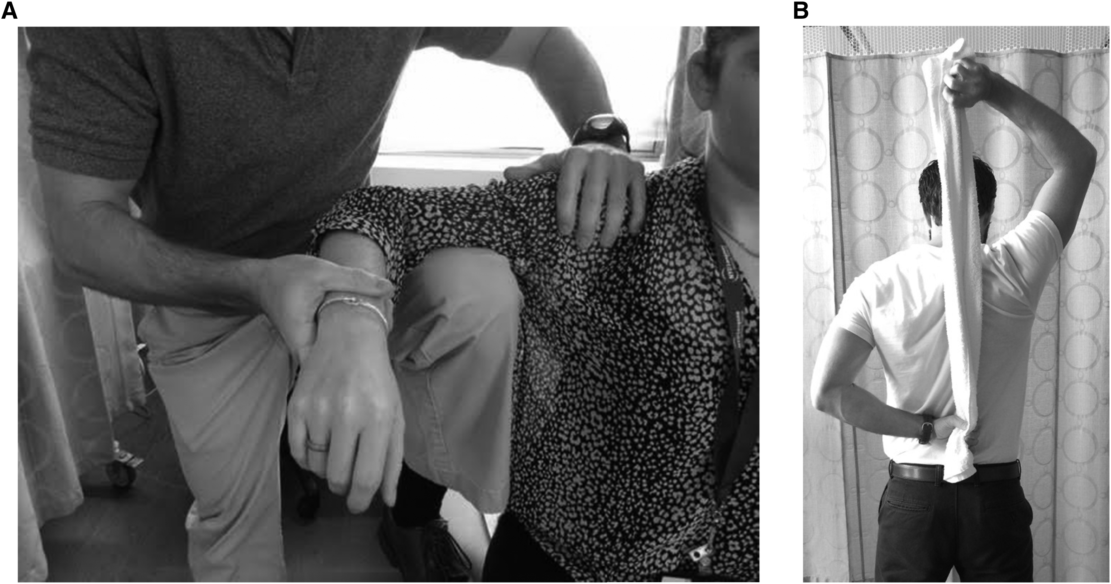
Passive internal rotation stretches (A) with clinician standing behind client using lower extremity and gravity assistance and (B) using a towel for leverage.
In Phase 4, the client had no strengthening precautions. In preparation for returning to overhead item retrieval for cooking, this client performed a combination of bicep curls and shoulder presses with eccentric lowering to simulate kitchen activity. Eccentric exercises allow the muscles to lengthen while producing force that simulates everyday activity. He also performed item retrieval and storage in diagonal movement patterns with progressive weights. Clients will be discharged without full ROM or strength because achieving a full return is unexpected; therefore, education on expectations should begin early in the rehabilitation process. Home exercises at discharge should continue to address stretching, particularly internal rotation (hand behind back), strengthening, and functional and diagonal movement patterns based on the client’s goals and prior level of function.
Results
The client was discharged from therapy without full ROM. At a 12-mo follow-up, the client reported moderate compliance, indicating approximately 50% compliance with the structured home exercise plan after discharge. He demonstrated a decline in active shoulder motion—most notably external rotation and abduction (Table 3). Manual muscle grades become functional at 3 mo, and he maintained strength throughout the 12-mo follow-up. The client reported 81.9% disability at evaluation and 36% disability at the 12-mo follow-up, indicating a 45.9% improvement using the QuickDASH. The client reported a decrease in pain from 7/10 to 3/10. This client demonstrated a gradual increase in independence from the start of care. He was able to complete all ADLs with modified independence within 2 wk and was able to return to his preferred occupations within 4 wk. He was highly satisfied with his outcomes because he was able to achieve his functional goals. He reported a full return to all ADLs and cooking, using the noninvolved upper extremity to reach for heavy items overhead, and a full return to leisure computer use.
Client Measurements
Note. MMT = manual muscle testing; NT = not tested secondary to precautions.
Discussion
Proximal humerus fractures are a common injury in the older adult population. Although not as numerous as other proximal humerus fractures, four-part proximal humerus fractures present as a challenging and complex clinical condition. This data collection, protocol development, and case study can be used by a clinician to properly treat, set goals, and manage expectations throughout rehabilitation.
Examination of the literature on conservative management of proximal humerus fractures demonstrates a lack of consistency. Variations in immobilization and initiation of ROM and strength were observed and outlined in Table 1. In construction of this protocol (Table 2), studies of four-part fractures were taken into consideration along with concepts in rehabilitation of one-, two-, and three-part proximal humerus fractures (Den Hartog et al., 2010; Nouraei, Majd, & Zamani, 2014). This protocol can act as an outline of milestone goals and functional usage for a clinician to incorporate into treatment plan development. As with all clients, multiple factors must be taken into consideration when designing and implementing a treatment plan to target client-specific goals. Continuous communication between the therapist and referring physician is also recommended because client progression can vary significantly.
According to Namdari et al. (2012), functional ROM of the shoulder is at least 121° of flexion, 46° of extension, 128° of abduction, 59° of external rotation, and 102° of internal rotation. This client’s shoulder flexion, abduction, and external and internal rotation would not be considered functional, but he reported a clinically considerable improvement according to the QuickDASH and NRS for pain. These improvements correlated with his self-reported full return to ADLs, cooking, and computer use. Measurements collected can aid a clinician in managing client expectations and developing appropriate goals. This particular client demonstrated a decrease in motion for flexion, extension, abduction, and external rotation between discharge and the 12-mo follow-up. This result may have been attributed to moderate compliance with the home exercise plan. It would be useful to see whether regression is a common trend within this population.
Limitations of this study include the case study design, which affects the generalizability of these findings to other clients. In addition, the unblinded nature of the data collection limits the scope of the data collected. This protocol, created by the authors, may guide future research in the form of randomized controlled trials to analyze this protocol. This research would be useful to determine timing for immobilization, functional goals, ROM progression, and strengthening.
Implications for Occupational Therapy Practice
The findings of this study have the following implications for occupational therapy practice:
A developed rehabilitation protocol could assist occupational therapy practitioners in treatment planning and appropriate occupational therapy goal setting.
A developed upper extremity rehabilitation protocol may improve occupational performance in this population.
Conclusion
Four-part proximal humerus fractures can be managed conservatively if the client is not a surgical candidate or refuses surgery. Client and clinician expectations and goals should be limited because this population will not regain full ROM because of the malunion of the fracture preventing proper anatomical alignment. Clients can experience considerable improvements in self-reported function and pain. Treatment planning should follow an outline of functional milestones but is a client-specific process. Continuous communication between the clinician and referring physician is recommended.