






Abstract
Purpose
To describe fundus autofluorescence (FAF) patterns in patients with type 2A idiopathic juxtafoveolar retinal telangiectasia (IJRT).
Methods
We reviewed FAF images, color photographs, and fluorescein angiography (FA) images of 30 eyes from 16 patients with type 2A IJRT. Eyes with presence of subretinal neovascularization or any other retinal pathology were excluded. All the imaging modalities were obtained with a Heidelberg Retina Angiograph (HRA) confocal laser scanning system.
Results
The mean age of the patients was 50.7 years, 68.5% female. At baseline, the median best-corrected visual acuity was 20/50. Loss of normal foveal hypoautofluorescence was noted in 93.3% of eyes. All the eyes showed hypoautofluorescence corresponding to intraretinal crystals and pigment clumps. Increased FAF around the pigments was noted in 93.3% of eyes. Increased FAF corresponding to the angiographic leakage from telangiectatic and nontelangiectatic areas was noted in 86.6% of eyes and 80% of eyes, respectively.
Conclusions
Loss of foveal hypoautofluorescence and increased FAF corresponding to the nontelangiectatic angiographic leakage areas were predominant features. Fundus autofluorescence patterns corresponding to color photography and FA findings may further add to the understanding of morphologic alterations in type 2A nonproliferative IJRT at early stages.
Introduction
Idiopathic juxtafoveolar retinal telangiectasis (IJRT) was first defined in 1982 by Gass and Oyakawa (1) as a unilateral or bilateral disease associated with incompetent retinal capillaries only in the perifoveal or juxtafoveal area. In 1993, Gass and Blodi (2) presented a revised classification, staging, and hypothesis on the pathogenesis of IJRT that was based largely on clinical examination and fluorescein angiography (FA).
Idiopathic juxtafoveolar retinal telangiectasis was classified into groups and stages. Among the 3 groups, type 2 is the most common and is further subdivided into 2 subgroups, A and B. Group 1 consists of young males with unilateral involvement with exudation and edema in the area of aneurysms. Subgroup 1A has more extensive telangiectasis, with 1 or more disc diameter areas involved, while subgroup 1B has more focal retinal involvement, with 2 clock hours or less of perifoveal capillary network affected. Group 2 is the most common form of IJRT, characterized by bilateral involvement and later onset than group 1, grouped into 2A, acquired, and 2B, congenital. Unlike in group 1 patients, group 2 patients show no hemorrhages, aneurysms, or lipid accumulation. Group 3 is rare and usually bilateral. Patients with related systemic vascular occlusive or inflammatory disease constitute subgroup 3A. Subgroup 3B, characterized by extensive occlusion of the juxtapapillary network without exudation seen later in life, is associated with several systemic diseases (2). The 3B subtype is associated with oculocerebral syndrome.
Herein, only the most common form, IJRT group 2A (also known as macular telangiectasia type 2) (3), is studied. Typical ophthalmoscopic findings in this subtype include a loss of retinal transparency, superficial retinal crystalline deposits, and right-angled venules. In later disease stages, there is intraretinal pigment migration and development of neovascular membranes. All of these alterations affect the juxtafoveolar area, predominantly the temporal side. Fluorescein angiography shows telangiectatic juxtafoveolar capillaries in the early phase with increasing diffuse hyperfluorescence at a deeper level in the late phase. In the later stages of the disease, both subretinal neovascularization and foveal atrophy may occur.
The pathogenesis of this disease is not known, though there is speculation that impaired transport and/or storage of lutein and zeaxanthin may play a role. A central depletion of macular pigment in patients with Type 2 IJRT has recently been established. This depletion of macular pigments has been studied noninvasively by confocal blue reflectance, autofluorescence, and macular pigment reflectometer (4-6). Anatomic alterations on optical coherence tomography (OCT) have also been reported (7, 8). Bottoni et al (6) described blue reflectance and autofluorescence changes in correlation to the OCT findings in type 2A IJRT. Functional deficit due to damage has been revealed by microperimetry and fine matrix mapping (9-13).
Autofluorescence has been found to be useful in the assessment of various macular diseases (14-16). The purpose of this study was to report various autofluorescence patterns in a consecutive series of patients affected by type 2A IJRT.
Subjects and Methods
In a prospective cross-sectional study, 30 eyes from 16 consecutive patients with type 2A IJRT according to Gass and Blodi (2) were examined from July 2009 to April 2010. Prior approval from the Institutional Review Board was taken (LEC 08061) and informed consent was obtained from each subject. Inclusion criteria were eyes with nonproliferative IJRT. Eyes with any other retinal pathology, proliferative IJRT, history of any prior anti-VEGF treatment in the same or the fellow eye, and poor-quality images were excluded. All patients underwent a complete ophthalmic evaluation, including best-corrected visual acuity examination using Early Treatment Diabetic Retinopathy Study (ETDRS) charts, slit-lamp biomicroscopy, and color fundus photographs. Pupillary dilation at least 6 mm with one drop of phenylephrine and one drop of tropicamide was achieved. Stereoscopic 30° photographs of the optic disc, macula, and temporal retina were captured with a mydriatic camera (Zeiss FF450, Carl Zeiss Meditec, Jena, Germany).
Fundus autofluorescence (FAF) imaging was performed with a confocal scanning laser ophthalmoscope (cSLO, Heidelberg Retina Angiograph 2 [HRA2]; Heidelberg Engineering, Dossenheim, Germany) and the same operator acquired all images. The FAF imaging was performed using 30-degrees field of view and a resolution of 1536 × 1536 pixels, using an optically pumped solid-state laser (488 nm) for excitation. A barrier filter at 500 nm suppressed the blue argon excitation light, so that reflectance signals did not contribute to the FAF image obtained from the posterior pole of the examined eye. For the acquisition of FAF images, a standard procedure was followed, which included focusing of the retinal image in the infrared reflection mode at 820 nm, sensitivity adjustment at 488 nm, and acquisition of 9-degree FAF images which encompassed the entire macular area and at least part of the optic disc. In order to improve the signal-to-noise ratio of the FAF signal, the 9 single images were aligned and a mean image was calculated after detection and correction of eye movements using the software provided by the manufacturer (Heidelberg Eye Explorer, Heidelberg Engineering). Images were digitized and saved on hard disc for further analysis and processing. Fundus fluorescein angiography (FFA) was performed using the same HRA2 as per the standard protocol. All the imaging procedures were performed by certified optometrists. The mean FAF image was compared manually with color fundus photography and early and late phase FFA pictures. Autofluorescence images of normal eyes of the same age group without any refractive error or any retinal pathology were taken as normal control images.
Results
Thirty eyes of 16 patients (11 women and 5 men) with type 2A IJRT were examined. The patients’ average age was 50.7 years (range 40-69 years). Four patients were diabetic and 8 were hypertensive. The median visual acuity was 20/50 (range 20/20-20/200). All eyes had type 2 IJRT confirmed by FFA.
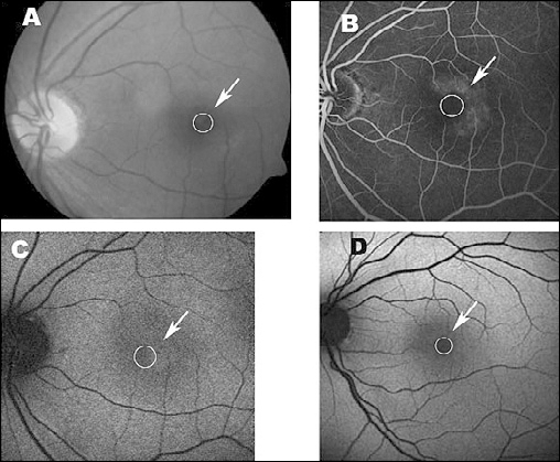
Complete loss of foveal normal hypoautofluorescence was noted in 28 (93.3%) eyes (Fig. 1). Two eyes showed incomplete disruption of foveal hypoautofluorescence (Fig. 2). Retinal crystals were present in 7 (23.3%) eyes, all of which had corresponding hypoautofluorescence.
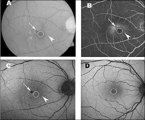
Intraretinal pigment clumping was present in 15 (50%) eyes, all of which showed corresponding hypoautofluorescence. Ninety-three percent (14/15) of these eyes showed hyperautofluorescence around the pigment (Fig. 1).
There was alteration in the pattern of parafoveal FAF in all the eyes. Twenty-two (73.3%) eyes showed hypoautofluorescence and 7 (23.3%) eyes showed hyperautofluorescence (Fig. 2).
On FFA, 25 (83.3%) eyes had diffuse leakage in the macula other than telangiectatic vessels. These areas had corresponding hyperautofluorescence in 20 eyes and hypoautofluorescence in 5 eyes. Telangiectatic leakage on FFA was present in all the eyes with corresponding hyperautofluorescence in 26 (86.6%) eyes (Tab. I).
Various autofluorescence patterns in type 2A idiopathic juxtafoveolar telangiectasia
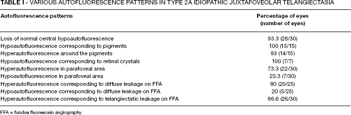
FFA = fundus fluorescein angiography
Discussion
The normal hypoautofluorescence at the fovea is believed to be due to masking by luteal pigments. The increase in foveal FAF or the loss of normal hypoautofluorescence is due to loss of masking of the FAF of the retinal pigment epithelium (RPE). Wong et al (10) speculated that the change in autofluorescence may not be solely because of loss of pigments but may occur due to compositional changes in the RPE, including endogenous fluorophores and melanin. This change in foveal and parafoveal FAF leads to almost uniform FAF at foveal and parafoveal region.
Loss of normal foveal hypoautofluorescence or increase in foveal FAF was noted in 93.3% (28/30) of eyes in our study. Clemons et al (17) reported this pattern in 89% of eyes. In our study, 2 eyes showed minimal disruption of foveal hypoautofluorescence; they showed minimal leakage on FFA, confirming the early stage of the disease (Fig. 2). Wong et al (10) reported no change in retinal sensitivity in areas with mild to moderate change in autofluorescence, but areas with significant increase in autofluorescence were associated with decrease in retinal sensitivity and with retinal atrophy on OCT. Hence, change of autofluorescence could be an early sign of the disease.
Chronic exudation from the telangiectatic vessels lying deep in the retina may cause atrophy of the outer retinal layers and stimulate pigment epithelial proliferative and metaplastic changes. The proliferating pigment epithelial cells may migrate into the outer retinal layers as well as into the subretinal space to form plaques of pigment in the vicinity of the right angle venules. Hypoautofluorescence corresponding to intraretinal pigment clumps can be explained by the blocking of normal autofluorescence of RPE. Hyperautofluorescence around the pigment could be due to actively proliferating RPE cells at the border of the pigment clumps.
The hyperfluorescence seen in late-phase FA at nontelangiectatic areas may be suggestive of breakdown of outer blood-retinal barrier. This contributes to increased FAF at corresponding area. This change in parafoveal FAF could be helpful to follow the progression of the disease.
Grayish retina in parafoveal area due to loss of retinal transparency can be found in IJRT. This also can be explained by the depletion of macula xanthophylls pigment leading to increased FAF (18).
Clemons et al (17) reported significant reduction in visual acuity in eyes with abnormal foveal autofluorescence in eyes with type 2A IJRT. Our study showed similar findings. Disruption of foveal autofluorescence has been reported to be associated with decreased visual acuity in various macular diseases like age-related macular degeneration (19-21). Foveal autofluorescence has been speculated as a predictor for treatment response in eyes with wet age-related macular degeneration (19). This may be applicable in eyes with type 2A IJRT.
Correlation of AF findings with OCT findings and retinal sensitivity on microperimetry would add to the understanding of the pathologic changes in the disease, especially at early stages (22).
In conclusion, our study demonstrates the changes in FAF patterns in type 2A IJRT. Loss of normal foveal hypoautofluorescence and increased FAF corresponding to the nontelangiectatic angiographic leakage areas were predominant features. Change in FAF pattern during the course of the disease would further add to the understanding of morphologic alterations. Changes in FAF in a longitudinal study would give insight into disease progression.