






Abstract
Purpose
To investigate the effect of the combination of photodynamic therapy with verteporfin and subconjunctival bevacizumab and triamcinolone acetonide on corneal neovascularization secondary to corneal graft failure.
Methods
A patient with extensive deep and superficial corneal neovascularization underwent same day triple therapy with subconjunctival bevacizumab (1.25 mg), subconjunctival triamcinolone acetonide (40 mg), and photodynamic therapy with verteporfin (fluence 50 J/cm2, irradiance 300 mW/cm2).
Results
A complete angiographic new vessel regression was obtained and was discernible starting 7 days after combination therapy. Three months after treatment, an initial new-vessel sprout was noted. Triple therapy was then repeated. No angiographic evidence of corneal neovascularization was detectable for the entire 6-month follow-up duration.
Conclusions
Triple therapy may offer a promising tool in the treatment of corneal neovascularization.
Introduction
Corneal neovascularization (NV) represents a serious clinical challenge whose consequences include reduction of visual acuity due to decreased tissue transparency and graft rejection as a result of the loss of immunologic privilege of the avascular cornea. Angiogenesis is the process of new vessel formation from established blood vessels and it is governed by a complex balance of positive and negative regulatory factors (1). Vascular endothelial growth factor (VEGF) is a critical proangiogenic factor and an inducer of vascular permeability. Additionally, it has a pivotal role in the immunoinflammatory response that accompanies corneal NV (2). Current therapeutic approaches for corneal NV include photodynamic therapy, subconjunctival or topical anti-VEGF agents, and steroids. Photodynamic therapy with verteporfin (PDT-V) has been shown to be effective in the obliteration of corneal NV (3). Subconjunctival injection of anti-VEGF monoclonal antibodies is associated with a partial regression of corneal NV at least in the short term (4). In an experimental model, triamcinolone acetonide (TA) was found to inhibit interleukin-6 (IL6) and VEGF-mediated corneal NV through the mifepristone-sensitive steroid receptor (5). Although favorable results have been published with these techniques, there is still the need for a therapeutic approach that induces a complete regression of corneal NV with long-lasting results. In the present article, we report our first experience with a combination therapy with PDT-V and subconjunctival bevacizumab and TA for corneal NV.
Case report
A 70-year-old man with a history of corneal transplant due to glaucomatous corneal decompensation presented with a painful, red right eye (RE) with extensive superficial and deep corneal NV unresponsive to topical treatment with steroids and cyclosporine. His left eye had been enucleated due to a traumatic event 40 years earlier. Best-corrected visual acuity (BCVA) RE was 20/400. Digital corneal fluorescein angiography was performed (Fig. 1). Proposed treatment consisted of off-label administration of subconjunctival TA and bevacizumab combined with corneal PDT-V. Written, informed consent was obtained. All procedures were carried out in accordance with the Declaration of Helsinki. The patient underwent same day triple therapy with subconjunctival bevacizumab (1.25 mg), subconjunctival TA (40 mg), and PDT-V (fluence 50 J/cm2, irradiance 300 mW/cm2). Three consecutive and partially overlapping laser spots were administered over the neovascular network. Feeder vessels were subsequently irradiated. The injections were made into the subconjunctival space at the limbus, adjacent to the root of pathologic blood vessel growth into the cornea. The patient had regular follow-up visits 1 day and 1 week after treatment and monthly thereafter. Digital corneal fluorescein angiography was scheduled at follow-up visits starting from day 7 after treatment. New-vessel obliteration was already evident during corneal PDT-V. A complete angiographic new-vessel regression was discernible at post-treatment day 7 (Fig. 2). Visual acuity improved to 20/200. Three months after treatment, corneal digital angiography showed an initial new-vessel sprout. At that time, treatment was repeated and no evidence of corneal NV was noticeable during a follow-up period of 6 months after the first treatment (Fig. 3). The BCVA at the end of the follow-up was 20/200. No significant changes in intraocular pressure or anterior chamber condition were observed during the follow-up.
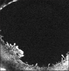
Baseline corneal fluorescein angio-graphy showing deep and superficial corneal neovascularization.
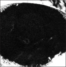
Corneal fluorescein angiography 1 week after triple treatment. A complete angiographic new-vessel regression is discernible. Whitish artifacts are present in the lower half of the corneal surface.
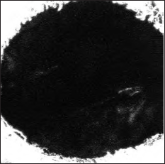
Corneal fluorescein angiography 6 months from baseline after 2 combination therapies. No evidence of corneal neovascularization is detectable. Whitish artifacts are present in the lower half of the corneal surface.
Discussion
One of the most common risk factors that can contribute to graft failure is corneal NV (6). The risk of an immune reaction in an avascular cornea is about 10%. This value rises to approximately 50% in vascularized corneas (7). Corneal NV is a complex disease in which chronic inflammation, hemangiogenesis, and lymphangiogenesis play a pivotal role. Hence, an effective combined treatment for improving corneal tissue architecture during corneal NV should be addressed at each of the above factors. Thereafter, a comprehensive therapeutic strategy should include a vaso-occlusive element to eradicate established corneal NV, an anti-VEGF drug to inhibit angiogenesis, and a steroidal drug to suppress chronic inflammation. The present report analyzes the potential role of triple therapy with PDT-V, subconjunctival bevacizumab, and TA. Photodynamic therapy with verteporfin is based on the interaction of laser light with a photosensitizing drug and oxygen. A major mechanism of PDT-V is thought to be occlusion of the vascular bed which occurs secondary to damage to endothelial cells and subsequent platelet adhesion, degranulation, and thrombus formation. Bevacizumab is a monoclonal antibody that binds to and inhibits all subtypes of VEGF-A. Neutralization of VEGF-A not only reverts hemangiogenesis and lymphangiogenesis but also interferes with recruitment of inflammatory cells into the cornea. In fact, anti-VEGF antibodies bind to VEGF receptor 1 and consequently inhibit neutrophil and macrophage chemotaxis (2). Neutrophils in inflammatory conditions produce VEGF, which perpetuates the cycle of corneal angiogenesis and influx of inflammatory cells that promote chronic corneal damage. The trigger to corneal NV is assumed to be chronic inflammation. The inhibition of VEGF alone has been reported to produce a partial regression of corneal new vessels. This is probably due to the fact that only new vessels not covered by pericytes mainly regress after VEGF inhibition (8). Moreover, the effect of VEGF inhibition when administered subconjunctivally is limited to 1 to 2 months (4, 9). Steroids have been for long time the treatment of choice to control corneal NV and reduce the risk of graft rejection. In an animal model, TA has been shown to inhibit corneal NV, suppressing IL-6 and VEGF-mediated molecular pathways (5).
The use of a comprehensive approach with triple therapy for the treatment of corneal NV brought about a complete regression of corneal NV within 7 post-treatment days, as evaluated by slit-lamp examination and corneal fluorescein angiography.
In conclusion, corneal NV is a chronic and progressive phenomenon and thus the vessels found at the time of diagnosis are a mixture of new and old vessels. Monotherapy alone offers only temporary alleviation because PDT-V fails to address the underlying biological mechanism of angiogenesis and subconjunctival anti-VEGF and steroidal injections are ineffective against previously established new vessels. We emphasize the importance of an appropriate comprehensive combination therapy for corneal NV and the role of triple therapy as a new promising strategy to avoid blinding sequelae in corneal diseases that lead to neovascularization and subsequent chronic inflammation. In the case reported here, triple therapy was found to be an effective alternative in treating corneal NV. Further investigations are needed to validate the results.