






Abstract
Purpose
To report on phototherapeutic keratectomy (PTK) in myopic patients who developed spontaneous recurrent corneal erosions (RCE) following IntraLASIK.
Methods
Recurrent corneal erosions developed spontaneously 3-6 months following IntraLASIK in 4 eyes of 4 myopic patients. Diffuse lamellar keratitis (DLK) subsequently developed. Initially, all patients underwent mechanical scraping of the loose epithelium. The erosion healed but recurred after 2-4 months. Phototherapeutic keratectomy was performed following scraping of the whole loose flap epithelium. The ablation diameter was 6.5 mm, transition zone was 2.7 mm, and the depth was 12 μm. This program was followed by a refractive laser treatment to correct +0.75 diopter at a zone of 6.0-9.0 mm.
Results
Recurrent corneal erosions healed completely following PTK within 8-10 days and DLK disappeared gradually within 9-12 days. Steroid drops were continued for 6-10 weeks according to degree of stromal haze. Final refraction revealed no significant change. Visual rehabilitation was noted to be slow. No patient lost best-corrected visual acuity. A +1-degree reticular haze was noted in all patients 4.5 years following treatment. The patients remained asymptomatic and corneal erosion did not recur during the follow-up period.
Conclusions
Phototherapeutic keratectomy was found to be an effective treatment for late spontaneous RCE after IntraLASIK and prevented recurrences. A mild stromal haze remained after laser treatment without loss of best-corrected visual acuity.
Introduction
Recurrent corneal erosion (RCE) following laser in situ keratomileusis (LASIK) has been described in several studies (1-5). Some of these cases were associated with a subclinical weakness of adherence of the corneal epithelium or undiagnosed anterior basement membrane dystrophy (ABMD) which resulted in RCE when the cornea was subjected to the shearing forces of the microkeratome during creation of the flap (6-8), even if the procedure was uneventful. In other cases, RCE developed late following an epithelial defect which occurred during LASIK, either due to a faulty microkeratome head, a damaged blade, older age, high hypermetropia, or overdosage of anesthetic drops (7, 9, 10). On the other hand, following IntraLASIK, RCE is less expected to occur since there are no shearing forces exerted on the corneal epithelium, but only an applanation force which is exerted by the applanation lens during flap creation by the femtosecond laser. Nevertheless, this applanation force in the presence of undiagnosed ABMD may rarely be associated with a complication of loosening of the epithelium during the procedure or RCE developing late following the procedure.
Additionally, following the femtosecond laser cut, stress forces may be exerted during the dissection of the flap by the IntraLASIK spatula, mainly in cases where it is difficult to open the flap. These mechanical forces can affect or loosen the undiagnosed dystrophic epithelium.
I describe 4 eyes of 4 patients who underwent uneventful IntraLASIK and developed RCE several months after the procedure. As these cases were found to be recalcitrant to conservative treatment by mechanical scraping of the loose epithelium and fitting a therapeutic contact lens (TCL), phototherapeutic keratectomy (PTK) was subsequently performed. The results of this treatment are presented.
Patients
Late RCE developed spontaneously in one eye of 4 patients with myopia, ranging between −1.75 and −5.25 diopters of sphere and −0.5 to −1.25 diopters of cylinder, who underwent uneventful IntraLASIK between 2004 and 2007. During this period, 10,000 IntraLASIK procedures were performed at Enaim Laser Center with the IntraLase system (model 1, IntraLase Corp., AMO, Irvine, California, USA), which was upgraded to 60 Hz in the last 5000 procedures. The rate of late RCE post IntraLASIK can therefore be estimated as 4 out of 10,000 eyes, or 0.04%. The patients’ age ranged between 20 and 37 years. Keratometry readings were found to be within normal limits: 42-46 D. Pachymetric data ranged between 505 and 562 μm. Their preoperative best-corrected visual acuity (BCVA) ranged between 6/6 and 6/7.5 and there was no history of corneal trauma, ocular discomfort, or other symptoms of recurrent corneal erosion syndrome in the past. All patients were daily-wear soft contact lens users and wore their contact lenses for at least 12 hours per day without any problems. Applanation tonometry readings ranged between 10 and 18 mmHg. Ophthalmologic examination including slit-lamp evaluation of the cornea and the anterior segment was normal.
Tear film evaluation was found to be normal and there was no lagophthalmos.
There were no clinical signs of ABMD and the epithelium revealed no pathology, even following applanation tonometry and repeat ultrasonic pachymetry. The funduscopic examination was normal. There was no history, nor any evidence of systemic disease in these patients.
The IntraLASIK procedure was performed in the conventional manner as previously reported (11, 12); the planned flap thickness was 100 μm and the diameter ranged between 8.8 and 9.00 mm.
Our routine planned flap thickness ranged between 100 and 110 μm, depending on the calibration measurements performed by our IntraLase technician. This thickness’ range yields a stable corneal flap with our IntraLase system, without any significant epithelial pathology.
Following surgery all 4 patients were treated routinely by nonpreserved artificial tears, such as Refresh (Allergan-Pharmaceuticals, Ireland), and there was no significant dry eye state.
Painful RCE developed twice in 2 patients and 3 times in the 2 other patients, 3-6 months following uneventful IntraLASIK surgery. At presentation, slit-lamp examination revealed a 3.5-4.5 mm corneal erosion with surrounding loose epithelium occupying the inferocentral area of the corneal flap created by the IntraLase, which was found to be well positioned and adherent to its bed. Moderate diffuse lamellar keratitis (DLK) subsequently developed within 3-5 days of presentation. All patients underwent scraping of the whole loose epithelium of the corneal flap, twice in 2 patients and once in the other 2, with subsequent fitting of a TCL; ofloxacin drops were instilled 4 times daily for 14 days. The DLK regressed within 9-12 days under local steroid treatment. The corneal erosion healed within 8-10 days but recurred in a more severe clinical form after 2-4 months. Therefore, it was decided to perform PTK within a short time following presentation.
The patients’ clinical data before PTK are presented in Table I.
PRE-PTK CLINICAL DATA
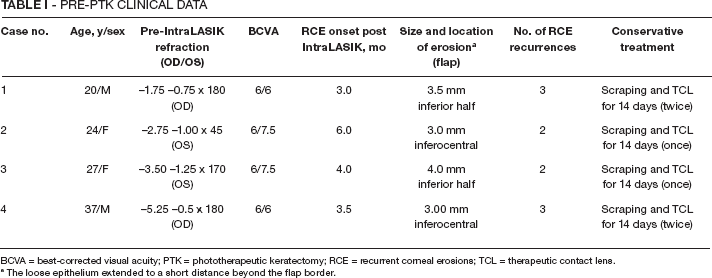
BCVA = best-corrected visual acuity; PTK = phototherapeutic keratectomy; RCE = recurrent corneal erosions; TCL = therapeutic contact lens.
The loose epithelium extended to a short distance beyond the flap border.
Methods
All patients signed an informed consent form to be treated. The research protocol of the study has been approved by the local ethics committee of Enaim Medical Center TA-Israel. Each patient underwent scraping of the whole corneal flap loose epithelium including its periphery following instillation of local anesthetic drops (oxybuprocaine HCl 0.4%, Fischer Pharmaceuticals, Tel Aviv, Israel). It should be noted that the flap epithelium was found to be loose up to a short distance beyond the flap circumference. It came out very easily except for the zone between the periphery of the flap and the limbus, where it was more tightly adherent and it was therefore left untouched. The epithelial scraping was performed with meticulous care in order to avoid any trauma to the flap itself.
Thereafter, excimer laser ablation was performed with the VISX-STAR-4 model laser system (AMO, VISX Company, Santa Clara, California, USA). Initially, a PTK mode of ablation was performed, reaching a depth of 12 μm and a diameter of 6.5 mm with transition zone of 2.7 mm. Subsequently, a refractive laser ablation treatment was performed, correcting +0.75 diopter sphere at a diameter of 6.00 mm to 9.00 mm. Finally, the ablated stromal surface was irrigated with cold balanced salt solution (4°C) and a TCL fitted and worn for 2 weeks. Ofloxacin 0.3% drops (Allergan, Ireland) were instilled 4 times a day for 2 weeks and Voltaren drops (0.1% diclofenac sodium, Novartis, Switzerland) were instilled twice daily for 4 days. All patients were warned about the possible corneal complications of Voltaren drops (13) and were instructed to use it not more than twice a day. Additionally, steroid drops (dexamethasone sodium phosphate [0.1%], Fischer Pharmaceuticals, Tel Aviv, Israel) were given 4 times daily for 8-12 weeks. All patients were followed for 4.5-5.0 years. Artificial tears (Refresh minims, Allergan Pharmaceuticals, Ireland) were instilled 4-6 times per day for 8-10 weeks post PTK. Additionally, Genteal-gel (Novartis Pharma AG, Basel, Switzerland) was used at night for 1 month.
Results
The large corneal erosion in all 4 patients healed completely following PTK within 8 to 10 days. The DLK regressed under local steroid treatment within 9 to 12 days. The TCL was removed after a minimum of 14 days in order to facilitate normal adhesion of the regenerated epithelium to the ablated flap stromal bed.
A diffuse reticular subepithelial stromal haze appeared in the central 6 mm zone of the corneal flap in all patients 4 weeks after PTK. The degree of haze ranged initially from +2 to +3 and therefore steroid drops were instilled 4 times daily for the first 6 weeks and subsequently tapered gradually according to degree of haze for another 2-6 weeks, until the haze reached a degree of +1.0. The visual recovery after laser treatment was relatively slow and it took 6-8 weeks until BCVA reached the preoperative level.
At the end of the follow-up period, final refraction revealed no change of BCVA compared to the data obtained 2 months after IntraLASIK surgery. The final subjective refraction was found to be within +0.25 to −0.25 diopters of spherical equivalent compared to the subjective refraction data obtained 2 months after IntraLASIK surgery (Tab. II). The final BCVA ranged between 6/6 and 6/7.5 (–) and there was no loss of BCVA compared to the visual acuity data before the refractive laser procedure.
POST-PTK CLINICAL DATA
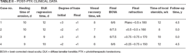
BCVA = best-corrected visual acuity; DLK = diffuse lamellar keratitis; PTK = phototherapeutic keratectomy.
A mild diffuse subepithelial haze (+1 degree) was noted in the central 6 mm zone of the IntraLASIK flap at the end of the follow-up period. The patients remained asymptomatic and corneal erosion did not recur during the follow-up period of 4.5-5.0 years after PTK. The post-PTK patients’ data are presented in Table II.
Discussion
During the IntraLASIK procedure, contrary to LASIK, there are no mechanical tangential shearing forces exerted on the corneal epithelium except for a vertical applanation force created by the smooth applanating lens of the IntraLase system cone which applanates the corneal surface during flap creation and stress forces created by mechanical dissection of the flap.
Additionally, there exists a light tangential friction force which is exerted on the epithelial surface while repositioning the corneal flap with a spatula, irrigating cannula, or a wet sponge at the end of both surgical procedures, IntraLASIK as well as LASIK. Therefore, corneal epithelial disinsertion or epithelial erosion are expected to occur much less frequently during IntraLASIK or thereafter compared to LASIK, in patients with undiagnosed ABMD, who were asymptomatic before surgery and had no clinical signs of this epithelial dystrophy. Our study confirms this postulation as the incidence of RCE after uneventful IntraLASIK was found to be very low (0.04%) and there was no case of intraoperative epithelial damage among our patients. In comparison, Kenyon et al (6) found an overall incidence of 10.2% of intraoperative LASIK flap epithelial defects, of which 6.2% were micro defects and 4.0%, macro defects. These authors also found a positive intraoperative epithelial adhesion test, performed during LASIK procedure, in 80% of the corneas having macro epithelial defects (>2 × 2 mm).
Chen at al (7) found an incidence of 1.66% of intraoperative epithelial damage which occurred during LASIK surgery performed on 1873 eyes included in their retrospective study.
The treatment of RCE after LASIK surgery was reported to be either conservative, with localized scraping of the loose epithelium and fitting a TCL, or more aggressive, including anterior stromal puncture (ASP) or excimer laser PTK (14-17).
Phototherapeutic keratectomy was reported by Rojas and Manche (16) to be a safe and effective treatment modality for recurrent corneal erosions and decreased visual acuity following LASIK in patients with ABMD.
Anterior stromal puncture is suggested to be the second line of treatment following epithelial scraping in this situation, due to its simplicity as an in-office procedure, low cost, speed of recovery which minimizes the extent of possible DLK, and no influence on spherical equivalent. If this second line of treatment fails, one can proceed with PTK.
As conservative treatment with epithelial scraping and fitting a TCL failed in all our 4 patients with recalcitrant RCE after IntraLASIK surgery, I decided to skip the option of ASP and to proceed with a more aggressive therapy, such as PTK. My thought was that PTK would be more effective than ASP in preventing recurrence of corneal erosion in these patients with recalcitrant RCE and loose epithelium of the whole corneal flap. This therapeutic laser treatment was performed on as large an area as possible of the existing IntraLASIK corneal flap, by combining a PTK ablation program with the largest transition zone and a secondary refractive hyperopic ablation program to correct +0.75 diopters. The aim of the refractive laser treatment was to prevent any possible laser-induced hyperopic effect and also to enlarge the zone of ablation to 9 mm. The mechanical epithelial scraping was also extensive, reaching a short distance beyond the borders of the IntraLASIK flap, in order to remove as much sick loose epithelium and abnormal basement membrane as possible, and perform PTK on a large area. This technique of PTK is based on the method described by Kremer and Blumenthal (18) regarding large area PTK and photorefractive keratectomy (PRK) treatment of myopic patients with RCE. I believe that the majority of the IntraLASIK corneal flap surface should be treated by excimer laser PTK in such cases, in order to prevent recurrence of the diffuse ABM pathologic changes. This diffuse laser treatment will prevent the recurrence of RCE after IntraLASIK and the subsequent development of DLK, which may be severe and lead to serious complications such as dense corneal scarring (19). The sick and loose epithelium of RCE following IntraLASIK or LASIK, probably releases cytokines and inflammatory mediators, which contribute to the development of DLK through keratocyte apoptosis and chemotaxis of polymorphonuclear cells. These inflammatory cells migrate through the surgical interface and finally into the stromal tissue of the flap, leading to necrosis and final subepithelial scarring (1, 19, 20).
In conclusion, our large area combined method of PTK and PRK laser ablation was found to be a safe and effective treatment of late spontaneous RCE post uneventful IntraLASIK surgery, which was recalcitrant to conservative treatment. The mild subepithelial haze of the corneal flap found in all 4 treated eyes did not affect the final visual results and was asymptomatic. Additionally, there were no significant final changes in refraction following PTK combined with PRK.
According to my data, RCE following IntraLASIK surgery was found to be a rare complication (0.04%). My opinion is that these rare cases had an undetected subclinical ABM corneal dystrophy and that the delayed onset, spontaneous recurrent flap erosion developed following the trigger inflicted by IntraLASIK surgery.
It should be pointed out that in those eyes with suspected epithelial adhesion problems or slit-lamp findings of ABM epithelial dystrophy, the state of the art is not performing LASIK of any kind but rather a surface laser ablation.