






Abstract
Purpose
We report a rare case of acute postoperative Staphylococcus schleiferi endophthalmitis following an uneventful cataract extraction.
Methods
An 83-year-old woman underwent uneventful phacoemulsification without receiving the usual intracameral cefuroxime solution due to cephalosporin intolerance. Two days postoperatively, she presented with acute ocular pain and blurred vision in the operated eye. Ophthalmic examination revealed signs of acute keratitis and endophthalmitis. Corneal scrapes and vitreous taps were sent for cultivation and analysis. The patient received intravitreal antibiotics at the same time.
Results
Cultivation grew S schleiferi, a pathogen commonly found in dogs. Following microbiological report, the patient received a repeated intravitreal injection of 1 mg/0.1 mL vancomycin combined with fortified topical vancomycin drops. One week later, the inflammation subsided completely while the corneal ulcer healed and visual acuity improved dramatically. Further examination revealed the existence of the same pathogen in a specimen obtained from the dog owned by the patient's family.
Conclusions
This description of S schleiferi endophthalmitis highlights the need to provide alternative intracameral antibiotic solution in the operating room and avoid direct contact with dogs during the first postoperative period after intraocular surgery.
Keywords
Introduction
As the elderly population grows and life expectancy increases progressively, cataract surgery has become one of the most prevalent surgical procedures performed worldwide. In the majority of cases, phacoemulsification is considered a successful operation, while advances in instrumentation and surgical techniques have dramatically improved the operating time and efficacy of the procedure. However, postoperative endophthalmitis, despite its low incidence due to perioperative and postoperative chemoprophylaxis, represents a devastating complication that can lead to significant and permanent loss of vision.
The Endophthalmitis Vitrectomy Study showed that coagulase-negative Staphylococcus is isolated in approximately 70% of endophthalmitis cases, followed by Staphylococcus aureus (9.9%) and Enterococcus (2.2%), and 5.9% of incidences are related to Gram-negative bacteria (1). More specifically, Staphylococcus epidermidis represents the commonest species of coagulase-negative Staphylococci related to postsurgical endophthalmitis and most of these infections resolve without permanent ocular sequelae (2). However, in clinical practice, there are some pathogens not frequently identifiable as causative agents of postoperative endophthalmitis.
We describe a case report of acute postoperative Staphylococcus schleiferi endophthalmitis following an uneventful cataract extraction. To our knowledge, this is the first report in the literature describing endophthalmitis caused by this specific pathogen. Furthermore, there is no other published ocular infection, of any kind, where S schleiferi has been isolated.
Case report
An 83-year-old woman presented to the emergency care unit 2 days following an uneventful cataract extraction carried out in our department. She was complaining of acute ocular pain and blurred vision in her left, operated on, eye. Visual acuity was counting fingers at 30 cm and intraocular pressure (IOP) 12 mm Hg. On slit-lamp examination, a corneal ulcer with epithelial defect and stromal infiltration was identified at the main cataract incision (Fig. 1) accompanied by an intense anterior chamber inflammatory reaction (cells >3+), hypopyon, and moderate vitritis (cells 2+). B-scan confirmed the diagnosis of vitritis. Postoperative endophthalmitis was considered as a possible diagnosis. As reported by her medical records, the patient was initially booked for cataract surgery due to a significant deterioration of vision OS with a nuclear cataract of NO4 and NC4 according to LOCS III grading system. On preoperative ophthalmic examination, best-corrected visual acuity (BCVA) was 5/10 OD and 2/10 OS. Age-related macular degeneration (AMD), dry type, stage II, had also been noted in both eyes. Best-corrected near vision was J2 OD and J3–2 OS. Otherwise, examination was unremarkable. Medical and family history of the patient revealed successfully treated systematic hypertension. No allergies were reported at that time.
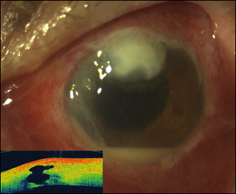
Slit-lamp photograph of anterior segment at presentation and respective optical coherence tomography (OCT) by means of Cirrus OCT (Carl Zeiss Meditec, Inc.). Note the advanced keratitis and corneal ulcer at 12 o'clock, showing large intrastromal cyst in the OCT picture. A dense anterior chamber reaction accompanied by hypopyon is also shown.
Subsequently, the patient underwent an uneventful cataract surgery by means of phacoemulsification (3-step 2.4-mm clear corneal incision at the superior limbus with Alcon disposable keratome, stop-and-chop technique, phaco time 00:48, cumulative dissipated energy 18.72 s, Infiniti Vision System, Alcon Laboratories, Inc., Fort Worth, Texas, USA). However, immediately before surgery, she mentioned intolerance to cephalosporin indicated by her general practitioner. Since allergy to cephalosporins was mentioned in the operating theater, the operation was completed without injection of the usual intracameral cefuroxime solution (0.05%). No other prophylactic intracameral antibiotic was available at that time and the surgeon decided to instill povidone iodine eyedrops (5%) and commence topical prophylaxis immediately after surgery. The regimen comprised a combination of tobramycin 0.3% and dexamethasone 0.1% (Tobradex, Alcon Laboratories, Inc.) and Nepafenac 0.1% (Nevanac, Alcon Laboratories, Inc.). On postoperative day 1, the visual acuity was 2/10 without any inflammation signs or leakage of the wound and IOP 14 mm Hg.
In the emergency department, and after keratitis and endophthalmitis diagnosis was considered, corneal scrapes taken with the use of #11 blade as well as vitreous taps were immediately sent for cultivation and microbiological analysis. At the same time, the patient received a pars plana intravitreal injection of 1 mg/0.1 mL vancomycin and 0.4 mg/0.1 mL amikacin and was started on topical antibiotics: moxifloxacin (Vigamox, Alcon Laboratories, Inc.) and fortified vancomycin eyedrops (50 mg/mL) on hourly basis.
Improvement in both pain and sight was noted 2 days following the initial management. Ophthalmic examination was in agreement with patient's symptomatology, revealing reduced inflammatory cells and hypopyon in anterior chamber (2+) and epithelialization of corneal ulcer. Visual acuity improved to 1/10 in the affected eye. For all the above reasons, therapeutic vitrectomy was not considered necessary at that time.
In the meantime, the cultivation of corneal smears and vitreous tap in several media (chocolate agar, Mannitol salt agar, and MacConkey agar) revealed the presence of S schleiferi, subspecies coagulans, bacteria commonly isolated in dogs and cats. The bacteria were found resistant to tobramycin, moxifloxacin, and ciprofloxacin, whereas they appeared to be very sensitive to vancomycin. We then decided to repeat intravitreal injection with vancomycin only (1 mg/0.1 mL). One week after the second intravitreal injection, the inflammation subsided completely, the corneal ulcer healed, and BCVA improved further, reaching 2/10 (Fig. 2). Topical vancomycin drops were continued until 4 weeks after the first event. On the last follow-up, 6 months postoperatively, there was a mild scar in the region of main cataract incision and no signs of active intraocular inflammation, while BCVA was 4/10, limited by the existence of postinflammatory and AMD-related changes in the macular area (Fig. 3).
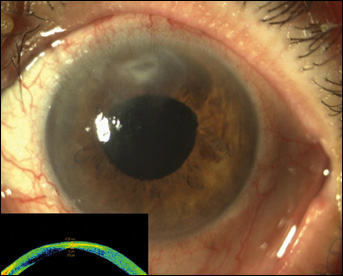
Slit-lamp photograph of anterior segment 1 week after the second intravitreal antibiotic injection. Anterior chamber appears quiet, hypopyon was resolved, while keratitis improved significantly. The respective optical coherence tomography (OCT) of the corneal lesion, by means of Visante AS-OCT (Carl Zeiss Meditec, Inc.), depicts fibrosis and corneal thinning (315 μm) on the phacoemulsification main incision site.
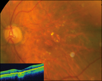
Fundus photograph and respective optical coherence tomography (OCT) of the macular region, using Cirrus OCT (Carl Zeiss Meditec, Inc.) of patient's left, affected, eye 6 months postoperatively. It shows age-related macular degeneration–related changes in the macular area.
Upon cultivation report, we were advised by the microbiologists to revisit the medical history, emphasizing possible possession of pets, especially dogs. The patient reported that she was consistently assisted by her daughter to instill eyedrops while she owned a dog. For scientific purposes, we then consulted a specialized veterinarian, who advised us to obtain a specimen from dog's external auditory meatus and send it for cultivation. The isolates taken from dog's ear confirmed the presence of S schleiferi.
Discussion
S schleiferi was first described in 1988 by Freney et al (3). It is commonly found in carnivores, but may be transferred from pets to their owners or handlers (4). Since its original description, few data have been published in the literature regarding its pathogenicity. In veterinary medicine, S schleiferi was previously assumed to be an inhabitant in carnivore skin; however, more recently, it has been repeatedly documented in the literature as both an inhabitant and pathogen. Nasal carriage of S schleiferi occurs in healthy dogs as well as dogs with otitis and pyoderma.
Methicillin-resistant and sensitive S schleiferi can be found in the nares of dogs with diseased ears and skin (5). S schleiferi is a coagulase-negative staphylococcus that has rarely been reported in human infections. The first description of the isolation of this microorganism from humans was reported in 1994 (6). In the largest series of human infections due to S schleiferi published, Hernández et al described the clinical course of 28 culture-positive patients (7). In that series, S schleiferi was frequently associated with wound infections, mainly surgical site infections, as previously reported in the literature (8). Definite clinical diagnoses included mostly wound and pacemaker-related infections as well as meningitis. Surgical procedures were performed in two-thirds of the patients, including the subgroup with solid tumors. In all cases, the infection developed postoperatively. To our knowledge, this is the first case of S schleiferi endophthalmitis and ocular infection ever reported in the literature. The unusual presence of S schleiferi in human flora could explain the low frequency of infections due to this microorganism. However, S schleiferi can be difficult to differentiate from common S aureus; as a result, this pathogen may have been underreported (9). Most probably, the presently described endophthalmitis has had a postoperative rather an intraoperative inoculation course. However, it is likely that the bacterium was already colonizing the ocular surface for this to occur rather than a direct contamination postoperatively. As such, one should consider prophylactic topical antibiotics as well.
In conclusion, the present report of S schleiferi endophthalmitis and coexistent keratitis is highlighting the need to provide an alternative prophylactic intracameral antibiotic solution when intolerance to commonly used prophylactic agents is reported and, at the same time, advise patients of the possible risks of direct contact with dogs and other animals during the first postoperative days after cataract surgery. Furthermore, early diagnosis and appropriate management decreases the need for surgical intervention and may result in favorable outcomes.