
Abstract
Purpose
To study the effect of a synthetic medium and compare it with a serum-based medium for corneal preservation in organ culture using an overall quality assessment system.
Methods
A randomized study with blinded observers was performed comparing parameters such as thickness, transparency, viable endothelial cell density (VECD), morphology, and overall quality (OQ) of the corneal tissues preserved in synthetic and a serum-based medium, respectively. Seven human paired corneas were randomly selected and assessed at day 0 (initial), day 2 (before organ culture), day 30 (before deturgescence/ deswelling storage), and 48 hours post deswelling. Thickness was determined with optical coherence tomography and transparency with a validated, custom device. The morphology and VECD were observed after treating the tissues with trypan blue and sucrose. Data were compared using paired t tests with p<0.05 deemed significant.
Results
Parameters were similar at the initial stage between the groups with no statistically significant difference. However, after preservation in the deturgescent medium, the corneas stored in a serum-based medium showed a higher and statistically significant OQ value (p = 0.0317).
Conclusions
The OQ of a serum-based medium was higher than that of the synthetic medium. A higher rate of transparency and reduction in thickness was observed in the serum-based medium at the end of the storage. Although complete synthetic media may have distinct advantages of being serum/animal-free, the quality of the cornea is of a reasonable concern when it is deemed for transplantation.
Introduction
Two major corneal preservation methods are cold/ hypothermic storage (2-6°C), which is mostly used in the United States and Asia, and organ culture (OC) (31-37°C), which is followed mainly in Europe. Organ culture is deemed for long-term preservation (up to a month), whereas hypothermic storage is usually used for short-term preservation (limited to 14-21 days). Organ culture has distinct features that include microbiological checks, quality assurance of the tissue before transplantation, and ease of surgical preparation due to longer preservation time.
Although most eye banks have their own internal standards, the European Eye Bank Association has minimum inclusion criteria (≥2,000-2,200 cells/mm2) of endothelial cell density (ECD) for penetrating keratoplasty (PK) (1). The ECD is considered as the most influential factor for corneal acceptance or failure; however, other parameters such as mortality and morphology are also accountable for tissue failure (2). Conventional OC is typically a 3-stage protocol: the initial medium that transports the cornea to the eye bank from the morgue, the OC medium (storage), and the final transport medium with a deswelling agent to reduce the thickness of cornea for ease of transplantation. This media contains serum of animal origin. Apart from serum, other nutrients of animal origin have also been investigated for prolongation of the endothelial metabolic activities, such as chicken feather, ovalbumin, and pig bone amino acids, usually used in combination with other sources of nutrient supplements (2-5). The significance of fetal calf serum (FCS) is not well-known; however, it was proved that a higher rate of endothelial cell damage occurred when the porcine corneal endothelial cells were preserved in the OC medium without serum.
It has also been found that FCS is responsible for resisting the stress levels of the cells in vitro(6-8), whereas other reports suggest that the serum-free medium causes higher endothelial cell loss (ECL) due to necrotic areas (9). A few studies report the effects of different deswelling compounds such as poloxamer or dextran-based medium (10, 11).
Animal-derived products have the potential to introduce animal viruses or prions that if assimilated in the human body could be hazardous. Animal viruses, especially retroviruses, could integrate into the human genome and activate human oncogenes or oncosuppressor genes, while prions could lead to human forms of bovine spongiform encephalopathy (BSE). This is why synthetic media have been developed. The aim of this article is to compare a serum-based medium with synthetic medium, both developed and used for corneal storage at 31°C, and to evaluate whether their efficacy is comparable.
Methods
Corneal Donor Characteristics
Seven pairs of human cadaveric donor corneal tissues unsuitable for transplantation and with the written consent from the donor's family were obtained for research. The average age of the corneal donors was 71.14 (±1.07) years, postmortem time was 10.26 (±5.86) hours, and male:female ratio was 5:2. All the tissues were preserved in Cornea Cold® (Eurobio, Paris, France) for <24 hours after retrieval before the first evaluation.
Media Description
The conventional OC protocol was followed for both the media. Each cornea from the same donor was placed in synthetic medium (Stem Alpha, Argentiere, France) and serum-containing medium (Eurobio, France) simultaneously. Stem Alpha 1 (SA1) and Cornea Prep II (CP) are the transport media that carry the cornea from the morgue to the eye banks. Stem Alpha 2 (SA2) and Cornea Max (CM) are the storage media in which the corneas are preserved for around 30 days at 31°C. Stem Alpha 3 (SA3) and Cornea Jet (CJ) are deswelling and the final transport media.
Experimental Stages
The corneas were checked and evaluated before preservation (initial: stage I). Each cornea from the same donor was preserved in SA1 and CP simultaneously for 48 hours at room temperature (before organ culture: stage II). These corneas were then transferred to SA2 and CM and preserved for 30 days at 31°C (before deturgescence: stage III). Here, SA2 is the synthetic medium and CM contains serum. Further, these corneas were transferred to SA3 and CJ and preserved for 48 hours at room temperature (after deturgescence: stage IV). Here, SA3 is the deturgescent medium containing poloxamer and CJ contains dextran as a medium for deswelling of the human corneas. The parameters mentioned below were checked at stages I, II, III, and IV to determine and score the cornea periodically.
Evaluation Parameters
All the corneas were prechecked (initial: stage I) using parameters such as thickness; transparency; ECD and mortality, incorporated together as viable ECD (VECD); and morphology. All the parameters were given values in a range of 0-4 as described in our previous publication (12). These values were further incorporated together to assess the overall quality (OQ) of the cornea in the range of 0-4 where a value of 0 was referred to as poor and 4 as the best quality tissue. The parameter checks were initiated from thickness measurement followed by transparency, VECD, and morphology, as described below.
Central Corneal Thickness
Central corneal thickness (CCT) was measured using optical coherence tomography (OCT SS-1000, Tomey Corporation, Nagoya, Japan). The final value was converted into the scoring system to define the OQ of the CCT (12).
Transparency
The corneas were analyzed objectively using a transparency device (13). The average of 3 transparency readings was considered suitable to reduce the error. The transparency readings were converted to the OQ scoring (12).
Viable ECD
Viable ECD incorporates both visual ECD and mortality. The endothelium was stained using Trypan blue (0.25%) for around a minute, washed with phosphate-buffered saline, and placed in a sterile petri plate containing sucrose solution (1.8%) with the epithelium facing the lid. A reticule (grid) (10 × 10 mm) inserted in the eyepiece of the microscope was used for manual cell counting and mortality at 100x magnification (14). Central (optic zone) and peripheral readings were recorded by 2 masked observers. These values were converted to the OQ scoring system (12).
Morphology
The morphologic parameters included intercellular borders, checked for disappearance, swelling, or irregularity (polymorphism, polymegathism, and pleomorphism); dystrophy (corneas without any dystrophy were selected and therefore all the values were marked as regular); and degeneration (disappearance of the intercellular borders and/or complete cells were recorded). Average of all the morphologic parameters gave the OQ score for morphology.
Overall quality
The final OQ score is an average of the individual parameter OQ scores. The valuation has been described in a previously published article (12). As all the parameters are based in a range of 0-4 (arbitrary value), the OQ value is also ranged from 0-4, where 0 is poor and 4 is regular.
Statistical Analysis
The data were analyzed using SAS 9.2 software for Windows (SAS Institute Inc., Cary, North Carolina, USA). Student t test was performed at each individual time point. The data were statistically significantly different between the groups when p<0.05.
Results
None of the corneas had microbial contamination. The parameters were evaluated periodically as follows.
Thickness
The average thickness after preserving the corneas at stage II showed an increase of 52.05% (±13.78) in SA1 and 52.55% (±18.21) in CP (p = 0.9548). Stage III showed an additional expected increase in thickness in both media, 27.38% (±11.03) in SA2 and 37.63% (±15.83) in CM (p = 0.1884), and the final preservation in a deturgescent medium showed an expected decrease in thickness of 28.00% (±5.31) in SA3 and 47.82% (±9.71) in CJ (p = 0.0009). Thus, increase in thickness was seen in both the media, but no statistically significant differences were found at stages II and III. However, after the corneas were preserved in the deturgescent media, the decrease in thickness was of interest, especially with the medium containing dextran (CJ), as it showed better results as compared to the one with poloxamer (SA3) (Fig. 1).
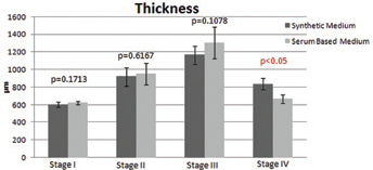
Thickness readings using optical coherence tomography at different phases of preservation in a synthetic medium and a serum-based medium. The readings are in the original scale in μm.
Transparency
At stage II, a loss of 39.38% (±9.98) in SA1 and 37.90% (±8.17) in CP (p = 0.7663) in transparency was observed. A total of 10.27% (±12.38) in SA2 and 6.21% (±8.32) in CM (p = 0.4861) was determined at stage III. However, the final preservation in a deturgescent medium (stage IV) showed an increase in transparency of 26.41% (±8.16) in SA3 and 68.98% (±19.93) in CJ (p = 0.0008), which was statistically significant. Thus, transparency loss was seen in both media, but had no statistically significant results at stages II and III. However, after the corneas were preserved in the deturgescent medium, transparency gain in the medium with dextran was observed as compared to the one with poloxamer (Fig. 2).
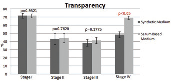
Transparency readings using a custom device to evaluate the degree of transparency at different phases of preservation in a synthetic medium and a serum-based medium. The readings are in the original scale in percentage transparency.
Viable ECD
The average VECD after preserving the corneas at stage II showed a loss of 0.51% (±7.18) in SA1 and 1.42% (±8.48) in CP (p = 0.8315). At stage III, higher damage due to mortality and therefore a lower value of VECD in both the media was observed, with 35.31% (±11.31) in SA2 and 34.51% (±11.92) in CM (p = 0.9002). However, the final preservation in a deturgescent medium showed a loss of 23.69% (±27.55) in SA3 and 40.72% (±30.94) in CJ (p = 0.2983). Thus, ECL and mortality (VECD) was seen in both media without any statistical difference (Fig. 3).
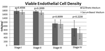
Viable endothelial cell density (VECD) readings using the formula that incorporates the visual endothelial cell density and mortality into a single parameter of VECD at different phases of preservation in a synthetic medium and a serum-based medium. The readings are in the original scale of cells/mm2.
Morphology
A total of 4.15% (±16.43) in SA1 and 5.50% (±20.21) in CP (p = 0.8934) was found at stage II. Further preservation at stage III showed higher morphologic damage in both the media, with 42.51% (±18.56) in SA2 and 44.52% (±16.57) in CM (p = 0.8344). However, the final preservation in a deturgescent medium showed a loss of 3.29% (±25.28) in SA3 and 17.55% (±26.52) in CJ (p = 0.3234). Although morphology loss was seen in both media, there was no statistical significance (Fig. 4). Endothelial cell morphology and damages are observed in Figure 5 for synthetic and Figure 6 for serum-based media series.
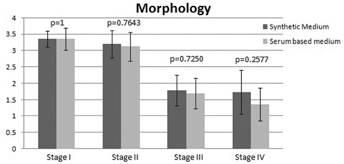
Morphology readings by masked observers at different phases of preservation in a synthetic medium and a serum-based medium. The readings are converted to a range of 0-4 (arbitrary units) using the conversion method from subjective to objective evaluation.
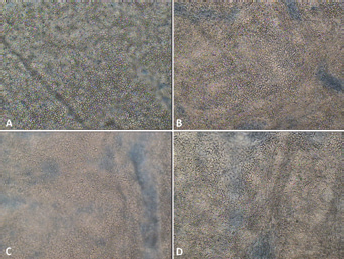
Endothelial cell morphology, integrity, and mortality found in the synthetic medium (100x magnification) at (A) stage I, (B) stage II, (C) stage III, and (D) stage IV of preservation.
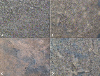
Endothelial cell morphology, integrity, and mortality found in the serum-based medium (100x magnification) at (A) stage I, (B) stage II, (C) stage III, and (D) stage IV of preservation.
Overall quality
After incorporating all the above parameters for determining the OQ of the cornea, it was found that the average OQ after preserving the corneas at stage II showed a loss of 41.62% (±4.42) in SA1 and 38.44% (±6.47) in CP (p = 0.3059). Further preservation at stage III showed an additional expected increase in thickness in both media, 33.93% (±11.00) with SA2 and 33.52% (±9.35) with CM (p = 0.9409). The final preservation in a deturgescent medium showed an improvement of the OQ at 3.26% (±14.07) with SA3 and 42.27% (±17.57) with CJ (p = 0.0007). The OQ deviation was seen in both media but had no statistically significant results at stage II and III. However, after the deturgescent preservation (stage IV), the value of OQ increased in the medium with dextran as compared to the one with poloxamer (Fig. 7). Thus, the final graft quality for transplantation was determined to be better in the serum-based media in combination with dextran as a deswelling agent.
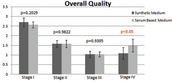
Overall quality readings after incorporating all the parameters together in a range of 0-4 (arbitrary units) at different phases of preservation in a synthetic medium and a serum-based medium.
Discussion
Although the tissues used in this study were unsuitable for transplantation, the results could be extrapolated to the transplantable corneas as the tissues were a high-grade research quality at stage I (relatively high VECD and better morphology). The inclusion criteria of our study were to use paired human corneas for each medium, i.e., synthetic versus serum-based, to allow better comparison. The subjective evaluation of corneal tissues between different eye banks makes it difficult to recommend a standardized method to evaluate a cornea and therefore select the best-suited medium for preservation. For this reason, there is a need for a well-defined way to characterize and measure the quality of the corneas before grafting. Therefore, a quality assessment system (OQ) was used that integrates all the necessary parameters required to accept a corneal graft (used for research purposes). This helps to make a baseline distribution of 0-4 and value the parameters almost equally.
The storage media SA2 and CM had no statistical differences when either of the parameters were considered; these storage media consist of synthetic ingredients and serum, respectively. Issues arise with synthetic medium after preservation of the corneas in deswelling medium with poloxamer, which is a crucial step. Dextran has been used widely in corneal preservation, be it cold storage or OC. It reduces the thickness of the cornea and reverts it back for ease of transplantation. Poloxamer has been used in medicine and pharmaceuticals. The first use of Poloxamer as a deswelling agent for corneal preservation was introduced by Zhao et al (11), in which the study included an investigation of efficacy and tolerance of different poloxamers for deswelling human corneas. Our study showed that the poloxamers did deswell the corneas but not as good as dextran. Dextran is also responsible for reducing the folds that are generated during the storage of the tissues, further leading to increased transparency. Therefore, it seems that a combination of serum-based medium with dextran-based deswelling medium results into a better quality cornea in terms of thickness, transparency and eventually a statistically significant overall quality. This further favors the use of a serum-based media.
In the literature, there is no document reporting the transmission of animal viruses due to the fact that animal serum was used and most of the safety concerns are ruled out. The potential transmission of BSE primarily comes from donors who have donated their corneas and were at risk of having BSE (from example, UK donors at the time of mad cow disease). Theoretically, there could be transmission of animal-derived viruses that could integrate in the genome and activate oncogenes; therefore, technically it would be better to develop a complete synthetic/animal-free media as it is safer overall.
The final quality of the graft is of primary importance and therefore tissue selection is of great concern. This study shows that the final quality of the tissue in serum-based media provides a higher maintenance standard (eye bank standards) of the quality of the tissue. However, development of a synthetic medium that could improve upon or show similar quality results as a serum-based medium would be of great benefit.
Footnotes
Financial support: None.
Conflict of interest: None.