
Abstract
Purpose
Elongation, thickening, and crowding of eyelashes are commonly seen after topical use of prostaglandin analog eyedrops for glaucoma treatment. The purpose of this study was to demonstrate the presence and characterize the location of prostaglandin analog F2α receptors (PGF2α) in human hair follicles.
Methods
In this observational clinical laboratory study, excised eyelid specimens following eyelid surgery were studied. Resected portions of eyelids were submitted for histopathologic evaluation. For immunohistochemistry evaluation, a polyclonal antibody directed against PGF2α was purchased from Cayman Chemical. The staining procedure was carried out on an automatic stainer.
Results
Out of 26 patients recruited, final analysis was conducted on 17 eyes of 15 patients. There were 10 men and 5 women (mean age 77 ± 14 years). Staining was detected only in hair follicles in the anagen stage (37 slices). No variation in pattern, distribution, or intensity of immunostaining was noted among sections of different individuals. Only the bulb and stem of the hair follicle stained positive. In the bulb, the strongest staining occurred in the matricular cells and in the inner sheath layer. In the stem, the strongest staining occurred in the Huxley layer of the inner sheath.
Conclusions
This immunohistologic study found that PGF2α receptors were located predominantly in the inner root sheath of the bulb and stem of eyelashes and expressed only in eyelashes in the anagen phase.
Introduction
Prostaglandin analogs have a prominent role in intraocular pressure reduction in ocular hypertension as well as glaucoma (1). Due to their potency and the lack of systemic side effects, they are often considered as first-line therapy (2). However, local side effects have been reported to a certain degree with all prostaglandin analogs. Elongation, hypertrichosis, and hyperpigmentation of the eyelashes are well-described side effects of topical prostaglandins and prostamide analogs that may sometimes reach blemishing levels (3-8). Iris and periocular hyperpigmentation, periorbital muscle atrophy, and both periorbital and orbital lipodystrophy may also occur (9-12). A variety of receptors for prostaglandin analogs in human tissues have been studied (EP1-4, PGE2, PGF2α, and others) (13, 14). Their presence throughout the human scalp hair follicle, specifically in the dermal papilla outer root sheath, has been proven (14, 15). The capability of PGF2α and FP receptors to introduce telogen to anagen transition in hair follicles has been documented (16).
Studies that have researched the EP and PF receptors in ocular tissues (cornea, trabecular meshwork, iris, ciliary body, choroid, and retina) documented a wider distribution of EP receptors (13). No studies have investigated and characterized the location of these receptors in human eyelid hair follicles. As the prostaglandin analog effect in the eyelid is mainly mediated via PGF2α (16), the purpose of this study was 1) to demonstrate the presence of PGF2α in human hair follicles; 2) to characterize their distribution along the hair follicle and its variation in different growth stages; and 3) to investigate the occurrence of heterogeneity in receptor expression among patients.
Methods
All data for the study were collected and analyzed in accordance with the policies and procedures of the institutional review board of Meir Medical Center and the tenets set forth in the Declaration of Helsinki.
A prospective consecutive design was used. The specimens studied were collected from patients undergoing lateral tarsal strip dissected as part of an ectropion repair procedure. When operated, none of the patients had a history of prostaglandin analog treatment or signs of inflammatory lesions or clinically appreciable signs of inflammation of the eyelids themselves. All the patients included in this study were Caucasian. For the sake of consistency, all the specimens were collected from the lower eyelid. Only specimens in which at least one hair follicle was identified were included in this study.
Histologic preparation
The resected portions of eyelids were immediately fixed in 4% buffered formalin and submitted for histopathologic processing and evaluation, according to routine protocols. Tissue sections of 4-μm thickness, stained by hematoxylin & eosin, were prepared from paraffin blocks.
Immunohistochemistry
The FP receptor polyclonal antibodies, directed against the PGF2α receptor, were purchased from Cayman Chemical (Ann Arbor, Michigan, USA; catalog no. 101802), as described and validated by previous studies (17-21). The staining procedure was carried out on a Ventana Benchmark automatic stainer (manufacturer: Ventana, Tucson, Arizona, USA). The FP receptor antibodies were diluted to 1:1100. The primary antibody was detected by streptavidin, which was conjugated to a biotinilated secondary antibody. The FP receptor and antibody complex was expressed as a red staining in the tissue sections.
Histopathologic review
All specimens were reviewed by the pathologist (D.K.) and staining was graded using a qualitative scale ranging between 0 and 2:0 = light staining, similar to baseline staining observed in the conjunctival epithelium; 1 = moderate staining; and 2 = intense staining.
Results
Overall, 26 patients were recruited for this study. Final analysis was conducted on 37 slices examining 17 eyes of 15 patients in which at least one hair follicle was identified. There were 10 men and 5 women. The mean ± SD age of the patients was 77 ± 14 years. The number of slices studied per patient varied from 1 to 7 with a median of 3. The slices were taken from the right eye in 7 patients, left eye in 6 patients, and both eyes in 2 patients.
All specimens contained hair follicles in the anagen phase while hair follicles in the catagen phase were detected in 4 specimens only. Staining was detected only in hair follicles in the anagen stage. No variation in pattern, distribution, and intensity of immunostaining was noted among sections of different individuals. Table I depicts the immunolocalization and grade of the PGF2α receptors in the hair follicles.
Location and degree of PGF2α receptor staining in hair follicles
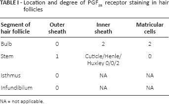
NA = not applicable.
Of the 4 parts of the hair follicle (bulb, stem/suprabulbar, isthmus, and infundibulum), only the first 2 stained positive for the receptors. In the bulb, the strongest staining occurred in the matricular cells and in the inner sheath layer. No staining occurred in the outer sheath of the bulb (Fig. 1).
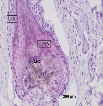
A longitudinal section through hair bulb and stem. There is strong cytoplasmic staining of the inner sheath (INS) and of the matricular cells (MC). Note that there is no staining in the outer sheath (OS).
The inner sheath divides into 3 apparent layers at the stem (suprabulbar) level (Henley, Huxley, and cuticle). The presence of PGF2α receptors was observed mainly in the Huxley layer, while no staining occurred in the Henley layer. Faint staining occurred in the outer sheath cells of the stem (Fig. 2).
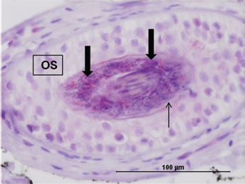
A transverse section of hair follicle at level of stem. Intense (grade 2) red staining in Huxley's layer of inner sheath (wide arrows). No staining in Henle's layer of inner sheath (thin arrow). Faint (grade 1) cytoplasmic staining of outer sheath cells (OS).
In general, whenever staining was detected, it occurred mostly in the cytoplasm of the cells with slight membranous staining. In the matricular cells, due to the paucity of cytoplasm, it was hard to determine whether this staining was solely cytoplasmic or also perinuclear.
Discussion
In this study, we demonstrated the presence of PGF2α receptors in human eyelid hair follicles. Immunohistochemical staining for PGF2α receptors was noted in the bulb and stem segments in hair follicles at the anagen phase. Staining occurred mostly in the cytoplasm of the cells. No heterogeneity in receptor expression was demonstrated among patients.
The PGF2α receptor is a membranous receptor. In our study, staining of the PGF2α receptor occurred mostly in the cytoplasm. This is in concordance with findings by Schlötzer-Schrehardt et al (13), who reported that staining of the receptor in human ocular tissues occurred in the cytoplasm. They reported that the most prominent localization of the staining occurred in the nuclear envelope and occasionally on outer mitochondrial membranes, on rough endoplasmic reticulum, and in cytoplasmic vesicles.
While there is abundant evidence on the effect of prostaglandin analogues on the growth of eyelashes, there is a paucity of histologic studies profiling the location of the receptors. To date, one cannot predict which patient will develop the adverse side effects. Even brief exposure to an ophthalmic prostaglandin analog can be associated with eyelash change (3). In fact, bimatoprost solution 0.03% (Latisse; Allergan, Inc., Irvine, California, USA) was found to be effective in enhancing eyelash growth (22).
This is the first study we are aware of to investigate the distribution of PGF2α receptors in the hair follicles of human eyelids. Schlötzer-Schrehardt et al (13) demonstrated the expression and localization of FP receptor proteins by immunohistochemistry in 10 normal human donor eyes. No comment was made on the expression of the receptor in the hair follicles. Colombe et al (15) demonstrated, on 3 scalp hair biopsies, that FP expression was essentially restricted to the outer root sheath companion layer and dermal papilla, and speculated that this could explain the biological effect of PGF2α on hair regrowth. Whether the difference in locations of staining (outer sheath versus inner sheath) between the 2 studies may be attributed to the fact that these are 2 different types of hair (scalp hair versus eyelashes) is unclear.
Our findings of staining of PGF2α receptors, preliminary in hair follicles in anagen stage, are in agreement with the understanding that hair growth occurs at this stage (23).
In this study, no apparent diversity in the degree of hair follicle staining among the specimens was noted. There are several possible explanations for this observation. This may be a result of the relatively small number of specimens. Another possible explanation is the lack of diversity in the patients; none was being treated with prostaglandin analogs and none had hypertrichosis (24, 25). The lack of variation may also be a result of the older age of our study group. Although the range was 42 to 94 years, the mean age was 77.
The hypertrichosis of eyelashes associated with prostaglandin and prostamide analogs usually occurs together with darkening of the eyelashes. This staining technique used did not efficiently evaluate the staining of the melanocytes as previous bleaching was not performed. Future studies should characterize the PGF2α staining of melanocytes.
The small number of specimens as well as the method of staining used precludes us from quantitative evaluation of PGF2α receptors.
In this study, all specimens were taken from lower eyelid specimens dissected as part of an ectropion repair procedure. Previous studies have demonstrated that the upper eyelashes are both longer and more numerous than their lower counterparts (26, 27). One study demonstrated nearly 2 times the amount of lashes in the upper eyelid versus the lower eyelid (28). Therefore, conclusions from this study may not fully apply to lashes from the upper eyelid. In addition, except for the mildest cases, ectropion is connected with exposure of conjunctiva, which causes inflammation. This was dealt with by only including specimens from patients with no clinically apparent signs of inflammation; however, subclinical inflammation may have affected the expression of prostaglandin receptors in this study (29, 30).
In this study, none of the patients was being treated with prostaglandin analogs. Future studies should compare the staining in eyes treated with prostaglandin analogs to those not being treated with prostaglandin analogs.
This study focused on the location of the receptors and did not offer insight into the function of PGF2α in human eyelid hair follicle growth. Further studies should focus on this issue.
In summary, we found that prostaglandin PGF2α receptors were located predominantly in the inner root sheath of the eyelashes and expressed in eyelashes at the anagen phase.
Footnotes
Financial support: No financial support was received for this submission.
Conflict of interest statement: None of the authors has conflict of interest with this submission.
Meeting presentation: Presented at the 23rd annual meeting of the American Glaucoma Society, San Francisco, California, USA, March 1, 2013.