






Abstract
Purpose
To present the case of a man with branch retinal artery occlusion (BRAO) and concomitant patent foramen ovale (PFO), which was first diagnosed during the evaluation of BRAO.
Methods
A 35-year-old man presented with blurred vision in the left eye for 2 days. His best-corrected visual acuity (BCVA) was 6/6 in the right eye and 6/12 in the left eye. He had no ophthalmic or medical history.
Results
Ophthalmologic examination revealed a left inferior BRAO, confirmed by fluorescein angiography. The patient underwent a thorough diagnostic workup. Cardiologic examination with transesophageal echocardiography revealed a right-to-left shunt across a PFO. The patient was treated with clopidogrel and at the 15-day review, BCVA in his left eye was 6/7.5 and the retinal edema was resolved, as detected on optical coherence tomography, but relative afferent pupillary defect was persistent. The patient had developed no other embolic event at the 9-month follow-up, having undergone an operation for PFO repair.
Conclusions
Patent foramen ovale, although usually asymptomatic, should be considered among the potential causes of BRAO, especially in young patients. Prompt diagnosis and liaison with cardiologists is important to help prevent ocular or systemic embolic events and associated morbidity.
Keywords
Introduction
Patent foramen ovale (PFO) is considered to be a benign condition in which the normal fetal interatrial communication of the heart persists after birth and fails to close normally within the first year of life, creating a potential right-to-left shunt (1). It occurs in approximately 25% of the general population, as detected by transesophageal echocardiography (1). Although PFO is usually asymptomatic and rarely accompanied by cerebrovascular episodes such as stroke, it predisposes the individual to embolic events, probably involving the eye (1, 2).
Branch retinal artery occlusion (BRAO) is a vascular occlusive disease in which the pathogenesis, clinical characteristics, and management differ from central retinal artery occlusion (3). In the literature, there has been reported an association between central retinal artery occlusion and PFO (3), as well as among retinal artery obstruction, migraine, and PFO (4), while only one case with BRAO and PFO has been described (2). We present the case of a young man with BRAO and concomitant PFO, which was first diagnosed during the evaluation of BRAO.
Case report
A 35-year-old man presented to the Accident and Emergencies Department of our hospital with blurred vision in the left eye for 2 days. He had no ophthalmic or medical history, including history of migraine or vasospasm. There was no history of cigarette or alcohol use.
His best-corrected visual acuity (BCVA) was 6/6 in the right eye and 6/12 in the left eye, not improving with pinhole. A left relative afferent pupillary defect (RAPD) was present and a left superior visual field defect on confrontation was demonstrated. Color vision, anterior segment examination, and intraocular pressures were normal in both eyes. Dilated funduscopy revealed a calcified white embolus at the third bifurcation of the inferior temporal artery in the left eye, as well as left inferior BRAO, which was confirmed by fluorescein angiography (Fig. 1), while it was unremarkable for the right eye. Optical coherence tomography (OCT) revealed mild retinal edema in the left eye (Fig. 1). As the history of blurred vision was more than 24 hours, the patient underwent no treatment, except oral aspirin, while he was referred to undergo a thorough diagnostic work-up.
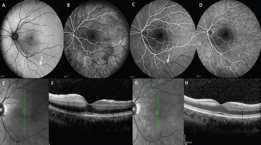
Autofluorescence (
Systemic neurologic, chest, and abdominal examination were unremarkable. Complete blood count, urea and electrolytes, clotting and inflammatory markers (erythrocyte sedimentation rate and C-reactive protein), as well as thrombophilia screen were within normal range. Blood pressure, pulses, heart sounds, electrocardiogram, and carotid Doppler ultrasound were all normal. Additionally, 24-hour Holter monitoring was done to exclude atrial fibrillation and had normal results. Cardiologic examination with transesophageal echocardiography revealed a right-to-left shunt across a PFO (Fig. 2), although the patient had never experienced any cardiac/systemic symptoms. He was treated with clopidogrel and at the 15-day review, BCVA in the left eye was 6/7.5, and retinal edema was resolved as detected on OCT (Fig. 1), but RAPD was persistent, as well as the visual field defect. The patient underwent an operation for PFO repair, after which he developed no other embolic event at the 9-month follow-up.
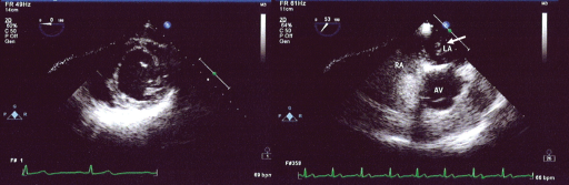
Transesophageal echocardiography illustrates abnormal passage of saline microbubbles (white arrow) through the patent foramen ovale.
Discussion
Retinal artery occlusion in young patients is a rare clinical entity and few cases with PFO as a cause have been described (3). As far as BRAO and its association with PFO, Shoeibi et al (2) published the case of a 29-year-old woman with superior visual field defect and diagnosed with BRAO in the context of PFO. Our case is the second in the literature reporting association between BRAO and PFO.
The patient received no treatment at baseline, as blurred vision was established in more than 24 hours. He underwent a thorough medical examination and PFO was detected. Most patients with PFO and no other systemic problems generally receive no treatment. When associated with otherwise unexplained ophthalmic or neurologic events, treatment may be medical or surgical (1). Our patient was treated with aspirin and clopidogrel and he underwent surgical repair of the PFO, presenting no other embolic events at the 6-month follow-up.
Branch retinal artery occlusion most commonly occurs secondary to an embolus, which typically originates within vessels upstream, where it dislodges and travels within the circulatory system to ultimately become lodged downstream in a vessel with a smaller lumen. The most common emboli are cholesterol from aorto-carotid atheromatous plaques, platelet-fibrin from thrombotic disease, and calcific emboli from cardiac valvular disease (5). The causal relation between PFO and thromboembolic events has not been established. The suggested mechanisms are likely complex, including paradoxical embolism from the peripheral venous system, embolization from thrombi formed within the atrial septum, and the formation of thrombus as a result of transient atrial arrhythmias (5). As a result, if calcific emboli were detected in funduscopy or fundus imaging, a thorough cardiologic examination using echocardiography is advised.
Transesophageal echocardiography plays an important role in the diagnosis of PFO in asymptomatic patients and should be considered for patients with BRAO, especially when they are young (6). Inatomi et al (6) referred that 2 out of 13 cases with retinal artery occlusion and cardiac abnormalities presented with PFO. In our case, transesophageal echocardiography revealed the PFO, while other examinations had normal results.
Another interesting finding in our case was the baseline reduced BCVA, which was the cause of the patient's visit to the hospital. As BRAO was far from the fovea, the decrease in visual acuity could be potentially attributed to retinal edema, which was confirmed on OCT, more in the inferior fundus. Histopathologically, BRAO reveals ischemia in the corresponding retinal quadrant, marked in inner retinal edema in the initial stages, which could affect a larger region than that defined by presence of the exact emboli (7).
In conclusion, the principal message of this case report is that ophthalmologists should consider PFO in patients with BRAO, especially if they are young, without any other problems or risk factors. It is essential to consider PFO in the differential diagnosis of BRAO causes and to refer the patient for further examination. Prompt diagnosis and liaison with cardiologists is important to help prevent ocular or systemic embolic events and associated morbidity.
Footnotes
Financial support: No financial support was received for this submission.
Conflict of interest: None of the authors has conflict of interest with this submission.