






Abstract
Purpose
To demonstrate that in case of absence of capsular support intraocular lens (IOL) scleral fixation is both effective and stable over years.
Methods
A total of 13 eyes from 13 patients who underwent an IOL scleral fixation according to Lewis suturing technique between January 2001 and December 2008 were studied. Patients underwent a complete ophthalmologic evaluation. The IOL stability was assessed using slit-lamp and anterior segment optical coherence tomography (AS-OCT) examination. The IOL stability was evaluated in terms of centration and tilting. All the knots were photographed and their integrity assessed.
Results
Follow-up was 60–129 months. Eleven knots appeared evident and undamaged, 6 knots were eroded, and 9 knots were not detectable. All IOLs were stable in the sulcus. Two patients presented a slight decentration of the IOL at the slit-lamp examination, while the AS-OCT demonstrated slight tilting of the lenses in 4 patients.
Conclusions
The Lewis technique for IOL scleral fixation is an optimal surgical technique in case of absence of capsular support. No IOL luxation in the vitreous chamber has been reported; only 2 of the 10 patients with at least one eroded knot presented a minimal decentration of the lens with no influence on visual acuity. Although knot erosion is not an uncommon occurrence, IOL remains stable in the long term, probably due to a fibrotic process around the suture, and the IOL haptics, which prevent IOL dislocation.
Introduction
Cataract surgery, with its microincision variants (coaxial microincision cataract surgery, bimanual microincision cataract surgery), is the most performed intraocular surgical procedure worldwide (1–6). Nowadays, owing to continuous technological innovation, cataract surgery has become more efficient and safe, offering excellent outcomes for patients.
Integrity of the capsular bag and of the zonular suspension are key for a successful procedure. Due to congenital (Marfan syndrome, homocystinuria, Weill-Marchesani syndrome) or secondary (ocular trauma, severe myopia, pseudoexfoliation, complicated cataract surgery, chronic uveitis, previous vitreoretinal surgery, elderly) weakness, the surgeon may not be able to perform in-the-bag implantation and needs alternative techniques for intraocular lens (IOL) positioning. Possible solutions are anterior chamber angle-fixated lenses, anterior chamber iris-fixated lenses, posterior chamber iris-sutured lenses, and posterior chamber scleral-sutured lenses (1).
Sulcus scleral fixation, first described by Malbran in 1986 (7), is an ab externo technique consisting of suturing the haptics of the IOL to the scleral wall, covering the sutures with 2 opposite scleral flaps in order to protect the knots.
Many authors have proposed variations of the original technique, which mainly differ in the surgical approach (ab externo or ab interno technique), in the haptic fixations, in the number of sutures needed to fix the IOL (ranging from 2 to 4), and in the method to avoid erosion of sutures over time (1).
In the early 1990s, James S. Lewis (8, 9) published a variation of the original technique in which the suture knots are buried and do not require scleral flaps, covering the suture only with conjunctival tissue.
Suture erosion, due to exposure, has always been considered the weakest point for long-term stability of the IOL and may cause rotation, tilting, and, in severe cases, luxation in the vitreous chamber (10–12).
The main outcome of this study was to demonstrate whether, in case of absence of capsular support, Lewis technique for IOL scleral fixation is effective and stable over time, evaluating the suture conditions and the IOL positioning using anterior segment optical coherence tomography (AS-OCT).
To our knowledge, no other study on the correlation of suture conditions and IOL position by AS-OCT in patient who underwent a Lewis technique scleral fixation has been published.
Patients and methods
Forty consecutive patients who underwent IOL scleral fixation according to Lewis suturing technique between January 2001 and December 2008 were considered. This retrospective observational study enrolled 13 eyes from 13 patients who agreed to take part to this study. All patients provided informed consent.
All patients were operated by the same experienced surgeon (G.M.C.) with the same surgical technique. All patients were examined at the Institute of Ophthalmology, University of Modena, and underwent a complete ophthalmologic evaluation (corrected distance visual acuity, endothelial biomicroscopy, slit-lamp examination, intraocular pressure calculation, anterior segment imaging at slit-lamp, camera fundus oculi examination).
The integrity of the suture knots was established by 3 independent operators observing the anterior segment photographs of the sutures assessing whether they were intact, eroded, or not detectable.
Assessment of IOL displacement was established through a slit-lamp examination and AS-OCT (Visante AS-OCT, Carl Zeiss Meditec, Jena, Germany), distinguishing well-positioned IOL from IOL malpositioning.
All data were recorded in an Excel database (Microsoft Excel 2010, Microsoft Office Professional Plus 2010, Redmond, WA, USA) and analyzed.
Surgical technique
A peritomy was made from 8 to 2 o'clock and a superior 7.0 mm corneoscleral wound was constructed.
A 10–0 polypropylene suture with a straight needle was passed from one scleral side to the opposite at 0.8–1 mm from the limbus. The needle was then turned around and passed back into the eye and emerged at the original scleral bed. Both sutures were withdrawn, cut, and sutured to the corresponding eyelet of the IOL (IOL Alcon® CZ70BD). The IOL was inserted through the corneoscleral wound. The sutures were tied and the knots rotated, buried, and covered with conjunctiva (8, 9).
Anterior segment optical coherence tomography examination
Noncontact AS-OCT examination (Visante AS-OCT, Carl Zeiss Meditec) was performed by the same examiner. To standardize the image capture, patients were asked to look straight at the fixation point so that the OCT beam would be as perpendicular as possible to the central corneal surface.
Four images from each eye on the vertical and horizontal axis were evaluated by 3 independent operators; the examiners looked for evidence of IOL displacement on the frontal plane and on the anteroposterior axis or IOL luxation in the vitreous chamber.
Results
The follow-up ranged from 60 to 129 months. Corrected distance visual acuity was 0.46 ± 0.42 with a mean astigmatism of 4.08 ± 2.66 D. Intraocular pressure was 15.31 ± 4.82 mm Hg (range 11–25 mm Hg). Mean endothelial cell density was 2446 ± 469.22 cells/mm2 (range 1709–3125 cells/mm2).
A total of 26 knots were analyzed: 11 knots (42.31%) were clearly evident and undamaged, 6 knots (23.07%) were evident but eroded, and 9 knots (34.62%) were not detectable (Fig. 1).
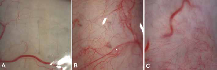
(A) Evident and undamaged knot. (B) Evident but eroded knot. (C) Not detectable knot.
At slit-lamp examination, the IOL was well-positioned in 11 cases (84.62%) and displaced in only 2 patients (15.38%) (Fig. 2).
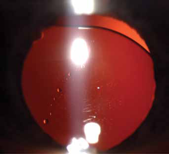
Intraocular lens slight displacement at slit-lamp examination.
At AS-OCT, the IOL was correctly positioned in the sulcus with no evidence of tilting or decentration in 9 cases. In 4 cases (30.77%), there was evidence of slight displacement in terms of misalignment between the IOL-optic plate and a hypothetical plane passing through the iris (Fig. 3).
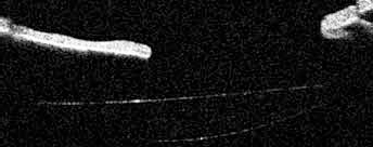
Intraocular lens slight displacement at anterior segment optical coherence tomography evaluation.
Discussion
It is well-known that the scleral fixation technique reduces the rate of complications associated with the iris-claw IOL. Numerous transscleral suturing techniques have been proposed through the years, but the most used approach is to implant a large diameter optic with a modified haptic for the suture.
One of the most discussed problems with the various suturing techniques is erosion of the sutures over time, which depends on the protection offered by the creation of scleral flaps or the conjunctiva, which may result in IOL dislocation or luxation in the vitreous.
To avoid this problem, knotless techniques have been developed, but it is still important to establish the risk of luxation in the vitreous chamber for the IOL implanted with the traditional technique, above all in case of suture erosion.
Strengths of this study include assessment of the correlation between knot erosion with slit-lamp pictures and IOL position using the AS-OCT to detect whether the implants are in the correct position in the posterior chamber or are subluxated or luxated, and the relatively long follow-up (from 60 to 129 months).
Several reports about postoperative erosion of polypropylene knots in the standard technique with scleral flaps have been published, but none has assessed the IOL position. In the literature, the rate of suture rupture is extremely variable, ranging from 0.58% to 26.2%, depending on follow-up, patient age, patient lifestyle, and suturing technique (9–12).
Our long-term follow-up shows that rupture or erosion of the suture knots are common findings in the Lewis technique. Our overall breakage rate is 57.69% for all types of sutures, normal, eroded, and not detectable, and is significantly higher than in other studies. Considering that the surgeon and the surgical technique were the same for all the patients enrolled, the erosion probably depends on suture damage over time due to exposure.
The 15.38% of displacement at the slit-lamp examination and the 30.77% of malpositioning at the AS-OCT were minor, only detectable through anterior segment imaging instruments and with a sensible degree of mydriasis. No IOL luxation in the vitreous chamber was reported. These findings were not clinically significant in terms of final corrected distance visual acuity, which was satisfactory for all the patients and overall high, considering that many of the patients had comorbidities such as glaucoma, prior retinal detachment surgery, or maculopathy.
Despite this evidence, in all our patients, IOLs were stable in the sulcus in the long term, probably due to a fibrotic process around the sutures and the IOL haptics, which prevents luxation of the lens. No patient needed IOL resuturing or an IOL exchange.
In conclusion, IOL scleral fixation by Lewis technique is an optimal surgical technique for patients with absence of capsular support; although knot erosion is not uncommon, the IOL remains stable in the long term.
Footnotes
Financial support: No financial support was received for this submission.
Conflict of interest: None of the authors has conflict of interest with this submission.
Meeting presentation: Presented as a free paper at the 18th ESCRS Winter Meeting, Ljubljana, Slovenia, February 14–16, 2014.