






Abstract
Purpose
To report a case of extreme corneal flattening after initially uneventful Descemet stripping endothelial keratoplasty (DSEK).
Methods
A 76-year-old man underwent uneventful DSEK for pseudophakic bullous keratopathy (PBK) in his left eye. Postoperative evaluations were performed using anterior segment optical coherence tomography and biomicroscopy.
Results
The DSEK graft showed full adherence to the host stroma. Two weeks after surgery, the transplanted cornea developed pronounced flattening of the central corneal curvature accompanied by graft folds and remained unchanged until the most recent follow-up 20 weeks after surgery.
Conclusions
Severe corneal flattening may be an undescribed complication occurring after uneventful DSEK, without any concomitant ocular pathology or detectable cause.
Keywords
Introduction
After its introduction by the Netherlands Institute for Innovative Ocular Surgery in 2003, Descemet stripping endothelial keratoplasty (DSEK) has been adopted worldwide as an alternative to penetrating keratoplasty (PK) for the treatment of corneal endothelial disorders (1–2–3). One of the main advantages of DSEK over PK is that the recipient anterior corneal surface is not compromised by corneal incisions or sutures, so that the preoperative to postoperative change in refraction and astigmatic error is minimized.
The aim of the current study is to document a previously undescribed complication: extreme corneal flattening of unknown cause after uneventful DSEK.
Case report
A 76-year-old man presented with pseudophakic bullous keratopathy in his left eye. Preoperative central pachymetry was 899 µm, best-corrected visual acuity (BCVA) was counting fingers, and topography showed a virtually spherical anterior corneal contour. The contralateral eye showed a mild cataract and was otherwise unremarkable. The patient denied previous corneal surgery, and the medical history did not reveal evidence of Down syndrome, Ehlers-Danlos syndrome, Marfan syndrome, cornea plana, or any syndrome associated with anterior segment dysgenesis. The patient had a medical history of hypertension and ischemic heart disease.
After the patient signed an institutional review board–approved informed consent, an uncomplicated DSEK procedure was performed under local anesthesia in the left eye in October 2014 at the Helmholtz Moscow Research Institute of Eye Disease.
Donor tissue preparation
The donor globe was harvested and processed at the eye bank of the same institute within 6 hours after death. Donor age was 67 years, and the preoperative endothelial cell count measured 2350 cells/mm2. A nonperforating limbal incision, 5.0 mm in width, was made 1-2 mm from the limbus. A corneal tunnel was dissected extending 1.0 mm into the clear cornea. Through a side port at 10 o'clock, the anterior chamber was completely filled with air to create an air-to-endothelium interface to optically visualize the depth of dissection. With dissection spatulas (Melles dissection spatulas; DORC International, Zuidland, the Netherlands), a manual stromal dissection was made at approximately 95% stromal depth using the air-to-endothelium reflex to monitor dissection depth through the operating microscope. The stromal dissection was extended up to the limbus over 360 degrees to complete the procedure. After the dissection was completed, a 16-mm corneoscleral rim was excised from the globe.
Thin-DSEK surgery
A descemetorhexis was performed under air with an inverted Sinskey hook through a 0.9-mm paracentesis at 10 o'clock. The manually dissected DSEK graft was trephined to obtain a 9.0-mm graft and was inserted as a taco through a self-sealing 5.0-mm scleral tunnel incision at 12 o'clock using a 30-G needle and anterior chamber glide. The graft was centered and stabilized with a complete air fill of the recipient anterior chamber. During the procedure, there was no hemorrhage and no sign of vitreous strands in the anterior chamber of the eye.
Postoperative medication included tobramycin/dexamethasone (Tobradex, Alcon, Fort Worth, TX, USA) 4 times daily and 3 times daily in the first and second week, respectively. Afterwards, dexamethasone was tapered by 1 drop every 2 months for 6 months from 3 times to 1 time daily.
Postoperative course
The immediate postoperative period was uneventful and the graft showed complete adhesion to the host posterior stroma. However, 2 weeks after surgery, the patient returned with a significant change in the corneal curvature. The BCVA was counting fingers and biomicroscopy showed large tissue folds converging vertically towards the inner lip of the main incision (Fig. 1A). The incision and side ports were fully sealed, there was no sign of intraocular inflammation, and the intraocular pressure was normal.
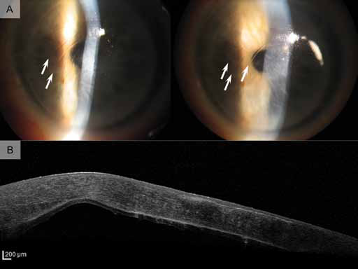
Slit-lamp and anterior segment optical coherence tomography images of the recipient cornea 2 weeks after Descemet stripping endothelial keratoplasty.
Anterior segment optical coherence tomography imaging (Spectralis, Heidelberg Engineering, Heidelberg, Germany) showed a deformation of the entire cornea, with pachymetry readings ranging from 618 µm to 1049 µm across the graft, but without any intraocular abnormalities (Fig. 1B). Postoperative topography readings were outside the Pentacam measurement range due to the extremely flattened corneal contour. Preoperative topography of the contralateral eye did not show any abnormalities. Twenty weeks after the surgery, the examination revealed no significant changes. The BCVA was 0.04 (20/500) and the cornea was clear with persistent tissue folds and a descending thickness in the range of 625 µm to 821 µm (Fig. 2A). Topography (Galilei G4, Ziemer Ophthalmic Systems AG, Port, Switzerland) showed an asymmetric and irregular corneal curvature with marked flattening of the inferior segment (Fig. 2B).
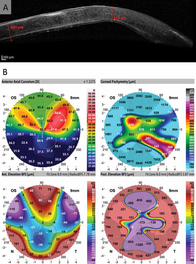
Anterior segment optical coherence tomography image and topography scans 20 weeks after surgery.
Discussion
Our study shows that uneventful DSEK can be complicated by severe deformation of the transplanted cornea, in the absence of intraocular hemorrhage, vitreous strands, or any other concomitant anterior segment pathology. Clinically, corneal flattening appeared to be associated with a contraction of the DSEK graft and prominent folds towards the main (superior) incision.
Graft folds have been reported in deep lamellar endothelial keratoplasty and DSEK grafts and have been related to variations in graft thickness secondary to manual dissection of the donor tissue or disparities in donor-host shape or diameter (4, 5). However, contraction of the graft inducing a cornea plana has not been reported previously. The most likely explanations could be the presence of an intraocular structure leading to local tissue contraction (like vitreous strands to the incision through which the graft was inserted) or incision leakage. However, both intraoperatively and postoperatively, no evidence for such conditions could be detected. Endothelial grafts may be subjected to a wound-healing response that may cause extreme tissue contraction in rare cases (6, 7).
Footnotes
Financial support: No financial support was received for this submission.
Conflict of interest: G.R.J. Melles is a consultant for DORC International BV/Dutch Ophthalmic USA and SurgiCube International.