






Abstract
Purpose
To report the safety and efficacy of intravitreal recombinant tissue plasminogen activator (rtPA) with gas for managing submacular hemorrhage.
Methods
Patients with submacular hemorrhage centered in or close to the fovea underwent hemorrhage displacement with intravitreal injection of rtPA (50 μg/0.05 mL) followed by gas injection (0.3 mL SF6). Anatomic and visual outcomes are described.
Results
Exudative age-related macular degeneration (AMD) (n = 4) and blunt trauma (n = 2) were the etiologies of submacular hemorrhage in this sample. Intravitreal injection of rtPA decreased the extent of submacular hemorrhage in all eyes, with complete hemorrhage displacement in 2 and partial displacement in 4. Visual acuity remained stable (n = 4) or improved (n = 2) after the procedure. Improvements in anatomic and visual outcomes were less evident in exudative AMD cases, which also had longer hemorrhage duration. Recurrence occurred only in 1 eye. No evident rtPA-associated retinal toxicity was observed.
Conclusions
Untreated submacular hemorrhage has poor visual prognosis. Our results suggest that rtPA injection is a minimally invasive, simple, inexpensive procedure with few related complications. Cost-benefit of this injection seems acceptable.
Introduction
Submacular hemorrhage may occur in several diseases and may lead to acute central vision loss. The most common causes are neovascular age-related macular degeneration (AMD) and retinal arterial macroaneurysm (1). Submacular hemorrhage causes irreversible damage to retina through several mechanisms, including direct toxic effects, limitation of passage of nutrients to the retina, and contraction of the outer retinal layers due to clot formation (2). Surgical removal of submacular blood through posterior pole retinotomy, proposed in the late 1980s, was abandoned due to poor visual outcome associated with large atrophy of retinal pigment epithelium and frequent retinal detachment (30%-37%) after surgery (3). Recombinant tissue plasminogen activator (rtPA) is a protease that converts plasminogen into plasmin and promotes the resolution of fibrin meshwork (4). Several modified approaches have been proposed with the main objective of submacular hemorrhage displacement. Less invasive approaches reported are intravitreal rtPA with or without gas injection and only gas intravitreal injection. Other studies also report the use of intraoperative subretinal rtPA as a vitrectomy adjunctive. However, it is not clear which method is generally most useful (1).
In this study, we report our experience with safety and efficacy of intravitreal rtPA with gas injection for managing patients with submacular hemorrhage due to neovascular AMD and trauma.
Cases
Patients with submacular hemorrhage centered in or close to the fovea were enrolled. After approval by the medical ethics committee, each patient signed an informed consent form. Intravitreal injections were done through a 30-G needle by pars plana in the inferotemporal quadrant, 3.5 mm to 4 mm posterior to the limbus. First, 50 µg rtPA in 0.05 mL was injected into the vitreous cavity. Patients were instructed to remain in a supine position for 30 minutes to allow the rtPA to set and then 0.3 mL gas injection (SF6) was performed. Anterior chamber paracentesis was done before gas injection and if needed at the end of the procedure. Patients were instructed to keep a face-down position until gas resorption, so that the submacular pressure would displace the submacular hemorrhage peripherally. One patient with submacular hemorrhage secondary to blunt trauma had a multilevel hemorrhage characterized by localization beneath the internal limiting membrane (ILM) as well as in the subretinal space. In this patient, an intravitreal injection of rtPA (50 µg/0.05 mL) was followed (30 minutes later) by pars plana vitrectomy (PPV) with ILM removal and gas tamponade with C2F6.
Six eyes of 6 patients (4 men and 2 women) were included. The average age of the patients was 74.3 years (range 46-86 years). All patients had at least 1 year of follow-up. They all had sudden vision loss with a large black spot blocking their central view. Table I summarizes relevant clinical information. Exudative AMD (n = 4) and blunt trauma (n = 2) were the etiologies of submacular hemorrhage in our sample. Patients with exudative AMD were also on treatment with intravitreal bevacizumab. All eyes showed extensive retinal hemorrhage occupying more than two-thirds of the macular area. After treatment, the extent of subretinal hemorrhage decreased in all eyes (Fig. 1). Submacular hemorrhage secondary to blunt trauma achieved better anatomic results such as complete hemorrhage displacement. In exudative AMD cases, hemorrhage displacement was only partial and fibrous scar was more prominent. Residual subretinal hemorrhage gradually disappeared 1 to 4 months after the procedure in all patients. Visual acuity remained stable in eyes with submacular hemorrhage secondary to AMD. Visual acuity improvement was evident in submacular hemorrhage secondary to blunt trauma. Six months after rtPA plus gas intravitreal injection, a submacular hemorrhage recurrence occurred in one eye (exudative AMD: patient 5) with additional vitreous hemorrhage. A PPV was performed. No major complications occurred intraoperatively. Moreover, no evident rtPA-associated retinal toxicity, such as exudative retinal detachment or retinal pigment epithelium lesions, occurred. However, one patient had retinal detachment with inferotemporal retina dialysis due to gas injection, 3 weeks after the procedure. No other complications were observed.
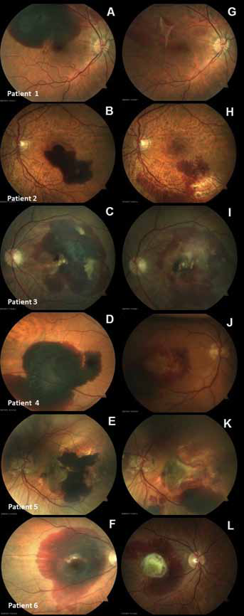
Preoperative (
Clinical characteristics of 6 patients with submacular hemorrhage before and after treatment with recombinant tissue plasminogen activator and gas
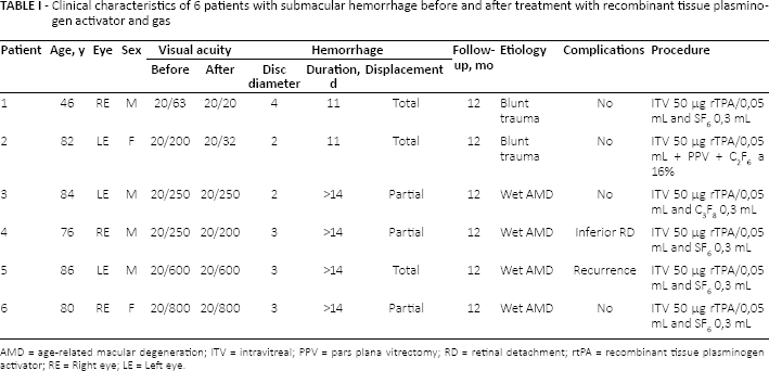
AMD = age-related macular degeneration; ITV = intravitreal; PPV = pars plana vitrectomy; RD = retinal detachment; rtPA = recombinant tissue plasminogen activator; RE = Right eye; LE = Left eye.
Discussion
Intravitreal rtPA and gas was found to be relatively effective for displacement of submacular hemorrhage. No evident rtPA-associated retinal toxicity was seen. Patients with submacular hemorrhage secondary to blunt trauma had complete hemorrhage displacement and significant visual acuity improvement. However, patients with hemorrhage due to exudative AMD had poorer anatomic and visual outcomes. The previous macular scar seems to act as an anatomic barrier to blood displacement and additionally to visual improvement. However, given the safety of using intravitreal rtPA and the fact that if untreated the risk of visual worsening is high, this procedure should be always considered in patients with submacular hemorrhage secondary to exudative AMD. Longer duration of hemorrhage (>14 days) could also have contributed to poorer treatment effect in exudative AMD cases. Based on previous studies that compare efficacy of rtPA considering the interval between the hemorrhage onset and the surgical intervention, it seems best to treat patients within 14 days. Liquefaction and displacement of the clot is most likely; moreover, irreversible retinal damage occurs after that period (1). The underlying disease and longer hemorrhage duration may be the reasons for nonimprovement in visual acuity and only partial displacement in patients with exudative AMD. Is difficult to dissociate these 2 facts (etiology and hemorrhage duration) in order to evaluate which contributed more to poor visual prognosis. Submacular hemorrhage has a low rate of spontaneous resorption and if untreated the prognosis is poor. The underlying disease has a strong impact on the visual outcome (5). The treatment options are varied and have evolved over the years, and comparisons are difficult due to lack of homogeneity between studies (1). The rtPA dose used is different between studies, varying between 25 µg and 100 µg. Based on available data, it seems that the lowest effective and safe dose of subretinal or intravitreal rtPA is 25 µg, and it seems best to treat patients within 14 days after the hemorrhage onset (1). This minimally invasive therapeutic approach is simple, inexpensive, and had few complications. Cost-benefit of this minimally invasive procedure seems acceptable.
Footnotes
Financial support: No financial support was received for this submission.
Conflict of interest: None of the authors has conflict of interest with this submission.