






Abstract
Purpose
Intravitreal implantation of dexamethasone implant (Ozurdex®) is being widely used for the treatment of macular edema (ME) after branch or central retinal vein occlusion, diabetic ME, and persistent ME associated with uveitis or Irvine-Gass syndrome. We describe a complication: migration of the dexamethasone implant into the anterior chamber through basal iridectomy (Ando) in a pseudophakic patient with development of corneal edema and secondary high intraocular pressure.
Methods
This is a case report of migration of Ozurdex® through basal iridectomy in a pseudophakic patient with iris claw lens.
Conclusions
Pseudophakic patients, with a history of vitrectomy, even those with a basal iridectomy (Ando), are at high risk for migration of the Ozurdex® into the anterior chamber and development of corneal edema.
Introduction
Ozurdex® 0.7 mg (dexamethasone 0.7 mg implant) is a 0.46 mm diameter and 6 mm length biodegradable implant to be injected into the eye (vitreous) for the treatment of macular edema (ME) after branch or central retinal vein occlusion and in cases of noninfectious uveitis affecting the posterior segment of the eye (1). Furthermore, its clinical efficacy has been documented in other diseases, such as diabetic ME and persistent ME associated with uveitis or Irvine-Gass syndrome (2, 3).
Compared with other routes of administration of dexamethasone analogues, intravitreal administration of this implant has been found to be more advantageous. Migration of such an implant into the anterior chamber has been described in the literature in aphakic cases and pseudophakic patients especially with iris claw intraocular lens (IOL). We report the case of the anterior migration of Ozurdex® implant through basal iridectomy (Ando) in a pseudophakic patient.
Case report
A 65-year-old pseudophakic man (iris claw anterior chamber IOL; Artisan®, Ophtec, Tokyo, Japan) was referred to the Eye Clinic San Raffaele in Milan in July 2015 with the complaint of decreased vision in his left eye. He had rhegmatogenous retinal detachment of the left eye in 2013 and a pars plana vitrectomy (PPV) was performed with C3F8 gas tamponade and cryocoagulation. Phacoemulsification cataract surgery was performed in 2014, but the operation was complicated by posterior capsule rupture, so the eye was aphakic.
Recurrent retinal detachment of the left eye developed 1 month after cataract surgery and PPV with silicone oil tamponade and basal iridectomy (Ando) was performed. Silicone oil was removed 4 months later and an iris claw anterior chamber IOL (Artisan) was implanted. Pseudophakic cystoid macular edema developed after the last operation (Fig. 1) and visual acuity of the left eye was 20/100 (Snellen). Treatment was started with topical nonsteroidal anti-inflammatory drugs in the ophthalmologic ambulance in the place of residence of the patient, but macular edema was still present 4 months later (July 2015).
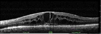
Cystoid macular edema on optical coherence tomography of the left eye.
Intravitreal injection of dexamethasone implant to the left eye was decided. After obtaining a signed informed consent, intravitreal Ozurdex® application was performed under the sterile conditions of the operation theater on July 6, 2015. A drop of 0.5% topical proparacaine hydrochloride with a drop of 5% povidone iodine was installed before the patient underwent an uncomplicated intravitreal dexamethasone implant application. The implant was seen inside the vitreous cavity soon after the injection.
Seven days after insertion, the patient presented with corneal edema and conjunctival injection. The intraocular pressure (IOP) was 40 mm Hg and the visual acuity was counting fingers. Slit-lamp biomicroscopy revealed severe corneal edema and the dexamethasone implant, which had migrated inferiorly into the angle of the anterior chamber (Fig. 2). Centration of the anterior chamber IOL was normal. Irrigation-aspiration of the anterior chamber was performed and the implant was removed. Seven days later, the corneal edema was decreased and visual acuity was 20/200 (Snellen). The IOP was normal and similar to the right eye. Further follow-up visits were performed in the place of residence of the patient, so we have no information on the evolution of the macular edema.
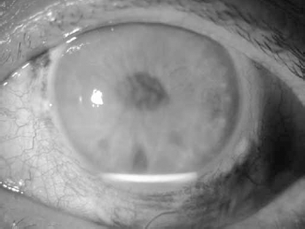
Dexamethasone 0.7 mg implant in the anterior chamber of the left eye.
Discussion
In aphakic eyes, with intact iris diaphragm, silicone oil has frequently caused a pupillary block. In this situation, aqueous humor accumulates behind the iris and forces silicone oil through the pupil into the anterior chamber. An iridectomy at the 6 o'clock position (Ando) can effectively prevent this pupillary block. The iridectomy allows free passage of aqueous to the anterior chamber, which remains free of silicone oil. No permanent contact with silicone oil and the cornea is established, and development of keratopathy is prevented. In our case, the patient has secondary implanted Artisan lens and Ando basal iridectomy. Dexamethasone intravitreal implants are known to settle to the bottom of the vitreous cavity according to Newton's law of gravity, especially after pars plana vitrectomy. It is most likely that the implant migrated through basal iridectomy with free passage of aqueous, but not through iris that was covered by Artisan lens. It was described in the literature that there is a possibility of passage of foreign bodies through the iridectomy from the posterior chamber to anterior (4), so in our case we can see the same situation.
Cystoid macular edema is a common cause of decreased vision following complicated or uncomplicated cataract surgery. This complication is an important cause of suboptimal postoperative vision. Spontaneous resolution of the edema is the most likely natural course in this pathology. However, up to 2% of patients will not have spontaneous resolution of the edema and must thus be treated (5). In this case, the patient had pseudophakic macular edema after secondary implantation of the iris claw anterior chamber IOL.
Glucocorticoids such as dexamethasone exert their anti-inflammatory effects by influencing multiple signal transduction pathways, including vascular endothelial growth factor. By binding to cytoplasmic glucocorticoid receptors, corticosteroids in high doses increase the activation of anti-inflammatory genes, whereas, at low concentrations, they play a role in the suppression of activated inflammatory genes. Therefore, in this case we decided to treat the pseudophakic macular edema by intravitreal injection of Ozurdex®.
Few complications have been reported after injection of intravitreal dexamethasone implants. Intraoperative complications include posterior capsular rent, vitreous loss, and zonular dehiscence. Fasce et al (6) described accidental injection of Ozurdex in the crystalline lens. Postoperative complications include elevated IOP, which may be associated with optic nerve damage, visual field defects, posterior subcapsular cataract formation, secondary ocular infection from pathogens including herpes simplex, and anterior migration of dexamethasone implant. Anterior migration of the dexamethasone implant has also been published in the literature as a rare complication. Migration of Ozurdex® into the anterior chamber was first described by Pardo-López et al (7) in a patient with iris-fixated IOL. This resulted from anterior migration of the implant with diffuse corneal edema in the postoperative 3rd week. The patient had to undergo corneal transplantation, since the corneal edema did not resolve even though surgical removal of the implant from the anterior chamber was performed. Vela et al (8) also reported the migration of Ozurdex® into the anterior chamber in a patient who had previously undergone cataract surgery with iris claw IOL implantation. We describe the migration of Ozurdex® through basal iridectomy 7 days after insertion of the implant in a patient with iris-fixated IOL. Corneal edema was resolved, so it was not necessary to perform corneal transplantation.
Conclusion
Pseudophakic patients with a compromised posterior capsule and a history of vitrectomy, even those with a basal iridectomy (Ando), are at risk for migration of Ozurdex® into the anterior chamber and development of corneal edema.
Footnotes
Financial support: No financial support was received for this submission.
Conflict of interest: None of the authors has conflict of interest with this submission.