
Abstract
Purpose
The neovascular or wet form of age-related macular degeneration is characterized by the growth of abnormal blood vessels in the retina stimulated by vascular endothelial growth factors (VEGF). In the last decade, several anti-VEGF drugs have been developed for treating neovascular diseases of the eyes. This study was conducted to compare the effects of 2 anti-VEGF-A drugs, ranibizumab and aflibercept, on the expression and secretion of VEGF family members in retinal pigment epithelial cells (RPE) in vitro.
Methods
ARPE-19 cells were exposed for 24 hours to ranibizumab or aflibercept at clinical dose concentration. Cell viability and expression and secretion of VEGF-A, VEGF-B, VEGF-C, and placental growth factor (PlGF) were evaluated respectively by real-time polymerase chain reaction and enzyme-linked immunosorbent assay.
Results
Ranibizumab and aflibercept did not affect ARPE-19 cell viability after 24 hours of treatment. Ranibizumab increased expression of VEGF-A and PlGF. On the contrary, expression and secretion of VEGF-C was decreased by ranibizumab. PlGF secretion was not affected by ranibizumab. Aflibercept strongly increased VEGF-A and PlGF expression but reduced their detection on the culture media, and decreased expression and secretion of VEGF-C. No effect on expression and secretion of VEGF-B was observed after exposure to these drugs.
Conclusions
Ranibizumab and aflibercept exert similar effects on VEGF expression and secretion, leading to establishing an antiangiogenic environment. Increased VEGF-A expression observed in RPE cells treated with these drugs suggests a compensatory response of the cells to the lack of VEGF-A.
Introduction
The neovascular or wet form of age-related macular degeneration is the major cause of blindness in the elderly population (1, 2). Members of the vascular endothelial growth factor (VEGF) family, such as VEGF-A, play a critical proangiogenic role by activating quiescent endothelial cells and promoting cell proliferation, migration, and vascular permeability (3, 4).
In the last decade, several anti-VEGF drugs have been developed for treating neovascular diseases of the eyes, such as ranibizumab and aflibercept, which have been approved by the US Food and Drugs Administration (5, 6). Ranibizumab (Lucentis®, Genentech, Inc., South San Francisco, CA, USA) is an affinity-matured antigen-binding fragment derived from the full-length anti-VEGF-A antibody bevacizumab (Avastin®, Genentech, Inc.) and specifically developed for intravitreal administration (5, 7). Ranibizumab acts against all isoforms of human VEGF-A, thus preventing binding of VEGF-A with its receptors VEGFR1 and VEGFR2. Aflibercept (Regeneron Pharmaceuticals, Inc., Tarrytown, NY, USA) is a recombinant protein that functions as a VEGF-soluble receptor; indeed it comprises the second immunoglobulin (Ig) domain of human VEGFR1 and the third Ig domain of human VEGFR2 expressed as an inline fusion with the constant region of human IgG1 (8). Therefore aflibercept, besides binding multiple isoforms of VEGF-A, also binds other VEGFR1 ligands implicated in pathologic vascular remodeling, such as VEGF-B and placental growth factor (PlGF) (3, 9, 10).
Recent studies suggest that VEGF-C, another member of the VEGF family, may play a role in ocular neovascularization, inducing mitogenesis and migration of endothelial cells, and promoting capillary-like formation by choroidal endothelial cells in vitro (11, 12). Furthermore, overexpression of VEGF-C in a model of diabetic retinopathy protects vascular endothelial cells from apoptosis, thus promoting neoangiogenesis (11). Recently we showed that treatment of retinal pigment epithelial (RPE) cells with bevacizumab markedly upregulated mRNA expression of VEGF-C and increased its secretion (13). These results suggest that blockage of secreted VEGF-A may be associated with increased expression and secretion of other VEGF family members, leading to intracellular signals that promote migration and proliferation of endothelial cells even when VEGF-A blockers are employed. Interestingly, several authors hypothesized that the chronic use of anti-VEGF-A drugs may have variable effects on retinal cells, therefore causing toxic effects despite their benefit (14-15-16).
The aim of this study was to investigate whether ranibizumab and aflibercept activate a functional response compensatory to the lack of VEGF-A in RPE cells.
Methods
Cell Culture and Experimental Conditions
Human RPE cell line (ARPE-19) cells from passages 22 to 28 (American Type Culture Collection, Manassas, VA, USA) were grown in a 1-to-1 ratio of Dulbecco Modified Eagle Medium: nutrient mixture F-12 (Cambrex BioScience, East Rutherford, NJ, USA) supplemented with 10% fetal bovine serum, 2 mmol/L glutamine (Sigma-Aldrich, Milan, Italy). Cells were grown to confluence, removed with trypsin-EDTA (Sigma-Aldrich, Milan), and then seeded in multiwell plates for all experiments. Before each experiment, confluent cells were washed with phosphate-buffered saline (PBS) (Cambrex BioScience) and cultured in the following media: standard medium (CTR) and medium containing 0.125 mg/mL ranibizumab (provided by Novartis Farma SpA, Origgio, Italy) or 0.5 mg/mL aflibercept (Bayer SpA, Berlin, Germany).
Cell Viability
To evaluate cell proliferation, ARPE-19 cells were plated in a 96-well plate (2 × 10⁴ cells/well) and cultured for 24 hours as described above. Viable cells were identified using the Cell Titer 96 Aqueous One Solution Cell Proliferation Assay (Promega, Milan, Italy) according to the manufacturer's instructions. Briefly, it is a colorimetric method that determines the number of viable cells via MTS [3-(4,5-dimethylthiazol-2-yl)-5-(3-carboxymethoxyphenyl)-2-(4-sulfophenyl)-2H-tetrazolium, inner salt] tetrazolium reduction into a colored formazan product directly proportional to the number of living cells in culture.
RNA Isolation, cDNA, and Quantitative Real-Time Reverse Transcription-Polymerase Chain Reaction
RNA was extracted from ARPE cells using the Quick-RNA Mini-prep Kit (Zymo Research, Irvine, CA, USA) according to the manufacturer's instructions. The amount and quality of RNA were determined spectrophotometrically and 1 microgram of RNA was reverse-transcripted to cDNA using GoScript Reverse Transcription System (Promega). The expression levels of the target genes, VEGFA (Applied Biosystems, Monza, Italy, assay ID: Hs00900055_m1), VEGFB (Applied Biosystems assay ID: Hs00173634_m1), VEGFC (Applied Biosystems assay ID: Hs00153458_m1), and PlGF (Applied Biosystems assay ID: Hs00182176_m1), were measured by quantitative real-time reverse transcription-polymerase chain reaction (qRT-PCR) amplification performed with TaqMan Gene Expression Assays products in an ABI PRISM 7900 HT fast real-time PCR system (Applied Biosystems). All measurements were performed in triplicate with the following qRT-PCR run protocol: activation Taq program (50°C 2 min and 95°C 10 min), amplification and quantification program repeated 40 times (95°C 15 s, 60°C 1 min). Gene expression was normalized using the housekeeping as control gene (β-Actin, Applied Biosystems assay ID: Hs01060665_g1). Comparisons in gene expression were done using the 2−ΔΔCt method (17).
Vascular Endothelial Growth Factor Secretion
To verify whether the changes in mRNA expression were coupled with altered VEGFs secretion, concentration of VEGFs in the conditioned media were determined by enzyme-linked immunosorbent assay (ELISA). ARPE-19 cells were cultured for 24 hours in standard conditions or in the presence of VEGF-A blockers. To quantify secretion of the VEGF family members, the conditioned media were collected and stored at −80°C until the assay was performed. Cells were then washed with PBS, lysed in radioimmunoprecipitation assay (RIPA) buffer, and protein content was determined with the BCA Protein Assay Kit (Pierce, Rockford, MD) according to the manufacturer's instructions. Secretion of VEGFs was assessed with ELISA (Bender MedSystem, Vienna, Austria), and concentrations were calculated from the standards curve and normalized to the total protein concentration of the respective lysate.
Immunoblot
At the end of the experiments, ARPE-19 cells were lysed in RIPA buffer supplemented with protease and phosphatase inhibitors, and protein concentrations were determined using the BCA Protein Assay Kit. Thirty micrograms of total cell lysate were separated on a sodium dodecyl sulfate polyacrylamide gel electrophoresis and transferred onto nitrocellulose. Filters were blocked in 5% bovine serum albumin and incubated overnight at 4°C with primary specific antibodies against hypoxia-inducible factor 1α (HIF-1α, Millipore, Billerica, MA, USA), the oxygen-regulated subunit of the transcription factor responsible for VEGF-A gene expression. Secondary specific horseradish peroxidase linked antibodies were added for 1 hour at room temperature. Bound antibodies were detected using an enhanced chemiluminescence lighting system (Luminata Classico, Millipore), according to manufacturer's instruction. To verify equal loading of the proteins, membranes were stripped, reblocked, and reprobed to detect glyceraldehyde 3-phosphate dehydrogenase (GAPDH) (FL335, Santa Cruz Biotechnology, Inc., Santa Cruz, CA, USA). Proteins of interest were normalized to total amounts of GAPDH, quantified by quantitative densitometry using Alliance imaging system, and expressed as percentages of control (defined as 100%).
Statistical Analysis
All statistical analyses were performed using GraphPad Prism 4.0 software (GraphPad Software, San Diego, CA, USA). Control was normalized to 100% in each experiment, and percentage changes for each drug were calculated. Data were expressed as mean ± SD and then analyzed by one-way analysis of variance. A p value <0.05 was considered statistically significant. The results are representative of at least 3 experiments.
Results
Ranibizumab and Aflibercept Do not Affect ARPE-19 Cell Viability
ARPE-19 cells cultured for 24 hours in media containing ranibizumab or aflibercept did not display any differences in cell morphology and viability compared to control cells (Fig. 1).
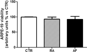
Ranibizumab (RA) and aflibercept (AF) do not affect ARPE-19 cell viability. Cells were cultured for 24 hours in the absence (CTR) or presence of 0.125 mg/mL RA or 0.5 mg/mL AF. Cell proliferation rate was determined by a colorimetric method based on the formazan product of the tetrazolium compound MTS. Results show the percentage of absorbance compared to CTR. Data were expressed as the mean ± SEM of 3 independent experiments.
Ranibizumab and Aflibercept Increase Expression of VEGF-A and PlGF
Treatment of ARPE-19 cells with ranibizumab or aflibercept induced a significant increment of VEGF-A gene expression compared to CTR (ranibizumab: +44.7% ± 2.333, p<0.05, vs CTR; aflibercept: +57.3% ± 12.81, p<0.01, vs CTR). Ranibizumab and aflibercept exerted opposite effects on VEGF-B gene expression; however, no significant difference compared to CTR was revealed. Both ranibizumab and aflibercept significantly decreased VEGF-C expression after 24 hours of treatment (ranibizumab: −27.33% ± 12.44, p<0.05, vs CTR; aflibercept: −38.33% ± 8.212, p<0.05, vs CTR). PlGF gene expression was significantly increased by ranibizumab and aflibercept (ranibizumab: +51.3% ± 11.56, p<0.01, vs CTR; aflibercept: +82.7% ± 28.67, p<0.001, vs CTR) (Fig. 2).
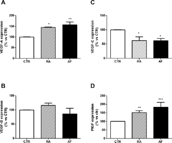
mRNA level of vascular endothelial growth factors (VEGFs) in ARPE-19 cells. Real-time polymerase chain reaction was performed using specific primers for (
Ranibizumab and Aflibercept Upregulate HIF-1α
Expression of VEGF-A is regulated by HIF-1, a heterodimeric transcription factor consisting of a constitutively expressed β-subunit and an oxygen-regulated α-subunit. Under normoxic conditions, HIF-1α is degraded by proteasomes; on the contrary, in hypoxic conditions, HIF-1α is stabilized and able to interact with its co-activators and the β-subunit to increase expression of its target genes (18). Here we found that culture with ranibizumab or aflibercept increased immunodetection of HIF-1α in lysates of ARPE-19 cells (Fig. 3).
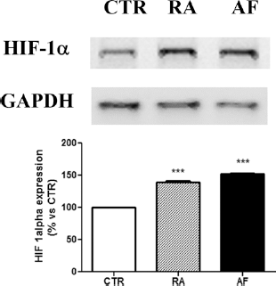
Ranibizumab (RA) and aflibercept (AF) increase hypoxia-inducible factor 1α (HIF-1α) accumulation. Representative Western blot analysis with quantification of densitometries of Western blot bands. Data were expressed as mean ± SEM of fold induction relative to glyceraldehyde 3-phosphate dehydrogenase (GAPDH) (n = 3).
Ranibizumab and Aflibercept Decrease Secretion of VEGF-C
Vascular endothelial growth factor-A was not detectable in media collected from cells treated with ranibizumab and aflibercept. The amount of VEGF-B secreted by ARPE-19 cells treated with ranibizumab or aflibercept was comparable to that of cells cultured under standard conditions (Fig. 4A). On the contrary, treatment with ranibizumab and aflibercept significantly reduced secretion of VEGF-C by ARPE-19 cells (ranibizumab: −36.67% ± 1.208, p<0.05, vs CTR; aflibercept: −54.33% ± 14.31, p<0.001, vs CTR) (Fig. 4B). Culture with ranibizumab did not affect PlGF secretion in comparison to CTR. Detection of PlGF in supernatants from ARPE-19 cells was significantly reduced after treatment with aflibercept (aflibercept: −29.33% ± 6.57, p<0.01, vs CTR; p<0.05 vs ranibizumab) (Fig. 4C).
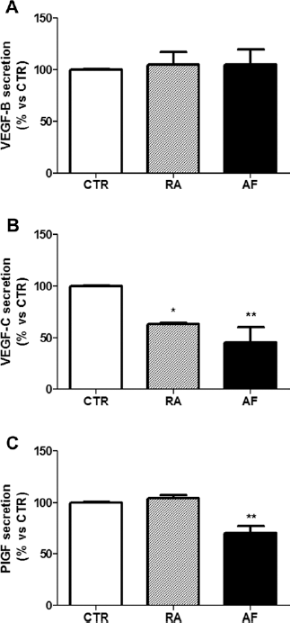
Secretion of vascular endothelial growth factor (VEGF)-B, VEGF-C, and placental growth factor (PlGF). ARPE-19 cells were cultured for 24 hours in the absence (CTR) or presence of 0.125 mg/mL ranibizumab (RA) or 0.5 mg/mL aflibercept (AF). The conditioned media were collected, and levels of VEGF-B (
Discussion
Although several studies have been conducted on the safety profile of anti-VEGF drugs on RPE cells both in vivo and in vitro (19-20-21), this is the first work that evaluates whether ranibizumab and aflibercept may activate a functional response compensatory to the lack of VEGF-A.
Retinal pigment epithelial cells play an important role in maintaining the homeostasis of the retina and choroid. Moreover, RPE cells are the major source of VEGFs in the eye. Besides being considered pathologic factors in the development of exudative age-related macular degeneration or diabetic macular edema, VEGFs have important functions for the survival and maintenance of the healthy retina and choroid (16, 22, 23). Therefore, any alteration of VEGFs secretion that may affect the homeostasis of the retina must be considered when designing administration of these drugs.
Our findings showed that in ARPE-19 cells, gene expression and secretion of important factors belonging to the VEGF family are affected by culture with ranibizumab or aflibercept.
Consistent with results reported by other authors (19-20-21), in our experimental model we did not find any differences in viability of ARPE-19 cells grown for 24 hours in presence of ranibizumab or aflibercept, confirming that no acute toxic effect is exerted by these drugs, and suggesting that modulation of mRNA gene expression was not related to an altered proliferation rate.
We found that culture with ranibizumab or aflibercept increased mRNA levels of VEGF-A, suggesting that ARPE-19 cells try to compensate the lack of VEGF-A in the medium upregulating VEGF-A transcription. Since expression of VEGF-A is regulated by HIF-1, this hypothesis is supported by the concomitant accumulation of HIF-1α during culture with ranibizumab or aflibercept. Furthermore, since HIF-1α directly activates transcription of VEGF-A (18), and is involved in the upregulation of PlGF expression (24, 25), our results suggest that transcription of VEGF-A and PlGF may be activated by the same compensatory pathway. However, the increased levels of mRNA for VEGF-A and PlGF were not coupled with upregulation of their secretion. Regarding VEGF-A, its detection in the culture media was prevented by binding with ranibizumab and aflibercept. The same is true regarding detection of PlGF in media from cells treated with aflibercept. Although ranibizumab has no affinity for PlGF, we would expected an increase of PlGF secretion as a consequence of the mRNA overexpression. Interestingly, the release of PlGF has been found comparable to that of control cells when cells were cultured with ranibizumab. The discrepancy between increased levels of mRNA and maintained secretion rate may be due to posttranscriptional mechanisms that occur after mRNA production.
A regulatory mechanism among the different VEGF isoforms has been reported (9, 12). For instance, VEGF-A upregulates VEGF-C mRNA expression and secretion (12), therefore the decreased expression and secretion of VEGF-C after treatment with ranibizumab or aflibercept might be attributable to the lack of VEGF-A signaling. Since synergistic action of VEGF-C with VEGF-A may play an important role in the development of choroidal neovascularization, decreased expression and secretion of VEGF-C induced by ranibizumab and aflibercept could be an additive beneficial effect in counteracting pathologic angiogenesis.
Among the VEGF family members, VEGF-B is probably the least studied. Although its proangiogenic role is debated, it has been reported that VEGF-B is required for blood vessel survival during pathologic angiogenesis (26). In our experimental model, ranibizumab and aflibercept did not modify the expression and secretion of VEGF-B, thus maintaining an environment comparable to that of control cells.
Considering that in clinical practice anti-VEGF injections are performed with intervals of 4 to 8 weeks, it could be of interest to extend the study period in future experiments.
In conclusion, these results confirm that ranibizumab and aflibercept significantly reduce the concentration of the VEGF-A secreted. Furthermore, we showed that ranibizumab and aflibercept reduce VEGF-C concentration, increasing the anti-angiogenic potential of RPE cells at least in vitro. More studies will be necessary in order to better understand the exact interaction between ranibizumab and PlGF so as to explain the clinical equivalence of ranibizumab and aflibercept in exudative age-related macular degeneration.
Footnotes
Financial support: This research was supported by an unconditional research grant from Novartis Farma SpA, which did not influence the planning, conduct, or analysis of the research, writing of the manuscript, or its submission for publication.
Conflict of interest: None of the authors has conflict of interest with this submission.