
Abstract
Purpose
To evaluate the concentration and molecular weight of hyaluronan (HA) polysaccharides as well as hyaluronidase activity in patients with rhegmatogenous retinal detachment (RRD).
Methods
Twenty vitreous samples from 20 patients with RRD and 19 samples from 19 patients with idiopathic epiretinal membrane, macular hole, or vitreomacular traction syndrome were collected during surgical management with pars plana vitrectomy. The molecular weight of various HA fragments was assessed using agarose gel electrophoresis. Enzyme-linked immunosorbent assay was employed for the measurement of HA (in μg/mL). Hyaluronidase activity was evaluated using substrate (HA) sodium dodecyl sulfate polyacrylamide gel electrophoresis.
Results
Agarose gel electrophoresis showed that the vitreous of the control group contained HA of high molecular mass, in contrast with the patient group. Mean HA concentration in the patient group was 50.96 μg/mL and differed significantly from that of the control group, which was 271.81 μg/mL (p<0.0005). Hyaluronidase activity was significantly higher in the vitreous of patients with RRD (p = 0.037).
Conclusions
The vitreous of patients with RRD is characterized by decreased HA concentration compared to controls of the same age and sex and shows higher hyaluronidase catalytic activity. Hyaluronan degradation could be associated with specific vitreous alterations that potentially contribute to retinal break formation and consequently detachment.
Keywords
Introduction
Retinal detachment occurs when the neurosensory retina separates from the underlying retinal pigment epithelium (RPE) and fluid accumulates within the subretinal space (SRS). Although no real anatomic junctions exist between the 2 layers, mechanical and metabolic forces promote and maintain adhesion between them. A retinal detachment can take place when these forces are overwhelmed (1).
Rhegmatogenous retinal detachment (RRD) is characterized by the presence of a full-thickness retinal break, which is held open by vitreoretinal traction that facilitates accumulation of liquefied vitreous in the SRS, thus separating the neural retina from the RPE. Consequently, prerequisites for this type of retinal detachment include liquefied vitreous, tractional forces that can produce and maintain a retinal break, and a break through which fluid gains access to the SRS (2). On the contrary, other retinal defects such as atrophic holes or retinal dialyses are each characterized by disparate pathophysiology and are not commonly implicated in RRD pathogenesis.
Vitreous liquefaction appears to play a key role in the pathogenesis of RRD. In order to understand the biological events leading to retinal break formation and eventually detachment, it is important to examine the structure of the vitreous and the molecular mechanisms leading to liquefaction. The vitreous is an extracellular matrix consisting mainly of water (98%) and macromolecules with complementary properties. The principal fibrillar macromolecule providing a functioning scaffold in the vitreous cavity is collagen. Glycosaminoglycans provide a swelling pressure that maintains fibrillar protein spacing and resists compressive forces by attracting water and counterions (3). Hyaluronic acid or hyaluronan (HA) is the predominant glycosaminoglycan of mammalian vitreous. It is composed of a disaccharide repeat of D-glucoronic acid and N-acetyl-D-glucosamine linked by alternating β(1→3) and β(1→4) linkages (4). Although several studies have described the alterations of collagen fibrils that contribute to structural changes of the vitreous (3), the role of HA in maintaining normal gel structure has been controversial (5).
The aim of this study was to investigate the role of HA in the maintenance of vitreous structural stability and the effects of its alterations in the pathogenesis of RRD. Moreover, potential associations between differential expression of HA in the vitreous with RRD clinical parameters (such as extent and duration) were investigated.
Methods
Patient and control groups
In this study, 39 vitreous samples from 39 eyes of 39 patients were collected for the quantitative and qualitative evaluation of HA fragments. The patient group consisted of 20 patients undergoing vitrectomy for the management of RRD. The control group consisted of 19 patients undergoing vitrectomy for the management of idiopathic epiretinal membrane, macular hole, or vitreomacular traction syndrome. Exclusion criteria included previous vitrectomy or intravitreous injections in the study eye, cataract extraction in the previous month, history of ocular trauma (penetrating or blunt) that could be associated with retinal break formation or detachment, and previous retinal break or subclinical retinal detachment management with laser photocoagulation or cryotherapy in the study eye. Exclusion criteria for the control group included secondary epiretinal membranes or macular holes.
Sample collection
Undiluted vitreous samples were collected according to the method described by Tsui et al (6). All patients were subjected to 25-G pars plana vitrectomy and all procedures were conducted by the same surgeon. Following priming, aspiration of air was conducted in order to remove any remaining balanced salt solution from the aspiration line, so as to avoid any sample dilution. Core vitrectomy was performed while the infusion cannula was closed and the aspiration rate was kept as low as possible. Prior to vitreous aspiration, instruments were removed from the eye and the 2 main ports were plugged. Vitreous was then extracted from the proximal end of the aspiration line using a 5-mL syringe. Typically, 1-1.5 mL of vitreous was collected. No significant hypotony was observed intraoperatively or postoperatively. Vitreous samples were then placed in sterile Eppendorf tubes of 1.5 mL volume (Eppendorf, Freemont, CA, USA) and rapidly frozen at −80°C until analyzed.
Ethics
The study was conducted according to the ethical principles of the World Medical Association Declaration of Helsinki (amended by the 64th WMA General Assembly, October 2013) (7) and was approved by the Research Committee of Aristotle University of Thessaloniki. All patients were informed about the vitreous sample collection and an informed consent form was signed.
Sample analysis
Hyaluronan molecular weight in the vitreous gel
The molecular weight of various HA fragments in the vitreous samples was assessed using agarose gel electrophoresis, as previously described (8). A 0.9% agarose gel was prepared by dissolving 0.9 gr agarose (Bioline, Taunton, MA, USA) in 100 mL 1× TAE buffer (40 mM Tris-CH3COOH, 1 mM EDTA, pH 8.3). The solution was boiled at 100°C until the agarose was fully diluted. A horizontal electrophoresis apparatus was used (Horizontal Gel Electrophoresis Apparatus, Horizon 11-14, GIBCO BRL, Life Technologies, Carlsbad, CA, USA). The solution was poured onto the electrophoresis gel tray, resulting in a 3-mm-thick gel, and a 14-tooth well-forming comb was used to create wells. The gel was allowed to set and then the unit was filled with 900 mL 1× TAE buffer; afterwards, the comb was removed for electrophoresis.
Samples contained approximately 8 μg of HA. Considering that HA concentration in human vitreous has been estimated at 240 ± 29 μg/mL (9), approximately 30 μL of vitreous should be added to the agarose gel. Due to this significant volume, samples were first lyophilized by centrifuging under vacuum. This procedure allows sublimation of water and dehydration of the samples, without affecting solid substances or their relative concentrations. The lyophilized samples were then diluted with 10 μL loading buffer, containing 0.5 M EDTA, 30% glycerol, and 0.025% (w/v) bromophenol blue and loaded at the agarose gel wells.
In order to estimate HA molecular weight in the vitreous samples, controls of known molecular weight (50, 250, and 1,000 kDa) were used.
Electrophoresis was carried out at room temperature for 3.5 hours with a constant voltage of 50 V. Each gel was then placed immediately in a solution containing 0.005% (w/v) Stains-All in 50% ethanol and was stained while being stirred overnight under light protective cover at room temperature. For destaining, the gel was transferred to distilled water. Finally, each gel was photographed over a translucent light box.
Hyaluronan concentration in the vitreous gel
Hyaluronan concentration of the vitreous samples was estimated using an enzyme-linked immunosorbent assay test kit (Hyaluronic Acid Test Kit, Corgenix Ltd., Lynch Wood, Peterborough, UK). This enzyme-linked binding protein assay uses a capture molecule known as hyaluronic acid binding protein (HABP). More specifically, the samples as well as hyaluronic acid reference solutions were incubated in HABP-coated microwells for 60 minutes, allowing HA present in the samples to react with the immobilized binding protein. After removal of unbound molecules by washing with 0.01 M phosphate-buffered saline (PBS), HABP conjugated with horseradish peroxidase solution was added to the microwells and left for 30 minutes at room temperature to form complexes with bound HA. After another washing step with 0.01 M PBS, the samples were incubated with a chromogenic substrate (3,3’, 5,5’-tetramethylbenzidine and hydrogen peroxide [TMB/H2O2]) for 30 minutes in room temperature to develop a color reaction. Finally, a stopping solution of 0.36 N sulfuric acid was added in the microwells to stop the enzyme reaction and the intensity of color was measured in optical density units with a spectrophotometer at 450 nm.
Hyaluronan levels in the samples were determined against a reference curve prepared from reagent blank (0 ng/mL) and the HA reference solutions of known concentration (50, 100, 200, 500, and 800 ng/mL) provided with the kit.
Since the concentration of HA in human vitreous gel has been estimated at 240 ± 29 μg/mL (9), a 1:150,000 dilution was performed in order to obtain optical density values within the reference curve.
Hyaluronidase activity in the vitreous gel
Hyaluronidase activity in the vitreous samples was evaluated using substrate (HA) sodium dodecyl sulfate polyacrylamide gel electrophoresis, as previously described (10, 11). Hyaluronic acid was copolymerized in 10% polyacrylamide gel to a final concentration of 0.17 mg/mL. The dimensions of the lower gel were 8 cm height and 7.3 cm width. Thirty microliters from each sample were lyophilized for the electrophoresis and the solid substances of each sample were diluted in 3 μL loading buffer consisting of 0.0625 M Tris/HCl, pH 6.8 with 10% glycerol and 0.5% bromophenol blue. One microliter of human plasma diluted in 4 μL loading buffer was used as a control marker for the run. Gels were run at 20 mA per gel for 90 minutes using Mini-Protean 3 apparatus (Bio-Rad Laboratories, Hercules, CA, USA). After the run, the gels were washed with 2.5% solution of Triton X-100 for 1 hour. The gels were incubated in 0.1 M formic acid buffer (pH 4.0) at 37°C for 20 hours. After washing with 20% ethanol and 10% acetic acid for 20 minutes, the gels were treated with 0.5% Alcian blue in 20% ethanol and 10% acetic acid for 1 hour to stain the HA, and then destained in 20% ethanol and 10% acetic acid for 2 hours. The region of the gel where HA had been degraded by hyaluronidase did not stain, and hyaluronidase activity could be detected as unstained bands. The gels were then stained with 0.04% Coomassie blue in 20% methanol and 10% acetic acid for 90 minutes. Unstained bands from nonenzymatic proteins were stained with Coomassie blue and appeared as dark blue bands. Finally, each gel was photographed over a translucent light box.
Hyaluronidase activity was quantified using the computer-assisted image analysis program EDAS (1D Image Analysis Software, Kodak Digital Science v.3.0, Eastman Kodak, Rochester, NY, USA).
Statistical analysis
Numerical data are presented using mean and standard deviation. Patient age and HA concentration between the 2 groups, as well as among patients with and without proliferative vitreoretinopathy (PVR), were compared using the independent-samples t test. Sex distribution between the 2 groups was assessed using the chi-square test. The relationship between HA concentration and age in each group, as well as the extent and the duration of RRD, was assessed using the Spearman rank correlation test. Finally, the Spearman rank correlation test was also employed in order to evaluate the relationship between hyaluronidase activity and the extent and duration of RRD as well as the presence of PVR. All statistical analyses were performed using SPSS (Chicago, IL, USA) 17.0 for Windows statistical software.
Results
Patients
The patient group consisted of 20 patients with RRD (14 male and 6 female, 70% and 30%, respectively). Patient age was 63.8 (10.1) years. Mean visual acuity in the patient group was 11.4 (9.0) Early Treatment Diabetic Retinopathy Study (ETDRS) letters (0.9 logMAR or 20/160) in the study eye and 38.2 (19.7) ETDRS letters (0.3 logMAR or 20/40) in the fellow eye. The extent of the detachment ranged from 3 hours to total retinal detachment and the macula was detached in 8 out of 20 patients (40%). The duration of the detachment ranged from 1 day to 4 months (mean 24.35 days). Proliferative vitreoretinopathy was observed in 10 out of 20 (50%) of the patients at the time of initial examination.
Controls
The control group consisted of 19 patients: 15 with epiretinal membrane, 3 with macular hole, and 1 with vitreomacular traction syndrome. Eight were male and 11 female (42.1% and 57.9%, respectively). Mean age in this group was 68.7 (7.0) years. Mean visual acuity was 23.2 (8.7) ETDRS letters (0.6 logMAR or 20/80) in the study eye and 41.8 (11.1) ETDRS letters (0.3 logMAR or 20/40) in the fellow eye.
Independent-samples t test revealed no statistically significant difference regarding age between the patient and the control group (p = 0.086). Moreover, regarding sex, there was no statistically significant difference between the 2 groups (χ2 = 3.08, p = 0.079).
Evaluation of HA molecular mass
Agarose gel electrophoresis showed that the vitreous samples of the control group contained HA of high molecular mass, greater than 3 × 10³ kDa. As the age of the patients increased, HA polysaccharides of lower molecular mass were also detected. However, mean molecular mass of HA polysaccharides was always above 2 × 10³ kDa.
Vitreous samples of patients with RRD contained HA polysaccharides with a more polydispersed molecular mass, which migrated on the agarose gel as a smear with molecular mass between 250 and 2 × 10³ kDa. Mean molecular mass of HA in the patient group was always lower than 2 × 10³ kDa, irrespectively of the age of the patients (Fig. 1).
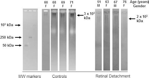
Agarose gels obtained from 4 representative patients with rhegmatogenous retinal detachment and 4 representative controls using 30 μL of vitreous samples. Hyaluronan molecular weight (MW) is estimated in kDa.
Hyaluronan concentration in the vitreous
Mean HA concentration in the control group was 271.81 (143.63) μg/mL. In the patient group, mean HA concentration was 50.96 (28.36) μg/mL. There was a statistically significant difference in the concentration of HA in the vitreous between the patient and control groups (p<0.0005).
There was no significant correlation between HA concentration and age in the control and patient group (p = 0.36 and 0.88, respectively). Furthermore, a significant correlation was not found between HA levels in the vitreous and the duration or the extent of RRD (p = 0.45 and 0.42, respectively). Regarding HA concentration and the presence of PVR, there was no significant difference observed (p = 0.40).
Hyaluronidase activity in the vitreous
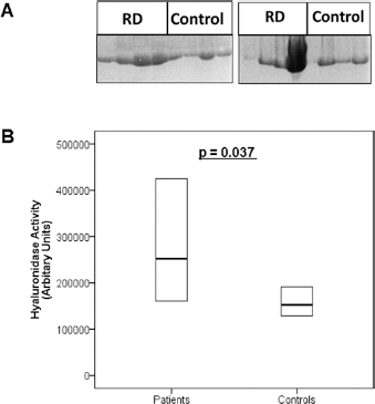
Hyaluronan substrate zymography from vitreous samples revealed that there was high hyaluronidase activity in the vitreous of patients with RRD and controls (Fig. 2A).
(
Quantification of enzymatic activity revealed that the activity of hyaluronidase was significantly higher in the vitreous of patients with RRD as compared to controls (p = 0.037) (Fig. 2B). These results may also explain the presence of HA polysaccharides of low molecular mass in the vitreous samples of patients with RRD that were observed on agarose gels. These fragments were not detected in the vitreous samples of controls (Fig. 1). Finally, a significant correlation was not found between hyaluronidase activity and the extent and duration of RRD or the presence of PVR (p = 0.35, 0.23, and 0.6, respectively).
Discussion
Several studies provide data regarding the biochemical alterations that potentially contribute to vitreous liquefaction and posterior vitreous detachment (12, 13). However, to our knowledge, an assessment of HA polysaccharides along with hyaluronidase activity in the vitreous of patients with RRD has not been performed in the past.
Vitreous liquefaction and posterior vitreous detachment are common states of the vitreous both in healthy individuals and patients with various retinal disorders, such as epiretinal membrane and macular hole. Eyes with epiretinal membranes and macular holes have been commonly employed as controls in similar studies focusing on vitreous biochemistry during RRD (14, 15). In this study, the observed mean HA concentration in the control group was in accordance with the relevant literature. Consequently, vitreous obtained from these patients could be considered as an appropriate control.
Regarding the distribution of molecular weight of HA polysaccharides in the control group, the incidence of lower molecular mass fragments in the vitreous increased with age. This finding may indicate that aging is characterized by a constant and progressive degradation of HA in the vitreous body. Similar age-related alterations have been described in several human tissues, such as cartilage (16), skin (17), and renal medulla (18), and have been attributed to either altered HA synthesis or increased enzymatic catabolism. A previous study has reported a negative correlation between HA levels in the vitreous and age (19), a finding not in accordance with the results of the present study. It should be noted that in the aforementioned study, patients with macular holes as well as diabetic retinopathy were included. As the pathophysiology of those clinical entities varies significantly, conclusions applicable to both cannot be drawn. In the present study, HA molecular weight was negatively associated with age in the control group. A similar age-related pattern was not found in patients with RRD. However, in these patients, the mean molecular mass of HA fragments was constantly lower than that of the control group, which could be attributed to the increased hyaluronidase enzymatic activity observed in the patient group.
Hyaluronan concentration in the vitreous of patients with RRD differed significantly from that of the control group. In order to investigate whether this difference was associated with increased degradation of HA polysaccharides, the activity of hyaluronidase in the vitreous of both patients and controls was assessed. Hyaluronidase activity has been detected in vitreous samples from postmortem eyes, as well as in nonhemorrhagic vitreous biopsy specimens from noninflamed eyes (20). However, HA degradation in vivo by hyaluronidase has not been demonstrated previously. In this study, we detected elevated hyaluronidase catalytic activity in the vitreous of patients with RRD, while vitreous levels of HA in this group were significantly lower than those of healthy individuals. These findings suggest that HA degradation by hyaluronidase could take place in vivo and may lead to structural and conformational changes, resulting in its dissociation from the collagen matrix.
Several previous studies have reported data regarding the concentration of HA in the vitreous of patients with retinal detachment. In a biochemical analysis of the subretinal fluid from patients with RRD, 30% of the samples did not contain HA polysaccharides, while hyaluronidase activity was observed in the majority of samples that did not contain HA (85%) (21). A similar study reported increased presence of chondroitin sulfate in the vitreous of patients with RRD (18% of total glycosaminoglycans as compared to 8% in normal vitreous). In addition, 10% were identified as undersulfated chondroitin and heparin sulfate (22). These findings provide indirect evidence of the lower relative concentration of HA in the vitreous of patients with RRD. However, to our knowledge, no absolute measurement of the concentration of HA in the vitreous of patients with RRD has been performed in the past.
It has been reported that total enzymatic depolymerization of vitreous ΗΑ resulted in a moderate reduction of vitreous wet weight but could not induce complete collapse of the gel structure, thus questioning the importance of glycosaminoglycans interactions for the maintenance of vitreous integrity (5). This study examined the immediate (after digestion for 48 hours) effects of ΗΑ degradation in vitro. Another study examining the long-term effects of hyaluronidase after intravitreal injection suggested that a dose of 10 IU or greater could induce posterior vitreous detachment over a period of 5 weeks in rabbits (23). In a more recent study, the effects of hyaluronidase injection in the vitreous cavity of rabbits were evaluated 3 and 6 months later using scanning electron microscope. According to the authors, although a 20 IU dose of the enzyme was capable of inducing vitreous liquefaction approximately 2 weeks later, it could not induce posterior vitreous detachment detected by scanning electron microscopy. The study concluded that hyaluronidase, by causing HA degradation, is sufficient for inducing vitreous liquefaction but cannot affect other molecules, such as laminin, fibronectin, and collagen, thought to bind the posterior vitreous cortex to the internal limiting lamina, and therefore does not induce posterior vitreous detachment (24).
According to these findings, it could be suggested that the decrease of HA concentration caused by hyaluronidase enzymatic activity in the vitreous is capable of reducing vitreous viscosity and may contribute to vitreous liquefaction without weakening vitreoretinal adhesion. Liquefied vitreous is more vulnerable to inertial mechanical forces exerted during eye movements, whereas at the same time the vitreous cortex remains attached to peripheral retina lacking the mechanical support provided by the vitreous body. These conditions potentially produce tractional forces capable of generating retinal breaks, allowing liquefied vitreous to accumulate in the SRS.
Finally, in order to explore whether increased hyaluronidase activity and the associated decrease in HA concentration constitute a repair process in the clinical course of RRD, the possible correlation between hyaluronidase activity and the extent and duration of RRD, as well as the presence of PVR, was investigated. However, a statistically significant correlation was not demonstrated based on the data provided by the present study. Further investigation is required in order to address this issue.
In conclusion, in this study the vitreous of patients with RRD contained significantly lower HA compared to controls of the corresponding age and sex. These findings can be attributed to increased hyaluronidase catalytic activity in the vitreous. Finally, it is conceivable that a possible pathogenic mechanism linking HA polysaccharides degradation with various vitreous alterations that potentially contribute to retinal break formation and consequently detachment may exist in these pathologic circumstances.
Footnotes
Financial support: No financial support was received for this submission.
Conflict of interest: None of the authors has conflict of interest with this submission.