






Abstract
Purpose
To compare complications after cataract surgery using 2 different phacoemulsification fluidic settings.
Methods
One cataract surgeon (M.K.) performed phacoemulsification cataract surgery in one eye of 43 patients in this randomized prospective clinical study conducted at St. Erik Eye Hospital, Stockholm, Sweden. The patients were randomized to phacoemulsification with low fluidic settings in one group and standard fluidic settings in the other group. Corrected distance visual acuity (CDVA), central corneal thickness (CCT), endothelial cell density (ECD), anterior chamber flare, intraocular pressure (IOP), and macular thickness were measured preoperatively and postoperatively, with a final evaluation at 3 months. Surgical time, ultrasound energy, and amount of balanced saline solution used intraoperatively were recorded.
Results
Twenty-one patients were included in the group with standard settings and 22 patients were in the group with low settings. There were no significant differences between the groups in CDVA, CCT, flare, IOP, macular thickness, or ECD at 1 day, 3 weeks, or 3 months postoperatively. The surgical time was significantly (p = 0.009) longer and cumulative dissipated energy was significantly (p<0.001) higher in the group with low fluidic settings.
Conclusions
Although the surgical time and ECD were significantly higher in the group with low fluidic settings, there were no differences in the CCT, ECD, macular thickness, or inflammation postoperatively between the low and standard fluidic settings during phacoemulsification.
Introduction
Phacoemulsification cataract surgery has been performed widely and has been the preferred technique in cataract surgery for at least the past 25 years. The main advantages of phacoemulsification are smaller incision sizes, reduced trauma, and fewer postoperative complications compared to earlier techniques.
Phacoemulsification uses ultrasound energy to emulsify the lens nucleus into smaller fragments and remove the fragments through the small phacoemulsification handpiece tip inserted into the corneal incision. Fluid enters and exits the eye intraoperatively, cooling the handpiece tip, which minimizes tissue damage, facilitates nuclear fragment movement to the handpiece tip, maintains the intraocular pressure (IOP) and the ocular structures, and hence contributes to a safer surgery.
Concerns have been expressed about the fluidics in the eye during phacoemulsification and the impact on long-term postoperative complications. However, few studies have investigated these issues (1–2–3).
Some parameters are associated with increased surgically induced trauma in phacoemulsification, such as older patient age, small pupillary diameter, high nucleus grade, large nucleus, greater infusion volume, type of intraocular lens (IOL) implanted, and a greater amount of total emitted ultrasound energy (4, 5).
Increased flare in the anterior chamber representing increased inflammation after cataract surgery has been studied (6–7–8). When phacoemulsification with a 3.2-mm corneal incision was compared with phacoemulsification using a 5.5-mm incision, more flare developed postoperatively in association with the larger incision with greater surgically induced trauma (9).
In patients with cystoid macular edema (CME), increased flare developed in the anterior chamber compared to patients without CME after cataract surgery (10, 11). Diagnosing macular edema recently has become easier and more reliable with the introduction of optical coherence tomography (OCT), which has become a standard ophthalmic diagnostic tool in most ophthalmic outpatient clinics in Sweden.
Increased central corneal thickness (CCT) on day 1 postoperatively is proportional to the degree of corneal endothelial cell loss leading to decreased endothelial cell density (ECD) at 3 months postoperatively and hence a predictor of the corneal status (12).
Baradaran-Rafii et al (1) and Vasavada et al (3) compared low settings (200 mm Hg vacuum and 20 mL/min flow rate and ≤400 mm Hg vacuum and 25 mL/min flow rate, respectively) with high settings (400 mm Hg vacuum and 40 mL/min flow rate and ≤650 mm Hg vacuum and 40 mL/min flow rate, respectively). The first study (1) found a strong relationship between total ultrasound energy and endothelial cell loss, but not between total infused fluid and endothelial cell loss. However, vacuum level did not have a significant effect on total ultrasound energy or total fluid consumed during phacoemulsification.
The second study (3) showed that low fluidics during phacoemulsification resulted in lower increases in CCT on 1 and 7 days postoperatively, clearer corneas postoperatively, and less intraocular inflammation on day 1 postoperatively compared to high fluidic settings.
The purpose of the current study was to identify any differences in several postoperative outcome parameters by comparing low with standard fluidic settings that mimic the common uncomplicated daily cases undergoing cataract surgery.
Methods
Patients from 50 to 85 years of age and living in Stockholm County, Sweden, with symptomatic senile cataracts, were randomized to either phacoemulsification with low fluidic settings or the same procedure with standard fluidic settings between 2012 and 2015, following approval from the regional ethical review board in Stockholm, Sweden, to conduct this study. The study followed the tenets of the Declaration of Helsinki. We obtained written informed consent before surgery from all patients in the study. The clinical trial is in the ISRCTN registry (DOI 10.1186/ISRCTN11821734; available at http://www.isrctn.com/ISRCTN11821734). The authors confirm that all ongoing and related trials for this intervention are registered.
When the patients met the inclusion criteria, they received a number generated by Microsoft (Redmond, WA, USA) Excel computer software that randomized them to one of the 2 groups. The patients were blinded during the course of the study, but the surgeon was not. Patients with a traumatic, extremely dense cataract, subluxated lens, macular or corneal disease, anterior chamber depth shallower than 2.1 mm, pupil dilated less than 5 mm in diameter with 0.2 mL solution of cyclopentolate 0.1% and phenylephrine 1.5%, diabetes, glaucoma, or continuous treatment with oral or nasal nonsteroidal anti-inflammatory drugs or steroids and those who had undergone a previous intraocular surgery or retinal photocoagulation were excluded from the study.
Preoperative reference values were obtained for the CCT using Visante OCT anterior segment imaging (Carl Zeiss Medical AG, Jena, Germany), macular thickness using the Stratus OCT macular thickness program (Carl Zeiss Medical AG), and anterior chamber flare using the laser flare meter Kowa FM-500 (Kowa Co. Ltd., Tokyo, Japan). Seven consecutive flare measurements were obtained with the flare meter. The highest and lowest values were discarded and the average value was calculated automatically in the flare meter from the remaining 5 measurements and used in subsequent statistical analyses. The IOP was measured using Goldmann applanation tonometry and corrected distance visual acuity (CDVA) was measured using the Early Treatment Diabetic Retinopathy Study chart. Endothelial cell density was determined by confocal microscopy (Nidek Confoscan 4, Nidek Technologies Srl, Padova, Italy).
Phacoemulsification parameters
Table I shows the fluidic settings in the 2 groups. The height of the infusion bottle is 30%-50% higher in the standard settings group during phacoemulsification.
Fluidic settings in the 2 study groups
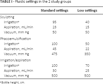
Bottle height, cm.
The total amount of balanced saline solution (BSS) used during phacoemulsification was measured by weighing the BSS bottle preoperatively and postoperatively with the Vetek HS-3000 scale (Vetek Weighing AB, Vaddo, Sweden) with a grading resolution at 2 g. The amount of BSS needed for hydrodissection, wetting of the cornea, and hydration of incision wounds was withdrawn from the BSS bag before weighing. The cumulative dissipated energy (CDE) was recorded from the display on the phacoemulsification machine at the end of surgery. The surgical duration was recorded from the time the incision was made to after injection of the antibiotic prophylaxis.
Surgical technique
One cataract surgeon (M.K.) performed standard torsional phacoemulsification using a 45-grade oval tip with the Infiniti Vision System (Alcon Laboratories, Fort Worth, TX, USA). The procedure began with creation of a 2.2-mm clear corneal incision temporally followed by injection of intracameral anesthesia and cohesive ocular viscosurgical device (OVD), 1.5% sodium hyaluronate (Z-HYALIN® Plus, Carl Zeiss Medical AG). A continuous capsulorhexis and hydrodissection with BSS and phacoemulsification with stop-and-chop technique in the capsular bag were performed for all patients. Irrigation and aspiration of the remaining lens cortex with BSS using an instrument tip were performed. An acrylic hydrophobic IOL, the AcrySof IQ SN60WF (Alcon Laboratories), was folded and injected into the capsular bag followed by OVD removal. The corneal wound was hydrated with BSS using a blunt injection needle. The procedure ended with an intracameral injection of 0.2 mg moxifloxacin as off-label antibiotic prophylaxis. This kind of off-label antibiotic prophylaxis has been widely used in most eye clinics in Sweden for many years. The patients instilled topical dexamethasone 3 times daily in a tapering dose, reducing the frequency of instillation once each week, during the first 3 postoperative weeks.
Postoperative examinations
Postoperative visits were scheduled for 1 day, 3 weeks, and 3 months postoperatively. The measurements obtained at the preoperative and the postoperative visits were performed using the same diagnostic tools mentioned earlier (Tab. II).
Study protocol
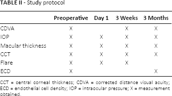
CCT = central corneal thickness; CDVA = corrected distance visual acuity; ECD = endothelial cell density; IOP = intraocular pressure; X = measurement obtained.
Statistical analysis
The data were normally distributed for CDVA and CDE but not for the other parameters. Normally distributed data were analyzed using the Student t test to compare the groups and the paired t test for comparisons within the groups. Nonparametric data analysis was conducted with the Wilcoxon rank sum test to compare the 2 groups, and the postoperative and preoperative values were compared within the group using repeated measures analysis of variance (Friedman) and the Wilcoxon signed-rank test.
The means and standard deviations of the parametric data and medians with lower and upper quartiles for the measured nonparametric data were calculated for both groups and logarithm of the minimum angle of resolution CDVA values was obtained for statistical analysis (Tabs. III–IV–V–VI–VII–VIII–IX).
Logarithm of the minimum angle of resolution corrected distance visual acuity (CDVA) values for both study groups
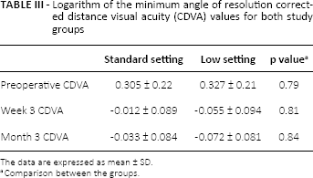
The data are expressed as mean ± SD.
Comparison between the groups.
Median intraocular pressure (IOP) values (mm Hg) in both study groups
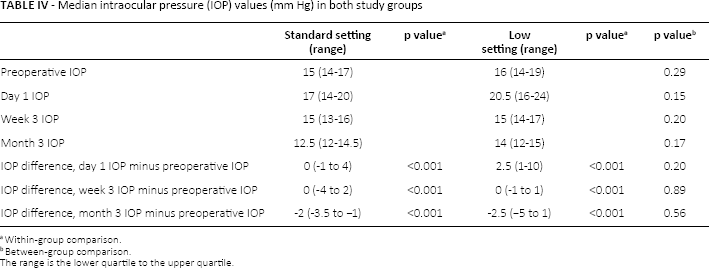
The range is the lower quartile to the upper quartile.
Within-group comparison.
Between-group comparison.
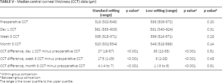
Median central corneal thickness (CCT) data (μm)
The range is the lower quartile to the upper quartile.
Within-group comparison.
Between-group comparison.
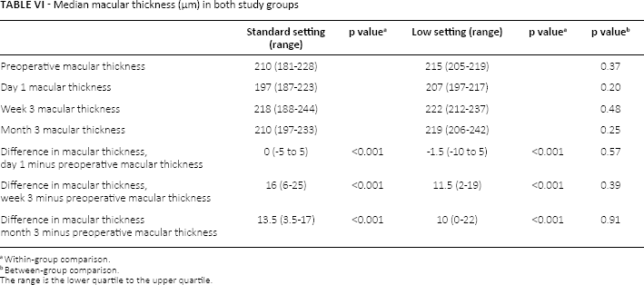
Median macular thickness (μm) in both study groups
The range is the lower quartile to the upper quartile.
Within-group comparison.
Between-group comparison.
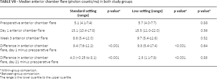
Median anterior chamber flare (photon counts/ms) in both study groups
The range is the lower quartile to the upper quartile.
Within-group comparison.
Between-group comparison.
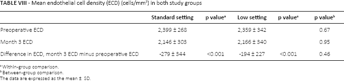
Mean endothelial cell density (ECD) (cells/mm2) in both study groups
The data are expressed as the mean ± SD.
Within-group comparison.
Between-group comparison.
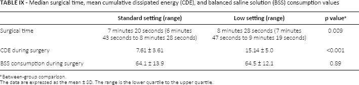
Median surgical time, mean cumulative dissipated energy (CDE), and balanced saline solution (BSS) consumption values
The data are expressed as the mean ± SD. The range is the lower quartile to the upper quartile.
Between-group comparison.
Results
Forty-three patients met the inclusion criteria in this study, 22 of whom were randomized to the low settings group and 21 to the standard settings group. The data from one patient obtained at 3 weeks and 3 months in the standard settings group were excluded because the patient needed treatment with a nonsteroidal anti-inflammatory drug for another disease from day 14 postoperatively. The mean age was 69.5 ± 8.3 years (range 50-85 years), 68.5 ± 8.0 years in the standard settings group and 70.5 ± 8.6 years in the low settings group.
No significant differences were seen between the groups in almost every measured parameter (Tabs. III–IV–V–VI–VII–VIII). However, the amount of ultrasound energy used intraoperatively was significantly (p<0.001) higher and the surgical time was significantly (p = 0.009) longer in the group with low settings (Tab. IX), although these results did not significantly affect differences in parameters representative of surgical trauma, such as the CCT, macular thickness, ECD, or intraocular inflammation (represented as anterior chamber flare) between the groups.
There were significant differences in the measurements during postoperative care in the groups. The median decrease in IOP was 2 mm Hg at 3 months (Tab. IV). The CCT increased the first postoperative day but returned to the preoperative values at 3 months (Tab. V). The macular thickness was unchanged on the first postoperative day but increased slightly at 3 weeks and at 3 months (Tab. VI). These tendencies were seen in both groups.
The CDVA, anterior chamber flare, and BSS used intraoperatively were similar in both groups (Tabs. III, VII, and IX).
Discussion
The lower fluidic settings in the current study were not associated with any significant advantage compared to standard settings regarding the postoperative impact on the CCT, IOP, anterior chamber inflammation, CDVA, ECD, or macular thickness. The lower settings used significantly more ultrasound energy and increased the surgical time but did not significantly affect the parameters. The surgical time increases with lower settings, because more time is needed to aspirate the lens nucleus, but the other parameters did not differ significantly, especially those related to surgical trauma.
This may have occurred because we only used standard settings and not higher settings, and there may be a threshold before significant differences due to surgical damage can be measured. Baradaran-Rafii et al (1) reported that increased CDE was correlated with decreased ECD postoperatively, but the amount of fluidics used intraoperatively was not. Those authors compared low versus high aspiration parameters in longitudinal phacoemulsification. The aspiration flow rate was almost the same, but the vacuum parameters were higher in the current study compared to their study. Nevertheless, we also did not see any significant differences in ECD between the groups at 3 months postoperatively.
One study (3) on aspiration parameters and the postoperative impact on the eye showed a lower increase in CCT on days 1 and 7, less inflammation in the anterior chamber, and no significant differences in ECD postoperatively with low compared to high aspiration parameters. In the current study, the aspiration/irrigation parameters in both groups were similar, but we had slightly higher vacuum settings compared to those reported by Vasavada et al (3). Another important factor was that we used torsional and they used longitudinal phacoemulsification. The torsional mode in phacoemulsification facilitates faster lens removal and uses less ultrasound energy (13). The CDE in the current study was lower. However, our results agreed with their study and showed that the CDE was higher and surgical time longer in the low settings group.
Previous studies have reported postoperative decreases in IOP after phacoemulsification in eyes without glaucoma (14). The long-term IOP decreases were 3.4 mm Hg (15) and 1.5 mm Hg (16) in those studies, respectively. The current decrease was 2 mm Hg in both groups 3 months postoperatively, which agreed with the 2 earlier studies.
In the current study that included uncomplicated cataract cases, we did not find any significant differences between lower versus standard fluidic and ultrasound energy settings regarding their impact on the eye. However, the current study had a small sample size, and significant differences may have been found if more patients had been enrolled. Studies with more cases are needed.
This study cannot conclude that either of the 2 fluidic settings was preferable to the other.
Footnotes
Financial support: Financial support was provided through a regional agreement on medical training and clinical research between the Stockholm County Council and the Karolinska Institutet, Stockholm. The study was also supported by grants from Ögonfonden. The funding organizations had no role in the design or conduct of this research.
Conflict of interest: None of the authors has conflict of interest with this submission.