






Abstract
Purpose
To compare the development of posterior capsule opacification (PCO) and survival rate without capsulotomy after implantation of a hydrophobic or hydrophilic acrylic intraocular lens (IOL) at the 9-year postoperative follow-up.
Methods
One of 3 experienced cataract surgeons performed standard phacoemulsification in one eye of 120 patients with cataract. The patients were randomized to implantation of either a hydrophobic acrylic IOL or a hydrophilic acrylic IOL. Both IOLs had sharp posterior edges. Retroillumination images of PCO were obtained with a fundus camera 9 years postoperatively and analyzed semiobjectively using POCOman computer software.
Results
Seventy-eight of the 120 patients completed the 9-year follow-up examination. Patients implanted with the hydrophilic IOL had significantly (p<0.001) more and denser PCO. The survival rate without Nd:YAG capsulotomy was significantly higher (p<0.001) in eyes with the hydrophobic IOL.
Conclusions
After 9 years, more and denser PCO developed in eyes with the hydrophilic IOL than the hydrophobic IOL. The survival rate without the need for capsulotomy was higher in eyes with the hydrophobic IOL.
Introduction
Posterior capsule opacification (PCO) is the most common complication after cataract surgery, with incidence varying from 10% to 50% among studies (1, 2). Surgical techniques with meticulous cortical cleanup and complete 360-degree coverage of anterior capsule over intraocular lens (IOL) optic have been known to decrease PCO rates (3–4–5–6–7–8). Newer IOLs with improved characteristics of known factors affecting development of PCO, such as the IOL material (9–10–11) and IOL design (12–13–14–15), have reduced the incidence of PCO below 10% (1, 2).
Capsulotomy is performed with a neodymium-yttrium-aluminium-garnet (Nd:YAG) laser to treat PCO during an outpatient visit. The laser creates an opening in the central hazy posterior capsule. The treatment has several complications, some of which can be severe, e.g., lens dislocation (16), cystoid macular edema (17), retinal breaks (18), retinal detachments (17, 18), and damage to the IOL (19). The Nd:YAG laser is not available everywhere, especially in rural areas in developing countries.
Posterior capsule opacification is a socioeconomic burden because of the need for extra visits to the surgeon and the increasing number of cataract surgeries annually. Patients with IOL implants tend to have their implants in their eyes even longer now due to the overall increased lifespan worldwide (World Health Organization 2015) combined with slightly lower age at surgery than before (Swedish National Cataract Registry; Kataraktregistret 2013). The probability to develop symptomatic PCO would then increase due to these facts.
Posterior capsule opacification develops postoperatively because remnant lens fiber cells in the capsular periphery proliferate and migrate toward the center of the posterior capsule over time (4), causing visual symptoms such as diminished visual acuity, glare, and diplopia (20, 21).
Hydrophobic acrylic IOLs, introduced in 1993, have become the most popular IOLs implanted during cataract surgery in most developed countries. The main advantage over hydrophilic IOLs has been a lower incidence of PCO (22, 23). Most studies conducted earlier have follow-ups that are not longer than 2 years postoperatively. This raises the following concern: Is the advantage of less PCO development associated with a foldable acrylic hydrophobic IOL compared to a foldable acrylic hydrophilic IOL still valid many years postoperatively?
The current study is a 9-year extended follow-up study. We published 2 previous follow-up studies of PCO at 1 and 2 years postoperatively (22, 23).
Methods
A total of 120 patients aged 60 to 90 years with senile cataract were included, of whom 74 (62%) were women and 46 (38%) were men; 19 men and 41 women were included in the AcryS of SA60AT IOL (Alcon Laboratories Inc., Fort Worth, TX, USA) group and 27 men and 33 women were included in the BL27 IOL group (Bausch & Lomb Inc., Rochester, NY, USA). There were no significant differences in the number of men and women between the groups (p = 0.13). The average patient age was 72.8 ± 6.7 years (range 60-84 years): 73.4 ± 6.8 years in the AcrySof SA60AT group and 72.1 ± 6.8 years in the BL27 group. The mean age was not significantly different between the groups (p = 0.29).
All patients lived in Stockholm County, Sweden. They underwent uneventful phacoemulsification from May 2002 to March 2004 at St. Erik Eye Hospital, Stockholm, after the ethical permission to conduct this study was granted by the regional ethical review board in Stockholm. The clinical trial is in the ISRCTN registry, DOI 10.1186/ISRCTN46164776, available at: http://www.isrctn.com/ISRCTN46164776. The authors confirm that all ongoing and related trials for this intervention are registered. The study followed the tenets of the Declaration of Helsinki. Written informed consent was obtained from all patients before surgery. Patients with dilated pupil smaller than 6.0 mm, ocular trauma, traumatic cataract, pseudoexfoliation syndrome, glaucoma, corneal pathologies, diabetes mellitus, uveitis, and moderate and advanced age-related macular degeneration were excluded, as were those receiving preoperative oral steroid therapy and those who underwent a previous intraocular surgery.
The patients were blinded throughout the duration of the study but the surgeon was not.
Randomization
The patients were randomized to 1 of the 2 IOLs by selection of an unmarked, opaque envelope from among 120 envelopes containing the name of either the hydrophobic SA60AT IOL or the hydrophilic BL27 IOL. Both IOLs are single-piece, square-edged acrylic lenses with a 6-mm optic diameter and no haptic angulation. The overall diameter of the SA60AT IOL is 13 mm; that of the BL27 is 10.75 mm.
Surgery
One of 3 experienced cataract surgeons performed phacoemulsification cataract surgery. A clear corneal incision was made, and topical and intracameral anaesthetic agents were administered. Sodium hyaluronate (Healon GV; Abbott Medical Optics Inc., Santa Ana, CA, USA) was used as a viscosurgical device. A continuous capsulorhexis was created, and hydrodissection and hydrodelineation with balanced saline solution (BSS) and phacoemulsification in the capsular bag were performed. Aspiration with irrigation and aspiration of the remaining lens cortex with BSS using an instrument tip were performed. One of the 2 IOLs was folded and injected into the capsular bag. The procedure ended with an intracameral injection of cephalosporin as prophylaxis against infection, and the corneal wound was hydrated with BSS using a blunt injection needle. The patients instilled topical dexamethasone 3 times daily in a tapering dose, reducing the frequency of instillation once each week, during the first 3 postoperative weeks.
Patient evaluation
Preoperative routine measurements evaluated if the patient was in need of cataract surgery and then if the patient was a candidate for the study.
Postoperative measurements were done at 1 week, 3 months, 1 year, and 2 years and are described in previously published articles (22, 23).
Each examination included logarithm of the minimum angle of resolution (logMAR) corrected distance visual acuity (CDVA) (2.5% and 100% contrast), glare disability with the brightness acuity test, intraocular pressure, laser flare, and the presence or absence of optic-capsulorhexis contact. A questionnaire asking about glare, eye sensibility, driving, unwanted images, and CDVA satisfaction rate was given to patients at the 3-month follow-up.
The patients were contacted for a follow-up evaluation 9 years (range 8.5-9.3 years) postoperatively. The CDVA was tested with Early Treatment of Diabetic Retinopathy Study chart.
The contrast sensitivity measurements and Scheimpflug images in glistening analysis were also obtained at the 9-year follow-up visit. Patients who developed corneal pathologies, macular diseases, and other eye diseases with the potential to affect CDVA at the 9-year follow-up visit were excluded from the analysis involving CDVA and contrast sensitivity and the correlation to the amount of glistenings. These analyses are part of another paper and are not discussed further in this paper.
Digital image evaluation of PCO
Digital retroillumination color images of PCO were obtained with a fundus camera (Nikon Photo Slit Lamp FS-3, Tokyo, Japan). The images in bitmap format were analyzed semiobjectively using POCOman (24) (Kings College, London, UK) computer software to evaluate the PCO. POCOman applies an overlay grid to the PCO images and divides PCO within capsulorhexis into approximately equal sector areas. The user marks any sector with more than 50% area affected by PCO and how dense the PCO are (i.e., PCO severity). The computer software then calculates the fraction of PCO affected area with total area within capsulorhexis and outputs a severity score between 0 and 3 (0 = no PCO, 1 = mild, 2 = moderate, and 3 = severe). POCOman comes with a package of reference images showing the different severity grades. The idea of POCOman is that the human eye is good at recognizing PCO, but poor in quantifying it.
Nd:YAG capsulotomy
At the 1-, 2-, and 9-year follow-up visits, the patients underwent Nd:YAG capsulotomy if the examiner could verify PCO at the slit-lamp and the patient had deterioration of CDVA and PCO-related symptoms such as glare and blurry vision.
Some patients already had Nd:YAG capsulotomy between the 2-year and the 9-year visits. Most of them had their Nd:YAG capsulotomies at our eye clinic, but some patients went to other eye clinics. We traced the exact dates the treatments were done by communicating with the respective eye clinics after approval from the patients.
It was noted if and when the patient had undergone an Nd:YAG capsulotomy. These cases received the highest scores for PCO fraction and severity. The survival rate without capsulotomy was calculated, since it was superior compared to only obtaining Nd:YAG frequency data, especially in this study with a long follow-up, in which many patients were lost to follow-up. Survival rate data considered all information in that the patients either underwent capsulotomy or were lost to follow-up.
The median survival time without Nd:YAG capsulotomy was defined as the time when 50% of the patients had undergone Nd:YAG capsulotomy.
Statistical Analysis
All statistical analyses were conducted with Stata® Data Analysis and Statistical Software release 10 (StataCorp LP, College Station, TX, USA).
The Student t test was used to compare the follow-up times and the average ages at surgery between the 2 groups. The Mann-Whitney U test was used to compare PCO development and severity and CDVA between the groups. Chi-square test compared if there was equal distribution of men and women in the 2 groups. The Gehan-Wilcoxon test and log-rank test were used to calculate the survival rate without Nd:YAG capsulotomy.
Results
Seventy-eight of the 120 patients completed the 9-year follow-up examination (mean 8.9 years; standard deviation 0.4 year) from February 2012 to May 2012, 42 in the BL27 group and 36 in the SA60AT IOL group. Twenty-eight of the original 120 patients died; 2 attended the examination but did not understand any commands the examiner was giving them during the examination due to severe dementia. These 2 patients were excluded from the study. One patient moved to another part of the country, 7 were unable to attend due to illness, 2 refused the examination, and 2 did not keep the scheduled appointment.
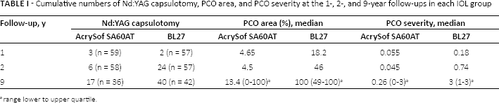
The hydrophilic BL27 IOL developed significantly more and denser (p<0.001 for both comparisons) PCO (Tab. I).
Cumulative numbers of Nd:YAG capsulotomy, PCO area, and PCO severity at the 1-, 2-, and 9-year follow-ups in each IOL group
range lower to upper quartile.
Nd:YAG capsulotomy had been performed in 40 out of 60 patients (67%) in the hydrophilic BL27 group and in 17 out of 60 (28%) in the hydrophobic SA60AT group. Table I shows the cumulative numbers of patients who had received Nd:YAG capsulotomy, PCO area fraction, and PCO severity at the follow-ups.
There were no significant differences in CDVA at the 9-year follow-up between the groups (p = 0.88). Twenty-nine patients from each group qualified for the analysis. Patients with other eye diseases that can affect CDVA were excluded. The median logMAR CDVA was the same in both groups: 0 (range Q1-Q3, -0.08 to 0.11) for the BL27 IOL and 0 (range Q1-Q3, -0.08 to 0.13) for the SA60AT IOL. Q1 and Q3 are lower and upper quartiles, respectively.
The hazard ratio for Nd:YAG capsulotomy was 3.3 (p<0.001) in favor of the hydrophobic IOL.
The survival rate without Nd:YAG capsulotomy for the 2 IOLs did not differ significantly regarding sex, operated eye, or patient age at surgery (p>0.05 for all 3 factors). The median survival time exceeded 9 years for the hydrophobic SA60AT IOL and was 2.6 years for the hydrophilic BL27 IOL (Fig. 1).
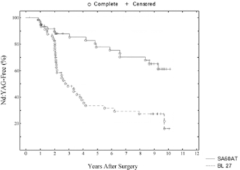
Kaplan-Meier curve shows the survival time in years before Nd:YAG capsulotomy after cataract surgery with the 2 acrylic intraocular lenses (IOLs). The hydrophilic BL27 IOL has a significantly shorter survival time compared to that of the hydrophobic SA60AT IOL.
Discussion
Few long-term follow-up studies exceeding 3 years have compared PCO development between hydrophobic acrylic and hydrophilic acrylic IOLs (25–26–27). To our knowledge, the current study had the longest follow-up.
Acrylic hydrophobic IOLs have been the most popular during the last 15 years in cataract surgery. Studies with short-term follow-up periods within 3 years postoperatively have reported less PCO development in hydrophobic IOLs compared to hydrophilic IOLs (22, 23, 28, 29).
The current study found that the patients implanted with hydrophobic IOLs were significantly more likely to not require Nd:YAG capsulotomy compared to patients with hydrophilic IOLs. This advantage of hydrophobic compared to a hydrophilic IOLs is still valid 9 years postoperatively (22, 23).
However, it is unclear if the IOL material or the sharpness of the posterior IOL edges was the main factor contributing to less PCO development in the hydrophobic acrylic SA60AT IOLs in this study. A previous study found that the sharpness of the edges varies among IOLs (14). Generally, hydrophilic IOLs have duller edges (14), which is believed to result from the manufacturing process, during which many hydrophilic IOLs are lathe cut while dry, thus damaging the sharp edge profile. However, in the same study, some hydrophilic IOLs with square edges were as sharp as most of the hydrophobic IOLs. The sharpness of the posterior IOL edges also varies among hydrophobic IOLs. The sharper the edges of the IOL, the less PCO that develops (30). The sharpness of the posterior square edge in the AcrySof SA60AT IOL has been studied (14), but the hydrophilic BL27 IOL has not been studied. It is therefore difficult to draw any conclusions from the current study if the IOL material or the sharpness of the posterior IOL square edge is the main factor preventing PCO development. For example, AcrySof IOLs have been associated with tighter adhesions between the IOL and the posterior capsule through fibronectin bindings, and one theory is that the tighter adhesions prevent migrating lens epithelial cells to invade the space between IOL and posterior capsule and cause symptomatic PCO (31, 32). The less dense PCO (i.e., severity) and smaller area affected by PCO in the hydrophobic AcrySof SA60AT IOL in this study might be partly attributed to this theory.
There were differences in overall IOL diameter between the lenses in this study, AcrySof SA60AT (13.00 mm) and BL27 (10.75 mm). Two earlier studies (33, 34) comparing PCO development in IOLs with different overall diameters with follow-ups 1-1.5 years after surgery show different results. One study on polymethylmethacrylate IOLs showed that IOLs with smaller diameter (<13.5 mm) required less Nd:YAG capsulotomy compared to IOLs with greater diameter (>13.5 mm) (33). The other study compared 2 hydrophilic IOLs from the same manufacturer but with different IOL diameters. The authors assessed PCO based on CDVA, subjective and automated image analysis software. They concluded that the use of IOL with variable total diameter seems not to influence the rate of PCO formation (34).
Hydrophilic IOLs have been observed in rare cases to develop opacifications, probably calcium and phosphate deposits on the lenses (35, 36). Primary calcifications have been addressed to factors during manufacturing and are now uncommon after altering the manufacturing process and development of new hydrophilic IOLs. However, secondary calcifications, often seen in eyes with preexisting eye diseases such as diabetic retinopathy, still appear. There have been case reports about opacified hydrophilic IOLs after intracameral injection of air or gas (37), and after vitrectomy in nonproliferative diabetic retinopathy (38).
Posterior capsule opacification development and Nd:YAG rates in a 5-year follow-up study (27) compared 3 IOLs: 2 hydrophobic IOLs, AR40e and AR40 (Abbot Medical Optics), with the hydrophilic BL27 IOL. The hydrophobic IOLs have a 3-piece design and the AR40 IOL has a rounded-edge design. The Nd:YAG rates were 52/300 (17.3%) for the AR40e IOL, 73/300 (24.3%) for the AR40 IOL, and 91/300 (30.3%) for the BL27 IOL, which did not differ significantly. The follow-up time in the current study was almost twice as long and showed an even stronger tendency for patients with the hydrophilic acrylic BL27 IOLs to undergo Nd:YAG capsulotomy more often.
The current study has provided more knowledge about how 2 different IOL materials behave in the long run regarding development of PCO, thus leading to continued improvements in the IOL designs and allowing surgeons to make better choices when selecting IOLs for their patients.
Footnotes
Financial support: Financial support was provided through a regional agreement on medical training and clinical research between the Stockholm County Council and the Karolinska Institutet, Stockholm. The study was also supported by grants from Ögonfonden. The funding organizations had no role in the design or conduct of this research.
Conflict of interest: None of the authors has conflict of interest with this submission.