






Abstract
Purpose:
Vision loss in Sturge-Weber syndrome (SWS), a rare congenital disorder, is primarily due to glaucoma.
Methods:
We reviewed the data of all consecutive SWS-associated glaucoma cases in patients who had undergone combined trabeculotomy-trabeculectomy (CTT) at a tertiary glaucoma facility between January 1993 and December 2015. We analyzed the preoperative and postoperative intraocular pressure (IOP), corneal clarity, visual acuity, success rate, need for repeat surgery, and number of topical antiglaucoma medications needed at last follow-up.
Results:
Twenty-six eyes of 20 patients with SWS (surgical age 0.7-96 months; mean 18.64 ± 29.74 months) had undergone primary CTT. The mean preoperative IOP was 32.76 ± 7.86 mm Hg (range 22-54 mm Hg) with medication (mean 3.11 ± 1.17; range 1-5). At the last follow-up (61-288 months); mean SD 134.73 ± 67.77 months), two eyes had IOP <6 mm Hg. Twenty-four eyes analyzed had an IOP of 13.63 ± 6.11 (mean ± SD; range 9-41) mm Hg. All these had an IOP <15 mm Hg at last follow-up except one, which had an IOP of 41 mm Hg. There was a mean reduction of 54.62% ± 31.33% in IOP from baseline. The antiglaucoma medication score at last follow-up visit was 0-3. No eye achieved predefined complete success or modified complete success. A total of 41.7% (10/24) of eyes attained both qualified and modified qualified success. Eleven eyes needed repeat surgeries. No intraoperative complications were noted. Visual acuity was below 6/60 in four eyes.
Conclusions:
Combined trabeculotomy-trabeculectomy showed promising results as a treatment for SWS-associated glaucoma in children. Long-term visual and surgical outcomes are encouraging.
Introduction
Sturge-Weber syndrome (SWS) is a congenital disorder with an incidence of 1 in 50,000 live births (1). Patients often present with facial port wine stain, ocular involvement including glaucoma, conjunctival, episcleral, choroidal, and retinal vascular malformations, and ipsilateral occipital leptomeningeal angioma (2, 3). Glaucoma occurs in nearly 30% of cases, but in those not presenting with facial nevi, glaucoma may be absent (4). If there is a port wine birthmark on both the upper and lower eyelid, the chances of glaucoma are nearly 50% (5). A combination of goniotomy and trabeculotomy has been the preferred treatment for SWS-associated glaucoma (6). Surgical interventions are often associated with complications, mainly attributed to rupture of vasculature in the choroidal hemangiomas (7).
We analyzed the clinical findings and long-term outcomes of combined trabeculotomy-trabeculectomy (CTT), a glaucoma filtering technique, in early-onset glaucoma in Indian patients diagnosed with SWS.
Methods
Preoperative data
We defined early-onset glaucoma as that which occurred either at birth or within the first 10 years of life, and reviewed the records of all patients with SWS who had undergone CTT over a period of 23 years between January 1993 and December 2015 at a tertiary glaucoma care center in India. The study adhered to the tenets of the Declaration of Helsinki and safeguarded patient confidentiality.
Baseline demographic data about the affected eye, surgical age, clinical presentations, extent and type of ocular involvement, pregnancy issues, family history, and history of systemic illness were noted. Extraocular manifestations of SWS such as facial angioma, facial hypertrophy, angioma on other parts of the body, and upper eyelid involvement were assessed. Changes in the conjunctiva, cornea, iris, and lens were noted. Preoperative visual acuity, cup-to-disc ratio, intraocular pressure (IOP), and antiglaucoma medication score were also analyzed. Radiologic imaging was done to detect calcification in brain tissues.
Operative procedure
Combined trabeculotomy-trabeculectomy, the primary procedure in all cases, was performed by one of the senior authors (NNS, DS). All patients received intravenous mannitol (100 mL) 1-2 hours preoperatively. Under general anesthesia and with aseptic technique, a limbus-based conjunctival flap was raised. A partial thickness triangular scleral flap was dissected. The Schlemm canal was then dissected and trabeculotomy ab externo (Harms trabeculotome) was performed on either side of the radial incision. Then, trabeculectomy was performed, followed by iridectomy. The surgeons emphasized quick closure of the scleral flap after the Schlemm canal had been cannulated. The scleral flap was sutured using three sutures (except two sutures in one eye). A drop of 1% atropine was instilled into the conjunctival sac. An eye patch or shield was applied.
Postoperative data
We reviewed the postoperative IOP, visual acuity, and number of topical antiglaucoma medications required at each visit. We assessed the need for additional glaucoma filtering surgery (GFS) and non-GFS after CTT, and when the repeat surgeries were done.
Success criteria
Complete success was defined as IOP <22 mm Hg without the need for any topical antiglaucoma medication at the last follow-up visit. Qualified success occurred when IOP was <22 mm Hg with the need for 1 or 2 topical antiglaucoma medications at the last follow-up visit. Modified success criteria were based on reduction in IOP. Modified complete success was defined as IOP reduction of 30% without the need for any topical medications. Modified qualified success was when this reduction occurred with the use of one or two topical medications. The surgery was a failure when IOP was ≥22 mm Hg, the IOP reduction achieved was <30%, the patient was receiving more than two topical antiglaucoma medications during the last follow-up visit or required repeat surgeries, and/or there were any complications.
Statistical analysis
All data were analyzed using MS Excel 2010 and SPSS statistical software, version 16.0, for Windows (SPSS Inc.).
Results
Demographic data and preoperative findings
The baseline demographic data are summarized in Table I. Combined trabeculotomy-trabeculectomy was done in 26 glaucomatous eyes of 20 patients at surgical age of 0.7 to 96 months (mean 18.64 ± 29.74 months). The commonest presentations were redness of the face and a hazy cornea. Three patients (15%) were born prematurely and one (5%) had blood incompatibility. There was no strong association with any systemic illnesses or family history of glaucoma (Tab. I). Facial angioma was present in all and was unilateral in 12 patients (60%). Radiologic imaging of the brain revealed that one patient had gyriform calcification on the left occipital cortex with surrounding focal atrophy and another patient had arteriovenous (AV) malformation. Preoperative visual acuity test was not possible in 22 eyes; two eyes recorded a visual acuity of 6/6 and two of 6/9. Twelve eyes (46.15%) had clear cornea. Other ocular and extraocular involvements are summarized in Table I. Glaucoma was bilateral in six patients (30%). Mean preoperative IOP was 32.76 ± 7.86 mm Hg (range 22-54 mm Hg).
Demographic data of patients who underwent CTT
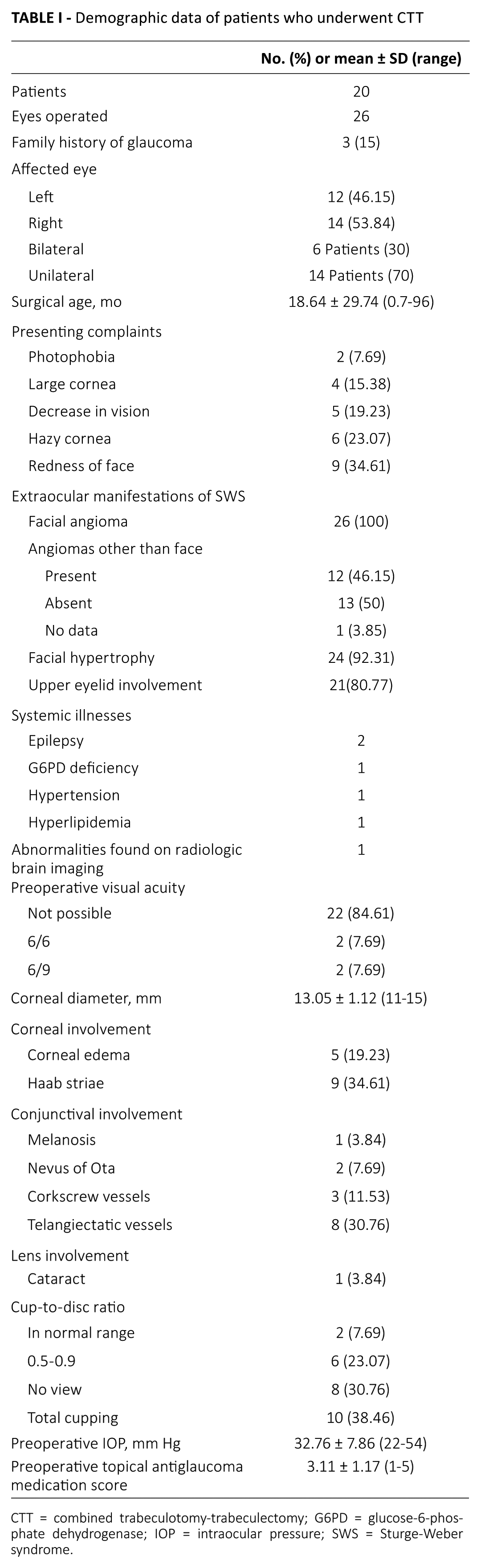
CTT = combined trabeculotomy-trabeculectomy; G6PD = glucose-6-phosphate dehydrogenase; IOP = intraocular pressure; SWS = Sturge-Weber syndrome.
Additional surgeries
The choice of additional surgeries was at the discretion of the operating surgeons (DS, NNS). The first additional GFS, as either trabeculectomy with adjunctive mitomycin C (TMMC) or TMMC combined with releasable sutures (RTMMC), was performed at a mean duration of 23.13 ± 23.41 months following CTT (Tab. II). Trabeculectomy with adjunctive mitomycin C, using 0.4 mg/mL mitomycin C applied for three minutes, was performed in seven eyes, while RTMMC using 0.2 mg/mL mitomycin C for two minutes was performed in eight eyes. A second additional GFS was required only in six eyes at a mean duration of 55.5 ± 29.43 months after the CTT. A third repeat procedure was required in only four eyes at a mean duration of 120.75 ± 61.68 months after CTT. Only one eye was subjected to a fourth GFS at 156 months after the primary CTT.
Repeat glaucoma surgeries performed after the primary CTT
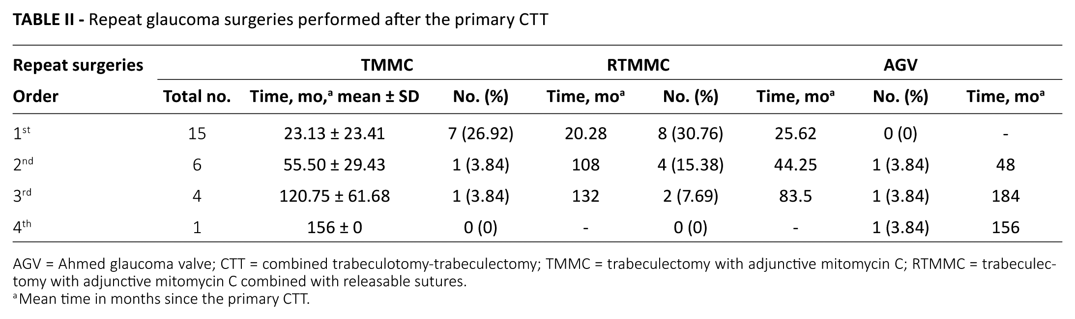
AGV = Ahmed glaucoma valve; CTT = combined trabeculotomy-trabeculectomy; TMMC = trabeculectomy with adjunctive mitomycin C; RTMMC = trabeculectomy with adjunctive mitomycin C combined with releasable sutures.
Mean time in months since the primary CTT.
Intraocular pressure during follow-up
Intraocular pressure was assessed at every postoperative visit. At the last follow-up, all 24 eyes analyzed (except one) had IOP ≤15 mm Hg (Fig. 1). The reduction in IOP in the 26 eyes at the last follow-up (61-288 months: mean 134.73 ± 67.77 months) was by 57.3% ± 31.54% (mean ± SD). In 1 eye, the IOP had increased from 22 mm Hg to 41 mm Hg at the last follow-up at 147 months. This eye also maintained an IOP ≤22 mm Hg for 10 years postsurgery. Two other eyes had hypotony (IOP <6 mm Hg) at last follow-up. One had IOP of 4 mm Hg at 288 months follow-up and had needed three additional TMMC surgeries. The other had IOP of 3 mm Hg at 81 months follow-up and had undergone an RTMMC at 11 months and an Ahmed glaucoma valve at 48 months. If we exclude the hypotonic eyes from the analysis, the mean reduction in IOP in 24 eyes at the last follow-up (mean 159.46 months, 61-288 months) was by 54.62% ± 31.33% (mean ± SD; range -86.36% to 83.33%).
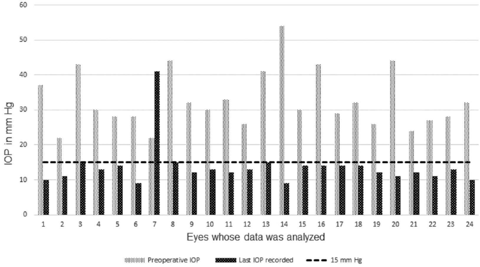
Change in intraocular pressure (IOP) following combined trabeculotomy-trabeculectomy in patients with Sturge-Weber syndrome (n = 24 eyes analyzed).
Postoperative visual acuity analysis
Postoperative visual acuity of all 26 eyes at the last follow-up visit is summarized in Table III. Only two patients had a visual acuity of <6/60 in the affected eye, out of which one had no light perception in the affected eye.
Visual acuity at the last follow-up visit (61-288 months)
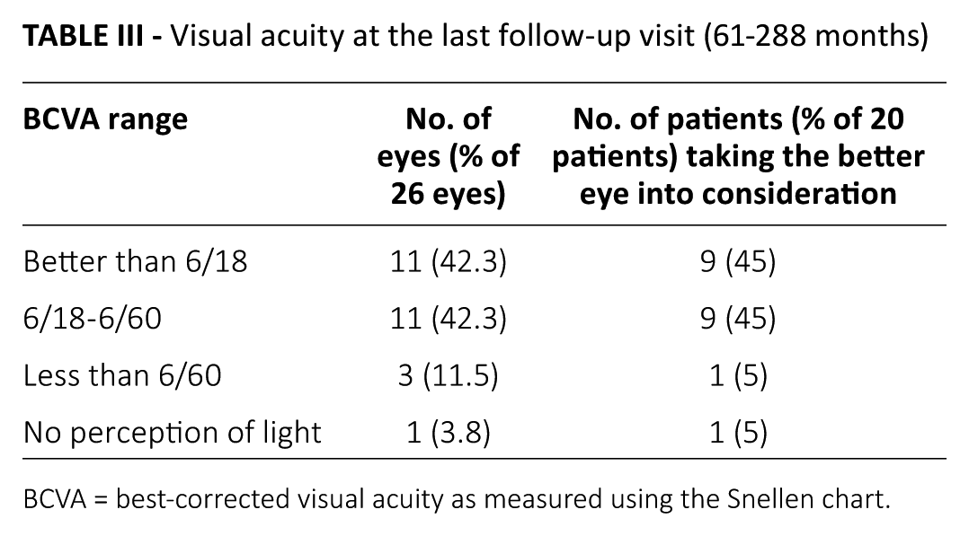
BCVA = best-corrected visual acuity as measured using the Snellen chart.
Topical antiglaucoma medication score
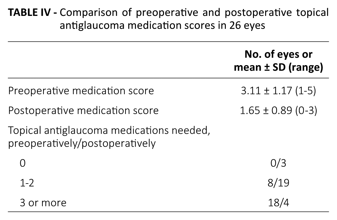
Preoperative and postoperative topical antiglaucoma medication scores of the eyes are compared in Table IV.
Comparison of preoperative and postoperative topical antiglaucoma medication scores in 26 eyes
Success rates
At the last follow-up, in two eyes IOP had dipped to <6 mm Hg (4 mm Hg and 3 mm Hg). We excluded these two eyes from the success analysis.
Of the 24 eyes analyzed, complete success or modified complete success was not achieved in any of the eyes. Qualified success and modified qualified success rates were the same. All the eyes that achieved an IOP <22 mm Hg at last follow-up also had an IOP reduction of >30%. Failure rates were determined based on topical antiglaucoma medication score at the last follow-up and the need for repeat surgeries after the initial CTT. Failure occurred in 14/24 (58.3%) eyes while qualified success and modified qualified success occurred in 10/24 (41.7%) eyes.
Of the six patients with bilateral glaucoma, only one achieved qualified or modified qualified success in both eyes and three achieved it in one eye each.
Complications
No surgical complications were reported in any of the eyes. One patient had calcification of brain tissue one year after initial surgery and presented with epileptic seizures and autism.
Corrective surgeries, other than those for glaucoma, were performed in four eyes. One patient developed cataract in both eyes and at 288 months, though one eye was hypotonic, the other had an IOP of 9 mm Hg. Cataract removal and intraocular lens (IOL) replacement was done at 15 years in one eye and at 17 years in the other. Another patient with bilateral glaucoma developed cataract in one eye. Cataract removal and IOL replacement was done at 11 years. One more patient with unilateral glaucoma had to undergo a lower lid entropion surgery at 13 months.
Discussion
Our study is the longest reported case series of Indian patients with SWS-associated glaucoma, spanning over 23 years with a mean follow-up period of over 14 years. Similar studies have been conducted to evaluate the success of CTT in SWS-associated glaucoma, but with shorter follow-up (6, 7). A more recent study analyzed the data of 27 eyes of 25 patients followed up for a mean of 20 months. However, the primary surgery done was TMMC, with only seven eyes undergoing CTT with or without mitomycin (8). Another study spanned a duration of about 25.5 years, but focused only on the clinical manifestations, disease course, and outcome of patients with SWS (9).
Early-onset glaucoma is believed to be due to trabeculo-dysgenesis (8). Anterior chamber anomalies and AV shunts in the conjunctiva result in episcleral venous pressure and increased IOP (6-10). Some other theories suggest there is fluid hypersecretion by either the ciliary body or the choroidal hemangioma (11). Early onset of glaucoma was evident in our study and the mean surgical age was 18.64 ± 29.74 months (range 21 days-8 years).
Like in most studies, the majority of our patients had unilateral glaucoma (6, 8, 11). Sturge-Weber syndrome, though congenital, is not inherited (5, 11) and we did not find a strong correlation with the family history. Congenital glaucoma is frequently associated with decreased vision, hazy cornea, nevus of Ota, melanosis, corkscrew vessels, and telangiectatic vessels in conjunctiva, all of which were noted in our study (11). Seizures, mental retardation, and gyriform pattern of calcification in the occipital and parietal area of the brain are other noted clinical signs of SWS (12). Seizures occurred in two patients while one showed gyriform calcification on the left occipital cortex with surrounding focal atrophy. One patient presented with AV malformation on radiologic imaging of the brain.
Facial angioma, the most evident symptom of SWS, was observed in all patients, with 60% of cases being unilateral. Facial hypertrophy (92.3%) and upper eyelid involvement (80.76%) were the other prominent symptoms, as reported in other SWS cases (11).
Although goniotomy and trabeculotomy have been considered to treat SWS-associated glaucoma, most Indian children, as in our study, present with cloudy cornea, which makes it difficult to use goniotomy as a treatment option (13, 14). Although both goniotomy and trabeculotomy relieve the obstruction caused by congenital angle deformity, neither effectively tackles the elevated venous pressure that increases the IOP (6). Combined trabeculotomy-trabeculectomy involves creating two exits to overcome anterior chamber anomalies and enable drainage of aqueous humor. Trabeculotomy establishes a communication between anterior chamber and Schlemm canal and trabeculectomy creates a fistula between anterior chamber and the subconjunctival space (15). This creates alternate outflow channels that bypass the episcleral venous system. Theoretically, this should provide long-term and better IOP control.
In a previous study by one of us (NNS), involving a series of 16 patients (19 eyes) with SWS, at a mean follow-up of 42 months (range 1-8 years), IOP was ≤22 mm Hg in 11 eyes (7). There was good IOP control without medications in 27.8%, control with medications in an additional 33%, and poor IOP control in 38.9% eyes. In another series of nine patients (10 eyes) with SWS, over a mean follow-up of 28 months, all eyes maintained a postoperative IOP <16 mm Hg without medication (6). The percentage reduction of IOP was 55.8% ± 12.6% (p<0.0001). In a more recently published series of 25 patients with SWS (27 eyes) with a median follow-up of 20 months (2-122 months), most patients were in the pediatric age group (mean age 10.1 years, range 1 month-48 years) (8). Surgeries done included TMMC (18 eyes), CTT with MMC (four eyes), CTT without antimetabolites (three eyes), trabeculectomy without antimetabolites (one eye), and transscleral cyclophotocoagulation (one eye). The overall success rate was 66.7%. Combined trabeculotomy-trabeculectomy had better success (85.7%) than trabeculectomy alone (63.2%).
Though the success rate in our study is not as high (41.7% qualified and modified qualified success), the percentage reduction in IOP at the last follow-up is encouraging (mean ± SD 54.62% ± 31.33%, range −86.36% to 83.33%). This difference from other studies may be because our study involved patients only in the pediatric age group, was associated with early-onset glaucoma, had a higher mean preoperative antiglaucoma medication score, and had a much longer follow-up period.
Some previous studies also involved young children below 10 years of age (6, 7). However, the percentage of IOP reduction is higher in our study. In the study by Agarwal et al (7) where NNS was a coresearcher, only 61.6% of the eyes had an IOP <22 mm Hg at last follow-up. In our current study, all eyes included in the success analysis (except the one where IOP increased to 41 mm Hg) had an IOP equal to or less than 15 mm Hg. Similar results were seen in the study by Mandal (6) but ours has a longer duration.
No intraoperative complications were noted in our study subjects. Many researchers report choroidal effusion or expulsive choroidal hemorrhage during various types of surgeries in patients with SWS (7, 16, 17). To reduce this complication, some surgeons suggest routine sclerotomy during the CTT and others administer intravenous mannitol an hour prior to the surgery (6). If the trabeculectomy part of the CTT is done quickly and the scleral flap is resutured without delay, this complication can be avoided. If there is a delay, the choroidal effusion can cause a shallow anterior chamber and dislocation of the lens-iris diaphragm into the trabeculectomy site. Agarwal et al (7) describe the use of postoperative subconjunctival injections of 5-fluorouracil for modulating wound healing and agree that using it in young children is extremely difficult. Mitomycin C may be a preferable option. Though we did not use any antimetabolite during the surgery, use of mitomycin C during CTT seems to be a viable option to modulate wound healing (8).
As this is a retrospective study, it relies on the accuracy of the record-keeping. However, conducting a prospective study for a rare disease like SWS may not be easy. Though this study does not have a very large number of subjects, the follow-up is considerably longer than most other studies reported.
Conclusion
A better understanding of the mechanism of glaucoma in SWS will help decide the optimum surgery for SWS-associated glaucoma. Combined trabeculotomy-trabeculectomy offers safe and effective long-term management of SWS-associated glaucoma.
Footnotes
Acknowledgements
The authors thank PFC Pharma Focus India Pvt Ltd. for data analysis and preparation of the manuscript.
Disclosures
Financial support: This study was funded by SK Glaucoma Care Foundation, New Delhi.
Conflict of interest: None of the authors has conflict of interest with this submission.