






Abstract
Introduction
The aim of this study was to investigate the influence of triple pelvic osteotomy on the internal pelvic dimensions and thus on the potential for normal vaginal delivery.
Methods
Data were acquired by processing fixed anatomical specimens of 19 female pelves with maintained sacrotuberous and sacrospinous ligaments after unilateral and bilateral osteotomy, respectively. The specimens were measured and x-ray images and photographs were taken.
Results
The dimensions in the plane of the pelvic inlet and the plane of the greatest pelvic dimension after unilateral osteotomy increased in 51%, remained unchanged in 34% and in 15% of cases they decreased. The most critical locations for the passage of fetus decreased in average from 0.016 cm in distantia interspinalis in the plane of the least pelvic dimension to 0.695 cm in distantia intertuberositas in the plane of the pelvic outlet. After bilateral osteotomy are decreases more significant from 0.226 cm (2.05%) to 1.00 cm (9.51%). Decreases we observed in the monitored dimensions were not so big in comparison to other published studies.
Conclusion
Results of this study confirm our hypothesis that unilateral triple pelvic osteotomy does not significantly narrow the bony birth canal and so it does not impede the ability to deliver per vias naturales in female patients with this surgical procedure in anamnesis. After bilateral triple osteotomy we would rather recommend Caesarean section.
Introduction
The consequences of developmental dysplasia of the hip (DDH) can influence the manner of delivery. This influence may be primary, i.e. pelvic deformity caused by abnormal development, or secondary, when alteration of the birth canal occurs as a result of surgical procedures conducted during childhood and adolescence increasing the risk to mother and fetus. There are many surgical procedures that can improve biomechanics and increase the load-bearing area of the hip joint (1–2–3–4–5–6, 11, 13–14–15–16). One of the treatment methods for residual dysplasia of the hip is triple pelvic osteotomy (TPO), which is based on osteotomy of the ilium, the ischium and the pubis and a subsequent change in the position of the acetabular fragment. In our department we have been using the Steel triple pelvic osteotomy (4) with our own modifications (1–2–3) for a long time (Fig. 1 a-d).
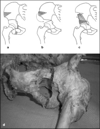
The scheme of triple pelvic osteotomy in our modification with a detailed view on the place of osteotomy of the ischium and the pubic with the resection of bone segment of 0.5 cm: a) resected fragments; b) rotation and tilting of acetabular segment; c) graft and osteotomy fixation with Kirschner wires (adopted with permission from Ortopedie (1)); d) cadaverous pelvis with resected fragments from the ischium and the pubic bone.
We chose the triple pelvic osteotomy for this investigation because the pelvis is interrupted in the plane of the least pelvic dimension and in the plane of the pelvic outlet, thus the possibility exists for the formation of an obstacle during the passage of the fetus in the course of spontaneous vaginal delivery. Very few articles have been published concerning this matter and their results are heterogeneous (7–8–9–10). We decided to investigate how triple pelvic osteotomy changes and influences internal and external pelvic dimensions and whether female patients undergoing this surgical procedure should be advised against normal vaginal delivery.
Our hypothesis is that our modification of the Steel triple pelvic osteotomy does not impede spontaneous vaginal delivery.
Materials and Methods
We used fixed anatomical specimens of the female pelvis. Pelves were obtained from cadavers of donors to the Anatomical Institute. The donors were mainly of advanced years and therefore may have sustained damage or injury to the pelvis with subsequent deformity over the course of their lives. Therefore, we included only non-deformed pelves in the study in order to avoid a decrease in the particular dimension or size below the average stated in the references (7, 10, 17). The Hospital's Ethics Committee approved the study. Soft tissues including muscles and tendons were removed from the pelves; the sacrospinous ligament, sacrotuberous ligament and sacroiliac ligament (structures important for the stability of the pelvis) were maintained, as well as the hip joint capsule with proximal parts of the femur and the 5th lumbar vertebra.
Measurements made in the following manner
Points were marked in coloured ink in order to ensure measuring at the same points. The following points of particular pelvic planes were measured (Fig. 2, a and b) – the pelvic inlet – promontorium, linea terminalis, the upper edge of the pubic symphysis; the plane of the greatest pelvic dimension – S2-3, the centre of the acetabulum, the centre of the symphysis; the plane of the least pelvic dimension – the ischial spine, the lower border of sacrum, the lower border of the symphysis; the pelvic outlet – the tip of the coccyx, the medial border of the ischial tuberosity, the lower border of the symphysis; the obstetrical conjugate (OC) – the distance between the promontory to the most bulging point on the back of the symphysis pubis; and the diagonal conjugate – the distance between the lower border of the symphysis pubic and promontory (17).

a) and b) Detail of cadaverous specimen with points for measurements marked in color. Pelvic inlet: A-A’ transverse diameter, B-B’ oblique diameter, C-C’ anteroposterior diameter; the plane of the greatest pelvic dimension: D-D’ transverse diameter E, F the beginning of oblique diameter and anteroposterior diameter; the plane of the least pelvic dimension: G-G’ interspinous diameter, I-the beginning of anteroposterior diameter.
Statistical analysis one-sided Student's t-test was performed by Stata statistical software, release 9.2, Stata Corp. LP, College Station, TX. The significance level was set at 0.05.
We measured all internal dimensions on particular pelvic planes, external pelvic dimensions, and dimensions used in obstetrics, and x-ray of specimens (anteroposterior view and inlet view). Photographs were also taken. Subsequently, a unilateral triple pelvic osteotomy was performed (with resection of a bone segment of 0.5 cm in the ischium and pubis). We inserted a bone graft, taken from crista iliaca with a base width of approximately 1 cm, into the iliac osteotomy and the osteotomy with the bone graft were fixed with three to four Kirschner wires. Pelvic dimensions were measured again and x-rays and photographs of specimens were taken. The osteotomy of the other side followed. After bilateral osteotomy, there was a problem with the stabilisation of the pelvic ring; therefore, to increase pelvic stabilisation we fixed the osteotomy of the pubis and the ischium with Kirschner wires which is not performed on living patients. X-ray and photographs of the specimens were taken.
In total we processed 19 specimens of female pelves and 38 hip joints. One specimen was removed because the sacrospinous ligament and sacrotuberous ligament failed to be maintained during preparation.
Results
After unilateral surgical procedures, pelvic dimensions changed as follows (Tab. I).
Pelvic Dimensions after Unilateral Osteotomy

not available.
There was an increase in 51% of all dimensions in the plane of the pelvic inlet and in the plane of the greatest pelvic dimension and 34% dimensions remained unchanged. Only in 15% of cases did they decrease (maximally in 0.7 cm in oblique dimension in the plane of the pelvic inlet). In the plane of the least pelvic dimension and the pelvic outlet the particular dimensions changed as follows:
anteroposterior dimension in the plane of least pelvic dimensions decreased in eight cases (up to 0.6 cm), increased in one (by 1.3 cm) and in 10 cases remained unchanged. Average change was −0.042 cm, which is 0.38% from average size of the given dimension (p = 0.311);
anteroposterior dimension of the pelvic outlet decreased in 10 cases, increased in two and in seven cases remained unchanged. On average, the dimension statistically significantly decreased by 0.132 cm, 1.54% (p = 0.015) (Fig. 3);
distance between the ischial spines increased in six cases (up to 1.2 cm), in 11 cases decreased (up to 0.5 cm) and in two cases remained the same with an average change of −0.016 cm, 0.15% (p = 0.445) (Fig. 3);
bituberous distance between the inner aspects of the ischial tuberosities remained the same in two cases and in 17 cases decreased (up to 1.4 cm) with an average statistically significant change of −0.695 cm, 6.6% (p<0.001) (Fig. 3);
external dimensions showed no statistically significant change (from the external conjugate with an average change in −0.033 with p = 0.376 to bispinous distance with an average increase in 0.237 with p = 0.959). Other dimensions used in obstetrics such as the obstetrical conjugate and diagonal conjugate remained unchanged in the majority of cases (the obstetrical conjugate in 10 cases and diagonal conjugate in eight cases) or slightly increased (the obstetrical conjugate in nine cases and diagonal conjugate in six cases) with an average change 0.07 cm in the obstetrical conjugate, resp. 0.15 cm in diagonal conjugate.
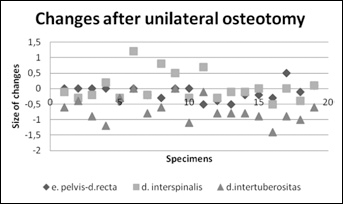
Changes of the most important dimension after unilateral osteotomy.
In 10 specimens it was impossible to measure the bicristal width after removal of the tricortical bone graft from the iliac crest.
In four specimens it was impossible to measure distantia externa, because three specimens were taken without the lumbar spine and without the fifth lumbar vertebra and one specimen demonstrated incomplete spinous processes of vertebra due to the previous removal of the spinal cord.
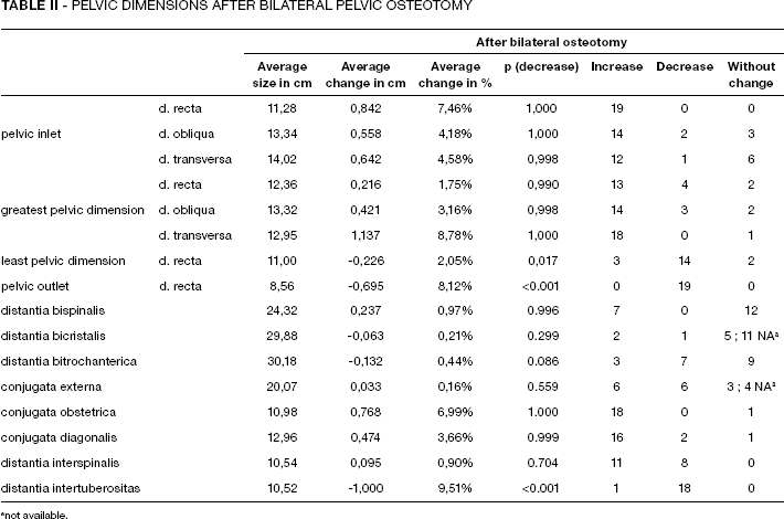
After bilateral surgical procedures, the pelvic dimensions changed as follows (Tab. II and Fig. 4).
Pelvic Dimensions after Bilateral Pelvic Osteotomy
not available.

Changes of the most important dimension after bilateral osteotomy.
All dimensions altered by unilateral surgical procedures also changed after bilateral triple osteotomy. Changes in the given dimensions were more noticeable after bilateral osteotomy, whether either an increase or decrease. The most noticeable and significant changes concerning a decrease were similarly observed in unilateral osteotomy – in the anteroposterior dimension in the pelvic outlet by 0.695 cm (8.12%, p<0.001) on average and in the distance between the ischial tuberosities by 1.00 cm (9.51%, p<0.001) on average. A detailed list of changes in all dimensions after bilateral surgery is in Table II and Figure 4.
Discussion
Triple pelvic osteotomy is one of the redirection osteotomies that enables improvement of the position of subluxed and dysplastic hip joint. It is based on the release of the acetabular segment and a change of its position with subsequent fixation with Kirschner wires. It is therefore logical that pelvic dimensions will change.
Our hypothesis that triple osteotomy does not significantly narrow the bony birth canal was only partially confirmed.
Dimensions in the plane of the pelvic inlet and in the plane of the greatest pelvic dimension increased in 51% of cases; 34% cases saw no change while 15% cases showed a decrease after unilateral osteotomy. The most critical locations for the passage of the fetus through the birth canal (transverse and anteroposterior dimensions in the plane of the least pelvic dimension and the plane of the pelvic outlet) decreased. After bilateral osteotomy, the dimensional decreases were even more noticeable. Only the intertrochanteric distance increased after bilateral osteotomy.
In spite of a relatively small group (19 specimens), statistically significant change was seen in the anteroposterior dimension of the pelvic outlet and in the intertuberous distance. After bilateral osteotomy, a statistically significant decrease was observed in three dimensions. These were the anteroposterior dimension in the plane of the least pelvic dimension, the anteroposterior dimension in the plane of the pelvic outlet and the intertuberous distance.
The point around which the acetabular segment rotates and which does not significantly change its position in the mediolateral direction during surgery is the ischial spine at the place of the attachment of the sacrospinous ligament. It is obvious that the dimensions in the pelvic planes which are above the level of the joint of both spines should remain the same or should increase in lateralisation of the given part of the acetabular segment; and the dimensions in the planes under the joint of both spines should remain the same or should decrease during medialisation of the given part of the segment. The largest part in the medial shift of the ischial tuberosity and the change in the given dimension represent the resection of the bone segment in the ischium medially from its tuberosity, which is the part of our modification of triple pelvic osteotomy.
After bilateral osteotomy, the changes in pelvic dimensions are not doubled as could be expected given the change in dimensions after unilateral surgery. This is due to the experimental method of performing bilateral osteotomy. During bilateral pelvic osteotomy, the pelvis is divided into four practically free parts whose attachment only by Kirschner wires is not sufficiently stable. During surgery on patients, fixation is much easier as osteotomy of the second side is only performed after healing of the first side on which a previous triple osteotomy was performed.
Dimensions in the plane of the least pelvic dimension and in the plane of the pelvic outlet are deciding factors enabling spontaneous delivery per vias naturales. Regarding the dimension of the foetal head, which is frontooccipitally 11 cm (12), the diminution of anteroposterior diameter in the plane of the least pelvic dimension and in the plane of the pelvic outlet in 0.04 cm (0.38%) and 0.13 cm (1.54%) should not be an obstacle for the passage of the foetal head. By comparing biparietal dimensions of foetal heads, which range from 9 cm (12) to 9,25 cm (9), with the distance between the ischial spines (10.54 cm) and between the ischial tuberosities (10.52 cm) it is apparent that an average decrease in the given dimensions of 0.016 cm (0.15%), and 0.695 cm (6.6%) respectively, should not have any influence on the passage of the foetal head, even if we include the thickness of soft tissues (about 4 mm) (12). It is known that hormonal changes before delivery lead to partial loosening of pelvic connective tissue and ligaments. On the basis of our measurements, we do not conclude that the given diminutions should have an influence on the passage of the foetal head through the birth canal during delivery.
The results of our study can be compared with similar results of other studies: Winkelmann (8) observed the narrowing of the birth canal in its middle part in female patients and cadaver specimens – up to 3.3 cm (26.6%) after triple pelvic osteotomy according to Steel (4), Carlioz (1) and Tönnis (5, 6). He recommended informing all female patients undergoing these surgeries, after termination of growth, of the possibility of the need for Caesarean section. In our measurements, we observed smaller changes in the distance between the ischial spines 1,2 cm to −0,8 cm after unilateral osteotomy and 1,0 cm to −1,7 cm after bilateral osteotomy, respectively.
Similarly, Loder (7) discovered a narrowing of pelvic dimensions after pelvic osteotomy according to Chiari (11), Steel (4), Salter (1) and Ganz (1). Steel's osteotomies saw a narrowing in the plane of the least pelvic dimension (d. interspinalis) an average of 15% and in the plane of the inferior pelvic aperture (d. intertuberositas) an average of 16.0%, in a plastic model of pelvis. This was the same in female patients. Loder recommended informing patients of a higher risk of Caesarean section delivery if the interspinous distance is smaller than 9.5 cm in an AP x-ray image of pelvis (7). This value was taken from the work of Christian et al (10) based on the research of the relation of pelvic dimensions and the possibility of vaginal delivery with breech presentation of the fetus. In our study, transverse dimensions in the plane of the least pelvic dimension and the plane of the pelvic outlet decreased after unilateral osteotomy on average less significantly (0,15% respectively 6,60%); and after bilateral surgery interspinous distance increased on average by 0.90% and intertuberous dimension decreased by 9.51%.
Abitbol's (9) study describes the relation between the cephalopelvic disproportion index (difference between the smallest pelvic dimension and biparietal dimensions of the foetal head) and the possibility of vaginal delivery. He measured pelvic dimensions and the bitemporal width of the foetal head on x-rays. He discovered that vaginal delivery with an index below 9 mm is impossible; with an index between 9 mm – 13 mm, delivery is either impossible or very difficult. With these values, the specificity of the index is 100%. When the index reaches 13 mm, specificity decreases to 51%; nevertheless, Caesarean section was necessary in some pregnant women with an index above 13 mm. Our measurements show that the cephalopelvic disproportion index is 24 mm and 30 mm in the plane of the pelvic inlet (after unilateral and bilateral surgery respectively) and 15.2 mm and 14.5 mm at the plane of the least pelvic dimension (after unilateral and bilateral osteotomy respectively) in considering biparietal the width of the foetal head at 9 cm (12). The delivery per vias naturales should be possible with such a high cephalopelvic disproportion index; but regarding the above given sensitivity of 51%, it will be necessary to use Caesarean section in some pregnant women.
Our study has some strengths and weaknesses. The advantage of the study is a relatively high number of processed specimens and the measurements of changes performed also after bilateral osteotomy.
The disadvantage is that the study originated in only one institution and it could possibly be influenced by the heightened interest of the institution to confirm the given hypothesis.
To conclude, unilateral triple pelvic osteotomy with our modifications does not produce a clinically significantly change in birth canal size and should not impede delivery per vias naturales in pregnant women who have previously undergone such surgery. In patients with bilateral osteotomy, pelvic dimensions significantly change and therefore we would recommend Caesarean section.
Footnotes
Acknowledgements
We wish to acknowledge the Institute of Anatomy First Faculty of Medicine Charles University in Prague for cooperation with obtaining suitable pelves, and RNDr. Marek Malý for statistical data processing.