






Abstract
Previous experience has demonstrated the importance of testing new bone cement in vivo before widespread clinical use. We performed a consecutive, radiostereometric (RSA) study comparing Refobacin Bone Cement (RBC) to the well proven Palacos with Gentamicin (PWG). According to the manufacturer of RBC it has the equivalent characteristics as PWG, and in vitro tests show good results. The purpose of this study was to evaluate whether RBC is safe to use in clinical practice for total hip arthroplasty (THA). Two consecutive series of patients with primary osteoarthritis received a THA using a highly polished, collarless, tapered stem with a hollow centralizer. The study comprises 21 hips with RBC and 30 with PWG. The patients were followed up for two years with repeated RSA examinations and clinical outcome questionnaires SF-12 and WOMAC. There were no statistically significant migratory differences between the groups. The mean subsidence after two years was 1.28 mm and 1.40 mm, and the mean retroversion was 1.03° and 0.99°, for the RBC and the PWG groups respectively. Almost all migration occurred in the interface between the stem and the cement. The WOMAC and SF12 clinical scores did not reveal any clinical differences between the groups. We conclude that, as previous in vitro tests indicate, RBC performs as well as PWG and seems to be safe to use in clinical practice for THA.
Introduction
All bone cements used in current clinical practice are PMMA-based. Even so their performance differs physically and clinically. Poor results with some low viscosity bone cements (1), and particularly with Boneloc cement (2, 3) have shown the importance of selecting the appropriate cement for clinical practice.
The long-term results of total hip arthroplasty (THA) using high viscosity cements are excellent (4). During the last decades cemented THA in Sweden has been dominated by the use of Palacos with Gentamicin (PWG) (Schering-Plough, Belgium), which is a high viscosity bone cement. The characteristics of Palacos and the antibiotic loaded PWG are well documented both in vitro and in vivo studies; long-term results show that it maintains good structural properties and thus often is used as a comparative agent and the gold standard in the analysis of untested bone cement (5, 6). In 2005 Palacos and PWG were no longer produced for the former distributors. It had been produced by Heraeus Kulzer GmbH (Hanau, Germany) and with the responsible manufacturer and distributor being Schering-Plough (Belgium). Furthermore, the cement was used under the name Refobacin Palacos R with Biomet Europe (Dordrecht, the Netherlands) as distributor and responsible manufacturer (Biomet Orthopaedics, Switzerland). As a replacement product Refobacin Bone Cement (RBC) was introduced by Biomet Europe in 2005 (produced by AAP Biomaterials GmbH, Dieburg, Germany), and they claim the characteristics are equivalent to those of the original PWG. At the same time Heraeus Medical GmbH (Wehrheim, Germany) started as distributor and responsible manufacturer of the follow-up product Palacos R+G, which they claim is produced with the same formula as the original Palacos (and the same producer, Heraeus Kulzer GmbH). Multiple in vitro studies have shown at least as good results with RBC as PWG (7, 8). However, even if they claim the equivalent characteristics, it should be considered as partly unproven bone cement and tested as such. To the best of our knowledge such a study regarding RBC has not yet been published.
The best method to evaluate and validate new bone cement in vivo is by using Radiostereometry (RSA), as it is the most accurate method for evaluation of prosthetic migration and predictive for later loosening (9, 10). The purpose of the present in vivo study was to use RSA to assess the micromotion of a femoral component cemented with RBC, using PWG as a reference. Henceforth, these cements will be referred to as RBC and PWG respectively.
Patients and Methods
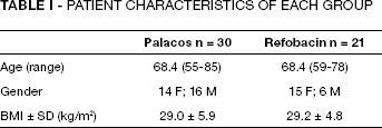
Patients with primary osteoarthritis (OA) of the hip necessitating THA, Charnley classification A (unilateral hip disease with no other disability) and classification B, (bilateral hip disease with no other disability) were included. A total of 21 consecutive hips were selected for THA using RBC for fixation of a MS-30 (Zimmer, Winterthur, Switzerland) femoral component, which is a collarless, highly polished and triple tapered stem with a hollow centraliser. The control group of 30 patients, who received a prosthesis cemented with PWG, was selected from a previous, yet unpublished study analyzing the same MS-30 prosthesis. These patients had been operated on shortly before the corporate reorganisation and had thus received the original version of PWG. Two surgeons (GF and CO) operated on an equal amount of patients in each group to avoid the influence of the surgeon on the results. Two patients in each group were lost to follow up. In the PWG group, one patient died before the two year follow up of causes not associated with the THA. The second patient suffered a stroke with sequelae and could not attend further follow-up. In the RBC group, one patient moved to another city and could not participate in the study any longer. She reported, however, great satisfaction with her THA. One patient was excluded due to bad visibility of RSA markers, resulting in a too high condition number. Total number of patients followed for two years were thus 19 in the RBC group and 28 in the PWG group. The age and characteristics of the groups are described in Table I.
Patient Characteristics of Each Group
Operative technique
The patients were operated on in a lateral position, with a postero-lateral approach. The MS-30 stem was available in two sizes (8 and 10) and two offsets (standard and lateral) and was used with a hollow centraliser. Duracul heads, 28 mm diameter (sizes S,M,L) were used in both groups. The acetabular component was a Weber-cup (46-58 mm) in the PWG group and a ZCA-cup (46-58 mm) in the RBC group. Third-generation cementing technique was used in all cases. Both PWG and RBC were used pre-chilled. Mixing was done with the closed Optivac vacuum mixing system (Biomet Cementing Technologies, Malmö, Sweden).
Radiostereometry (RSA)
The proximal femur was marked peroperatively with 7-9 tantalum markers of 0.8 mm size. Three to four were inserted into the lesser trochanter, and 4-5 markers were put in the greater trochanter. The cement was marked with 6-7 tantalum markers of 0.8 mm size; 3-4 at the tip of the cement-gun just before filling out the femur, and 3-4 markers in the proximal cement mantle after the stem had been introduced. The stems were pre-marked by the manufacturer with three tantalum markers of 1.0 mm size.
The RSA migration results were analysed for translation and rotation along and about the three axes of the orthogonal coordinate system. As primary effects variables we selected the Y-translation (subsidence) and the Y-rotation (retroversion) of the stem. Signed values were used.
The initial RSA examination, i.e. the reference examination, was performed on the first postoperative day, before mobilisation. The follow-up examinations were done at three, six, 12 and 24 months postoperatively, with a time tolerance of ± 5% each time. The patient was placed in a horizontal position and a uniplanar technique was used with a type 41 calibration cage beneath the patient (Tilly Medical AB, Lund, Sweden). The 3-D position of the markers and the translation/rotation of the stem in comparison to the cement and the bone were computed using the software UmRSA (version 6.0, RSA Biomedical, Umeå, Sweden). The upper limits for the condition number and the mean error of rigid body fitting were set at 120 and 0.3 respectively. The precision of our RSA-technique was validated by double examinations of all patients at one time during the follow up period (Tab. II).
Precision of Rsa Based on Double Investigations of Patients in the Study
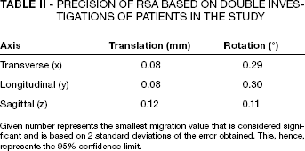
Given number represents the smallest migration value that is considered significant and is based on 2 standard deviations of the error obtained. This, hence, represents the 95% confidence limit.
Clinical follow-up
Clinical evaluation, including self administered questionnaires, the hip specific Western Ontario and McMaster Universities Osteoarthritis Index (WOMAC) (11) and the general Short-Form Health survey (SF-12) (12) were performed preoperatively and at 12 and 24 months postoperatively in correlation to RSA follow-ups.
Statistical analysis
Repeated measures ANOVA and Students T-test were used to analyse migration and rotation data. For analysis of the outcome questionnaires we used the Friedman test over time, and the Mann-Whitney U test for comparison between the groups.
The primary effect variable, used for the power analysis, was subsidence of the stem (y-translation). Based on our results from the baseline study of 30 patients with PWG (SD 0.43 mm) and assuming a difference in means of 0.4 mm, a power of 0.80 and a risk of 0.05 for type-1 error, 16 patients were needed in the RBC group.
We used the IBM SPSS statistics software (version 19.0, IBM, New York, USA), and p<0.05 was considered significant.
Ethics
The trial was approved by the ethics committee of Lund University and performed in compliance with the Helsinki Declaration of 1975, as revised in 2000, and all patients had given their informed written consent.
Results
The overall migrations measured with RSA were small for both groups. We did not find any statistically significant difference in migration along any of the three cardinal axes between the two groups over time (Tab. III, Figs. 1 and 2). The majority of the migration occurred before the six months follow-up. Regarding y-translation, i.e. subsidence, at six months the PWG group had subsided 1.02 mm and the RBC group 0.96 mm, which is 75% and 73% of the total two year subsidence respectively.
Translations and Rotations Measured by rsa at the 2 Years follow-up. Signed Values
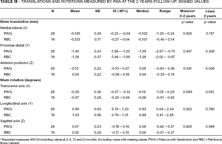
Repeated measures ANOVA including values at 3, 6, 12 and 24 months. Excluding cases with missing values. PWG = Palacos with Gentamicin and RBC = Refobacin Bone Cement.

Observed mean values of subsidence of the femoral component with the two cements (Error bars represent standard error of the mean). Solid lines represent subsidence between the stem and femur and dotted lines between the stem-cement interface. PWG = Palacos with Gentamicin and RBC = Refobacin Bone Cement.
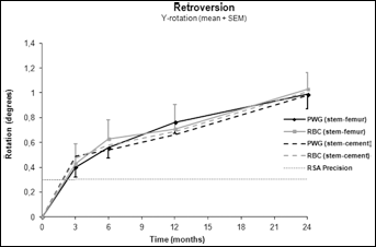
Observed mean values of retroversion of the femoral component with the two cements (Error bars represent standard error of the mean). Solid lines represent retroversion between the stem and femur and dotted lines between the stem-cement interface. PWG = Palacos with Gentamicin and RBC = Refobacin Bone Cement.
X-rotation at the two-year follow-up showed a statistically significant difference (p = 0.03) between the two groups, but with migration values below the RSA precision.
The absolute majority of the migration in both groups occurred between the stem and the cement mantle (dotted lines in Figures 1 and 2).
At two years no stems had been revised and there were no obvious signs of radiolucent lines or other radiological signs of loosening in any stems.
Clinical results
There were no statistically significant differences between the two groups preoperatively or at one or two years postoperatively, neither regarding the results from WOMAC nor SF-12 (Fig. 3). The two groups taken together as a whole, however, improved significantly between the pre- and postoperative assessments. Hip function as measured by WOMAC for all three subscales improved significantly (p<0.001). The general health questionnaire SF-12 also improved significantly (p<0.001).
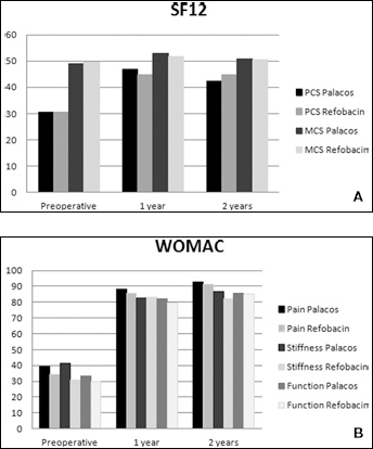
A) Above mean values of WOMAC hip specific scores as subgroups Pain, Stiffness and Function. B) Below the general SF-12 Health survey with mean values for PCS (Physical Component Score) and MCS (Mental Component Score).
Discussion
Regardless if RBC or PWG was used we found no difference in migratory behaviour for the stems during the first two years of follow-up. Both the retroversion and the subsidence of the stems are comparable to published RSA-results from stems of similar type (i.e. collarless, tapered and highly polished). Stefansdottir et al (13) studied the movement pattern of the Exeter stem cemented with PWG. They showed mean subsidence at two years of 1.34 mm and retroversion of 1.2o. Alfaro-Adrian et al (14) reported very similar values with the Exeter stem cemented with CMW bone cement (DePuy International Ltd, Leeds, UK). A similar publication studying the movement pattern of the C-stem (DePuy International Ltd, Leeds, UK) cemented with PWG showed mean subsidence at two years of 1.35 mm and retroversion of 1.9o (15).
We found that the vast majority of the migration occurred between the stem and cement whilst the cement mantle in relation to the bone remained very stable. When using a tapered, collarless and highly polished stem, subsidence between the stem and the cement mantle is to be expected, as the stem adapts to creep in the cement (16). Clinical outcome according to WOMAC and SF-12 showed excellent results and improved significantly from pre- to postoperatively.
The current standards for preclinical testing of new bone cements include In vitro studies of their structural properties in order to predict in vivo behavior (ISO 5833:2002 and ASTM F451-76). Previous experience has shown the importance of clinically testing new bone cements before widespread use, (e.g. Boneloc- which showed good results in in vitro testing, but disastrous results clinically) (2). Furnes et al (17) showed that an Exeter type prosthesis had a seven times higher risk of revision if cemented with Boneloc, in comparison to a high viscosity cement. Charnley type prostheses had an astonishing 14 times higher risk of revision if cemented with Boneloc. Breuch et al (18) showed that bone cements can differ in their structural properties from in vitro to in vivo testing. This shows the importance of performing studies like ours, even though the cements are structurally equal in the laboratory.
There has been a widespread discussion over the last decade regarding different cements in the fixation of THA. Among the topics discussed are weather to use high or low viscosity cements and what type of mixing system should be utilised. Soderlund et al (19) published 10 year results showing no statistically significant RSA measured differences between PWG and the low viscosity cement Cemex Rx (Tecres S.p.A., Italy). Nivbrant et al (20), which was the five year publication of the same patient material, compared the Cemex Rx with PWG, using an air-pressure mixing system in comparison to the vacuum-mixing system used for PWG. They showed less mechanical strength and more radiolucent lines in the Cemex group compared to PWG. Although neither the five-year or ten-year results showed clinical or RSA differences, they concluded that closed vacuum mixing is to be preferred. It is likely that when comparing two cements, which are as similar in structure as e.g. RBC and PWG, the mixing system used is important. It has been shown that by using a vacuum mixing system, the porosity of the cement is reduced and thereby the cement strength increased (21). Our study compares RBC and PWG, using the same vacuum mixing system (Optivac). If different mixing systems had been used, the results might have been different.
A potential weakness with our study is that it is not randomised. We did, however, reproduce the characteristics of the reference study as far as possible. Thus the same two surgeons operated on an equal amount of patients in both groups, with the same technique, environmental conditions and stem type. We have small patient numbers in our study, but as it is an RSA study the numbers should be adequate, and based on our power calculation and earlier RSA-experience we aimed at having a minimum 20 patients in the groups (22).
In conclusion, RBC performed as well as the proven PWG. We conclude that Refobacin Bone Cement, which is claimed to have the equivalent characteristics as Palacos with Gentamicin, and has been well tested in vitro, also seems to be safe to use in clinical practice in THA. Longer term follow-up and future results from national registries are of course desirable.
Footnotes
Acknowledgments
We thank Håkan Leijon at the RSA laboratory in Lund, for help in computerising the RSA pictures and data, and Philippe Wagner at RC Syd, Clinical Sciences, Lund University for statistical advice.