






Abstract
Purpose
A comparison was made of the clinical and radiological results of cylindrical fully porous-coated femoral stems (Group A) and Zweymüller-type femoral stems (Group B) used for the treatment of hip osteoathrosis, secondary to Crowe III and IV dysplasia, with total hip arthroplasty combined with femoral transverse shortening osteotomy.
Method
This study is a retrospective evaluation of 86 hips in 50 patients. Group A comprised of 43 hips and Group B comprised of 43 hips. During final follow-up evaluation, patients were clinically assessed with Harris Hip Score (HHS), Merle d'Aubigne-Postel scale (MAP), and SF-36 scale. For radiological examination the Gulman THA score was used. Femoral osteotomy union, osteolysis around the components and component migration were also recorded and evaluated.
Results
In Group B, nonunion of the osteotomy site was found in 18.6% and delayed union in 20.9% of the hips. In Group A, delayed union was found in 7% of the hips. Patients’ mean daily walking distance was found to be lower in Group A when compared to Group B. When the postoperative clinical HSS, and MAP and the radiological Gulman scores were compared, no statistically significant differences (p = 0.275) were found.
Conclusions
Patients with hip osteoarthritis secondary to Crowe III and IV dysplasia, who were treated with THA and transverse osteotomy showed a higher incidence of osteotomy complications when the Zweymüller femoral stem was used. However, these complications did not affect clinical outcomes.
Introduction
In developmental hip dysplasia (DHD), the development of secondary osteoarthritis due to dynamic mechanical changes caused by the anatomy of the hip joint requires total hip arthroplasty (THA) in young adulthood (1). Different factors such as abnormal centre of hip rotation, proximal femoral deformity, soft tissue contractures, abnormal muscle development and inadequate bone stock in this patient group render complex THA operations and increase the risk of both intraoperative and postoperative complications (2, 3). Particularly in severely dysplastic hips, such as Crowe types III and IV, THA is a difficult surgical procedure (4).
The incidence of DHD in Turkey has been reported at the level of 1.34% (5). Due to this high incidence and the fact that it is often overlooked in children, it is often encountered late, and many comparative studies related to the various surgical techniques of treatment applied to this patient group have been published in literature. 1 of the most important factors for the reduction of surgery-related complications to a minimum is the selection of the most appropriate implant for the patient (6). However, to the best of our knowledge, there has been no study comparing current implants of different design and properties and their effect on clinical and radiological results. The hypothesis of this study was that implants of different femoral design could have different effects on clinical and radiological results. To examine this hypothesis, a clinical and radiological evaluation was made of 2 different uncemented femoral stem designs used in the treatment of osteoarthritic hips, secondary to Crowe type III and IV DHD, with THA and transverse subtrochanteric osteotomy.
Patients and methods
In this study, 50 patients (86 hips) with secondary osteoarthritis, due to Crowe type III and IV DHD, who were treated with THA and transverse subtrochanteric osteotomy at the Ankara Ataturk Training and Research Hospital between January 1999 and September 2010 were included. Patients with active infection, neuromuscular arthritis, abductor muscles’ pathology, obesity or psychiatric disease were excluded from the study.
Patients were randomly separated into 2 groups regardless of the proximal femoral morphology. Group A comprised of 24 (55.8%) Crowe type III hips and 19 (44.2%) Crowe type IV hips. Group B comprised of 21 (48.8%) Crowe type III hips and 22 (51.2%) Crowe type IV hips. In both groups there were 23 females (92%) and 2 males (8%). In both groups, a total of 43 hips were operated on as 18 bilateral (72%) and 7 unilateral (28%) cases. In the unilateral cases, which were 1 male and 6 female in both groups, 2 (4.6%) were right side and 5 (11.6%) were left side.
Preoperative evaluation of the Harris Hip Score (HHS) and the Merle d'Aubigne-Postel scale (MAP) was carried out for all patients. After completion of preoperative consultations, surgery was carried out under general anaesthesia with the patient in the lateral decubitus position. Patients of both groups were operated on by the same surgical team consisting of 1 senior surgeon and 2 assistant doctors. Exposure was obtained with a posterolateral incision extended distally. Following excision of the femoral head, circular multiple drilling was done 2-cm distal from the level of the trochanter minor, then with the help of a wire osteotome, a subtrochanteric transverse femoral shortening osteotomy was performed. The acetabular component was then placed in the true acetabulum. Following femoral stem implantation, stabilisation of the osteotomy line was achieved with a low contact dynamic plate and screws in all patients (Fig. 1A, B). On postoperative day 2, all patients were mobilised and rehabilitation was started in the Physical Therapy Unit. Weight-bearing was not permitted on the operated side for 6 weeks. At the end of the 6-week period, partial weight-bearing was permitted for 2 weeks with crutches, then, after 8 weeks, full weight-bearing was permitted. The postoperative evaluation of all the patients was made by the team which had carried out the surgery.
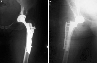
(
Early postoperative radiographs, at monthly intervals for the first 6 months and every 2 months thereafter were taken using AP pelvis and lateral radiographs.
The THA operations were carried out using a standard uncemented fully porous-coated cylindrical total hip prosthesis (Corinium® Corin) in Group A and an uncented Zweymüller-type (SL-Plus® Smith & Nephew) in Group B.
In all patients, an attempt was made to ensure that the femoral stem end bypassed the osteotomy level for at least 5 cm.
The mean follow-up period was 69 (range 58-124) months in Group A and 73 (range 58-122) months in Group B. At final follow-up evaluation, clinical evaluation of the patients was done using the Harris Hip Score (HHS) and the Merle d'Aubigne-Postel scale (MAP), and quality of life was also evaluated using the SF-36 scale. The union of the femoral subtrochanteric shortening osteotomy and acetabular component migration was radiologically evaluated. The presence of osteolysis around the stem was also recorded according to the Gruen criteria (7). Further radiological evaluation was carried out using the Gulman radiological THA score. The preoperative and postoperative length of the extremity was evaluated by measuring the distance between the anterio-superior iliac spine and the tip of the medial malleolus.
Statistical analysis
Statistical analysis of the study data was done with SPSS for Windows 11.5 software. The normality of the distribution of continuous variables was assessed using the Shapiro Wilk test. Continuous variables were presented with descriptive statistics as mean ± standard deviation (SD) or median (minimum-maximum) and categorical variables as number (n) and percentage (%). The significance of the difference of the means between groups was evaluated with the Student's t-test and the significance of the difference between median values with the Mann-Whitney U-test. Categorical variables were evaluated with the Pearson chi-square or the Fisher's exact test. A value of p<0.05 was accepted as statistically significant.
Results
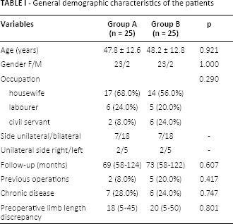
The demographic characteristics of all patients are shown in Table I. In both groups there was a similar distribution of age (p = 0.921), gender (p = 1.00), occupation (p = 0.290), THA side (p = 0.752) and the side of the unilateral prosthesis (p= 0.554). The median postoperative follow-up was 69 (range 58-124) months in Group A and 73 (range 58-122) months in Group B (p = 0.607). History of previous surgery was found in 8% in Group A and in 20% of the hips in Group B (p = 0.417) and the presence of chronic disease was recorded in 28% in Group A and 24% of the patients in Group B (p = 0.747) (Tab.I). No statistically significant difference was seen between the 2 groups in respect of preoperative limb length discrepancy (p = 0.801).
General demographic characteristics of the patients
Patients who were sexually active were questioned (within 1 month, the number of times of sexual relations, the number of positions used, the presence of any feeling of tension during sexual relations) and problems were reported subjectively by 20% in Group A and approximately 10% of the patients in Group B. No statistically significant difference was found between groups (p = 0.661). The preoperative mean HHS scores were 38 (31-48) and 37 (29-44) for Groups A and B (p = 0.052) and the postoperative values raised to 86 (71-96) and 87 (69-96) respectively (p = 0.836). The pre- and postoperative MAP values are shown in Table II. No statistically significant difference was found between the groups concerning the Gulman radiological THA score (p = 0.275). Osteolysis was recorded around the femoral stem in 11 (25.6%) hips of Group A and in 12 (27.9%) hips of Group B and no statistically significant difference was found between groups (p = 0.808). Vertical migration of the femoral stem was not found in any hip in Group A, but was recorded in 4 hips of Group B (p<0.001). Change in position of the femoral component was observed in 1 (7%) hip in Group A and in 9 (20.9%) hips in Group B, a finding which was proved to be statistically significant (p<0.007).
Clinical scores and radiological findings of the groups
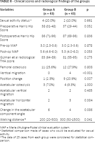
Statistical comparison made of cases who could be evaluated for sexual activity.
The data of 25 cases from each group were considered for statistical comparison.
No statistically significant differences were found between groups concerning vertical and horizontal migration of the acetabular component and the acetabular cup index (p = 0.485, p = 0.384, p = 0.086). When the parameter of walking distance was compared between the 2 groups, Group B showed statistically significantly higher values (p = 0.041). Within Group B, the preoperative HHS values of the patients with postoperative union (p<0.001) or delayed union (p = 0.038) were found to be statistically significantly higher compared to those with postoperative nonunion (Tab. II). This result shows a direct correlation between preoperative clinical scores and postoperative osteotomy line union.
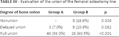
When Groups A and B were compared postoperative Q angles were found to be higher in Group B (p = 0.018). The union rate of the subtrochanteric femoral shortening osteotomy area was 93% in Group A and 60.5% in Group B (p<0.001). Delayed union was found in 7% of the hips in Group A. Delayed union was observed in 20.9% and nonunion in 18.6% of the hips of Group B (p = 0.003) (Tab. III). In all patients with nonunion, revision surgery was carried out with iliac autografts on the osteotomy site and union was achieved in all cases.
Evaluation of the union of the femoral osteotomy line
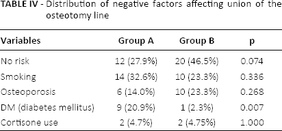
Assessing factors which could have a negative effect on femoral osteotomy healing, apart from a higher rate of DM in Group A, no other factors developed statistically significant differences (p = 0.007) (Tab. IV).
Distribution of negative factors affecting union of the osteotomy line
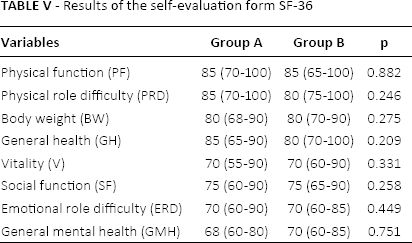
No statistically significant difference were seen when quality of life scores were compared between Groups. In Group B, statistically significantly higher scores were found concerning physical activity parameters in patients with union compared to those with nonunion (p = 0.002), general health scores of patients with union compared to those with nonunion (p = 0.006) and those with delayed union (p = 0.036), and social functional scores in patients with union (p<0.001) and those with delayed union (p = 0.027) compared to those with nonunion (Tab. V). Quality of life scores of patients with nonunion and delayed union were lower in Group B when compared to the rest.
Results of the self-evaluation form SF-36
When complication rates were compared between groups, the overall complication rate was higher in Group A, with intraoperative proximal femoral fissure rate being also higher when compared to Group B, although no patient required reoperation for this reason. During the postoperative period of follow-up, all postoperative dislocations were reduced with closed manipulation under anaesthesia, and stability was retained without any need for component revision.
Discussion
The treatment of hip osteoarthritis secondary to DHD is always more difficult than that of primary cases and the complication rate has been reported as higher (2, 3). Surgical techniques and complications in this specific patient group have been described based on the evidence of the results of studies which, to date, have compared different patient groups. However, there has been no study comparing different implant designs and their effects in this complex patient group. In this study, by comparing the clinical and radiological results obtained with the use of 2 different designs of uncemented femoral stems combined with transverse subtrochanteric femoral osteotomy for the treatment of DHD osteoarthritic hips with THA, we aimed to determine the effect of implant design on clinical and radiological outcomes. In contrast to primary THA patients, in most cases in our patient group there is a need for additional procedures. In THA carried out on Crowe type III or IV dysplastic hips, there is generally a need for subtrochanteric shortening osteotomy as a technique to provide both joint reduction and to avoid neurological complications. Different forms of osteotomy have been described, the most widely used are transverse, oblique, z-subtrochanteric and double chevron. In a biomechanical study comparing these 4 osteotomies, it was reported that no single technique carried out for osteotomy stability had any superior effect and that the application of external graft did not make any significant contribution to stability (8). In a meta-analysis which examined a total of 37 studies, nonunion in the osteotomy area was determined at 3.79%. When subtrochanteric transverse osteotomy was compared with other modified techniques, while no significant difference was found, transverse osteotomy was recommended due to the simplicity and ease of reconstructing femoral anteversion (9). In some studies, the addition of plate and screw fixation, as it was used in the current study, has been recommended for rotational stability in the straight osteotomy site (3).
Sofu et al (10) applied transverse femoral shortening osteotomy together with THA in a series of 73 cases and reported a mean time to union of the osteotomy area of 5.4 months and a nonunion rate of 5.4%.
Encouraging clinical results have been reported with different designs of implants in THA carried out for the treatment of hip osteoarthritis on a dysplastic basis in various studies (11–12–13). Kato et al (14) reported higher osteotomy union rates when implants with extended porous-coated distal cylindrical tip were used. This observation confirms the results obtained in the current study. In the same study, despite stress shielding being reported in the proximal femur in 19% of cases at a mean 12.1-year follow-up, survival of the femoral stem was found at the same follow-up time in all patients. Stress shielding encountered in the femur following THA resulting in bone loss is proposed as an unwanted iatrogenic event. In THA cases where a cylindrical fully porous-coated femoral stem for distal fixation is used, this phenomenon is seen more, in contrast with femoral stems designed for proximal fixation. In a study carried out using 4 different designs of custom-designed femoral stems, stress-shielding was determined in all the designs (15). In 2 different studies, successful results have been reported after treatment with cylindrical, fully-coated and modular design S-ROM femoral design implants combined with transverse femoral shortening osteotomy in dysplastic hips (16, 17).
In a study by Petis et al (18), fully porous-coated cylindrical femoral stems were used, as in the current study, and the 10-year survival rate was reported as 98.8%. Using the same design of femoral stem, in a series where subtrochanteric femoral shortening osteotomy was performed in 20 hips (16 patients), 6.2% nonunion was reported and 20% stress-shielding. For external fixation, rather than the plate screw combination, cable fixation of the excised femoral segment was used (19).
Delayed union was observed at the low rate of 7% in Group A and there were no cases of nonunion in that group. In Group B, nonunion was observed in 18.6% and delayed union in 20.9% hips. When the factors were examined which could have caused this overall high rate of 39.5% of union failures, no difference was determined between the groups. This shows that the groups consisted of patients with similar characteristics in respect of comorbitities which could negatively affect bone union and indicates that there were no differences that could affect the results. When it was shown that personal characteristics had no statistically significant effect on rates of union, it was considered that the reason for the difference could be related to the design of the femoral stem that was used. However, in contrast to the findings of the current study, in another study in which the Alloclassic–SL stems were used in a series of 7 patients, union of the osteotomy site was reported in all the cases (20). However, the low number of patients in that series is a weak point.
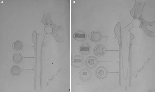
The surface of the stems used for Group A patients was processed and roughened and the coating was applied over this surface. In this form, a Press Fit stem implantation is very efficient, which is a significant advantage. The shape of the stems, used in Group B patients, was wider at the proximal end and became narrower and thinner towards the distal end. The stem had no collar and the overall shape was square in cross-section. The difference between stems in geometric shape and design may be the cause of delayed union and nonunion because the femoral stem, besides transferring the load, is a fixator for the fracture site created by the osteotomy and as this provides endosteal fixation and behaves like an intramedullary nail. Thus, successful results were obtained from the femoral stem used in Group A patients (Fig. 2A, B). In total hip replacement operations, the factors determining the selection of the femoral component are the femoral anatomic morphology and the requirement for osteotomy (21, 22). Some authors suggest that it is not necessary to use a custom-made prosthesis in DHD hips (23). In the current study, successful results were obtained both clinically and radiologically with the standard cylindrical fully porous-coated femoral design. Thus, there can be considered to be no need for patient-specific femoral stem design.
(
Conclusion
According to the results of this study, in the mid-term follow-up of cases where THA has been applied together with transverse subtrochanteric femoral shortening osteotomy in Crowe type III and type IV dysplastic hips, the femoral stem design was not seen to have any effect on the radiological results and no significant differences were seen in the effects on the clinical results in that time period. The Zweymüller type of design was found to negatively affect osteotomy union rates.
Footnotes
Financial support: None.
Conflict of interest: None.