





Abstract
Large ureter damages are difficult to reconstruct. Current techniques are complicated, difficult to perform, and often associated with failures. The ureter has never been regenerated thus far. Therefore the use of tissue engineering techniques for ureter reconstruction and regeneration seems to be a promising way to resolve these problems. For proper ureter regeneration the following problems must be considered: the physiological aspects of the tissue, the type and shape of the scaffold, the type of cells, and the specific environment (urine).
This review presents tissue engineering achievements in the field of ureter regeneration focusing on the scaffold, the cells, and ureter healing.
Introduction
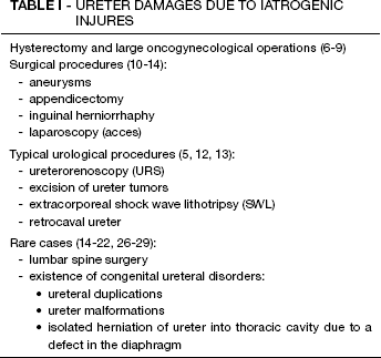
Reconstruction of the ureter is still an unresolved task in urology. The regeneration of the ureter is a widespread problem due to an increasing number of injuries and a highly specific one due to its complicated character (1–4). Ureter regeneration is hampered mainly by inappropriate blood supply, necrosis, and urine toxicity. Iatrogenic surgical complications are responsible for the largest number of ureter injures (about 75%) apart from ordinary ureteral trauma (5). Iatrogenic ureteral injures are related to different surgical procedures (Tab. I). The crush, ligation, intersection, devascularization and ischemia, obstruction of the ureter, or cutout portion of the ureter wall may be a side effect of such procedures (6–10). The result of the injury may be patient's pain or obstruction of the urine passage that can lead to severe complications, frequently associated with renal damage.
Ureter Damages Due to Iatrogenic Injures
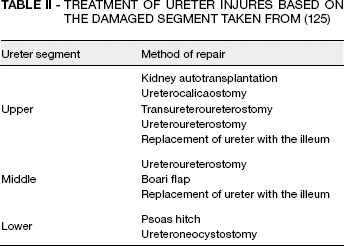
The current methods of ureter regeneration mainly focus on urine transport and protection against urine leakage. The main difficulty in ureter reconstruction is to perform anastomosis and repair of the separated segments of the ureter. The current methods of ureter reconstruction are presented in Table II. The selection of proper reconstruction methods strictly depends on the damaged part (Fig. 1 and Tab. II) Shoemaker was the first who reconstructed the ureter with the ileal segment. This method was popularized in the 1950s by Goodwin, Turner, Winter, Goldstein, and Wells (11–14). Surgeons have to pay attention to the antiperistaltic and isoperistaltic arm of the ileal segment. Proper urine passage is important for proper function of the graft. Unfortunately, despite the frequent use of this method, which is sometimes used as the only choice, it involves the long-term danger of the possible numerous complications and disorders. Problems with the substitution of the ureter are concerned with the lack of proper translucency, backflow of urine that causes nephritis, kidney stones, stenosis, hydronephrosis, infections, kidney failure, renal resection or uremia and hyperchloremic metabolic acidosis (15–17).
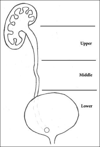
Ureter division.
Treatment of Ureter Injures Based on the Damaged Segment Taken from (125)
Other methods of ureter reconstruction are mainly the modifications of method introduced by Shoemaker, e.g. in the laparoscopic variant (18). None of these methods have been perfected yet (19). Therefore, the use of tissue engineering techniques in ureter reconstruction and regeneration present a promising way to resolve these problems (20–22).
Ureter Histology and Function
The ureter is built from two distinct tissue components, inner epithelium and outer muscle covered by tunica adventitia. It begins at the ureteropelvic junction lying posterior to the renal artery and vein. The inner layer is transitional epithelium. The most important layer for ureter regeneration is the muscle layer, because of its function. Ureter muscle tissue is arranged into two patterns: inner longitudinal and outer circular. The outer layer mainly covers only one-third of the lower ureter part and is an extension of the bladder muscle. The muscle layer is richly interwoven with the connective tissue, also containing elastic fibers. The ureter muscle layer plays a significant role in urine transport form the renal pelvis to the bladder due to peristaltic movements. Peristaltic waves run approximately every 15 to 60s at a rate of 2 to 3 cm/s. These movements are generated in the pacemaker, which is probably localized in the renal pelvis. Peristaltic waves occur even during the absence of urine and they increases when urine appears. When the bladder is filled up, peristaltic waves are stopped. Ureter regeneration is hampered by difficulties in rebuilding complete and functional muscle layer, which in consequence disrupts the peristaltic movement of ureter (23, 24).
Urine and Inflammation
The main problem in urinary tract reconstruction that includes the ureter is urine leakage. It occurs mainly as a result of urothelial iatrogenic injures and can lead to the patient's debility, nephrectomy, and even death. The toxic influence of urine on neighboring tissues triggers inflammation and, in consequence, tissue fibrosis. Fibrous tissue leads to ureter stenosis which in consequence inhibits urine transport to the bladder with subsequent renal damage. Urine leakage from the kidney, ureter, bladder, or urethra can lead to the development of urinoma. Urinoma can be found in three forms (confined, encapsulated urine, or as a free fluid). All these forms of urinomas can lead to urinary tract trauma (25). Urinoma leads to fibrotic tissue formation similar to retroperitoneal fibrosis (RPF). RPF is disease affecting the retroperitoneal space, characterized by inflammation and development of fibrotic tissue. A fibrotic mass wraps around the ureters causing hydronephrosis through extrinsic compression on the ureters or interference with ureteral peristaltic. RPF leads to compression of the retroperitoneal structures, including ureters. Compression of the ureter leads to peristaltic wave inhibition resulting in urine transport disorder, even when the ureter is not damaged and its lumen is patent. The same can be observed as result of urine leakage and urinoma formation. This shows how important urine transport is for proper ureter regeneration (23).
Scaffolds
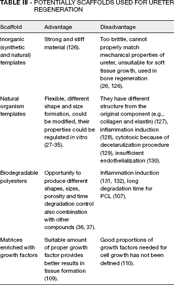
The ideal graft material should be impermeable, protective against urine leaks in the long term and should allow appropriate cell growth and tissue remodeling. It should inhibit inflammation or possible tissue irritation. Four types of polymers can be used for regeneration purposes: synthetic, semi-synthetic, natural, and hybrid materials (Tab. III) (26). Not all types of tested materials were suitable for ureter regeneration. Synthetic and natural inorganic materials such as hydroxyapatite (HA) or tricalcium phosphate have been found to be unsuitable because they cannot properly match the mechanical properties of the ureter because of their brittleness. Natural materials are more appropriate, including collagen, elastin, alginate, agarose, chitosan, fibrin, and hyaluronic acid. These polymers have the proper flexibility and are thus more appropriate. Additionally, natural polymers can be modified and their properties can be regulated in vitro (27–35).
Potentially Scaffolds Used for Ureter Regeneration
The Use of Polyesters for Scaffold Formation
Polyglycolic acid (PGA), polylactic acid (PLA), their copolymers and polycaprolactone (PCL) are commonly used to construct scaffolds for experimental ureter regeneration. The greatest advantage of these materials is biodegradability. It occurs by natural metabolic pathways (36, 37). These polyesters seem to be the most suitable compounds for scaffold preparation. High plasticity and the ability to be combined with other compounds enable the production of matrices with different shape, size, and porosity. Polyester application allows the time of the scaffold degradation after implantation to be regulated.
The crucial issue related to proper ureter regeneration is cell and scaffold exposition on urine. Our in vitro experiments on mesenchymal stem cells (MSCs) and the studies of Davis et al on urothelial cells indicated that urine caused death of both cell types (38, 39). This problem may be solved by the use of scaffolds with well-developed epithelium or by using an appropriate scaffold. Such a scaffold should be created from materials which will not react with urine components and prevent crystallization of urine components. This scaffold should also be biodegradable in a time span sufficient to allow self-regeneration of the epithelium.
The most suitable scaffold for ureter regeneration seems to be produced from polyesters. Polyesters are created de novo; they can be combined with other compounds; and it is possible to regulate their shape, size, porosity, cell attachment, or biodegradability (40–46).
Electrospun Scaffolds for Tissue Engineering – Nanotechnology Mimics Nature
Electrospinning of nanofibers is a new perspective and a very promising method for preparation of scaffolds for tissue engineering. The structure of nanofibers resembles an extracellular collagen matrix (it is built of fibers 50–500 nm in diameter). Electrospinning of bioresorbable polymers gives a unique opportunity to produce a graft from material treated by cells as native. The graft is degraded in a human body in a controllable manner leaving no foreign material behind, which is very important to the regeneration process.
Copolymers of lactic acid have been known for decades and used in medicine as surgical sutures, drug delivery systems, and other applications (47). Despite the fact that it is far easier to harvest ram intestines to manufacture catgut than to produce poly(lactide-co-glycolide) as material for surgical sutures, it is noticeable that the latter completely captured the market. One can assume on this basis that nanostructured biodegradable polyesters will be the future of urinary tract medicine (compared to current experiments that use acellular collagen matrix of animal origin) (48, 49). Application of a biomedically approved polymeric material for production of nanofibrous grafts seems to be a good perspective as only a new form is used to shape the old material (Fig. 2). Polyesters have advantages of over products with animal origins because they offer the use of a well-defined, standardized material free of natural product deviations, absent from xenoproteins and possible problems related to infections from animal material (viral, prion). Polyesters are degraded to natural metabolites such as lactic acid. There have already been attempts to use polyesters alone for bladder regeneration (50).
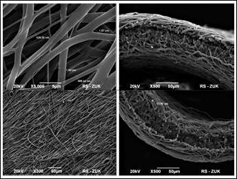
Electrospun PCL triple-sheet material of cross-aligned fibers. Fibers thickness ca. 0.6–1.7 μm, membrane thickness ca. 55 μm.
It has been common practice to use electrospun scaffolds for cell culturing. Many types of cell lines have been cultured on electrospun scaffolds, among them fibroblasts, neural cells, muscle cells, osteoblasts, and stem cells (51, 52). There have also been many attempts to use nano-fibers in regenerative medicine for nerve guides, or vascular grafts to mention a few (53, 54). Electrospun scaffolds seem to be ideal candidates for reconstruction of a thin tissue (skin), or thin tubular organs (blood vessels or ureter), but they can also help reconstruction of thick organs as thin coatings on bulky implants (e.g., bioresorbable bone implants or cartilage implants). They can also serve as scaffold to guide proper healing (55, 56).
Electrospinning used for bioresorbable polymer processing allows production of material with many controllable (and tunable) properties, e.g., polymer digestion time, single fiber and membrane thickness, fiber alignment, and surface modification. Also special features of the scaffold are possible: the use of native polymers (i.e., human serum albumin), controllable drug delivery from fibrous matrix or in situ cell seeding during graft preparation.
A very important property from the medical point of view is the possibility of arrangements made with controllable fibers. This solution enables proper alignment of cells seeded on a scaffold and thus proper regeneration that can potentially restore the peristaltic movement of the ureter. An application of this sort was used in vascular grafts and nerve regeneration. Controllable fiber arrangements are crucial for ureter function restoration, due to its physiological movements.
Control over the Fiber Diameter and Matrix Thickness
The fiber diameter can be controlled and varied by the conditions of the electrospinning process. The type of solvent used for the process depends on the type of material. Also, the more polar the solvent, the thinner the fibers that are produced through the electrospinning process. Poly(3-hydroxybutyrate) p(3HB) is a valuable material of piezoelectric properties that are very important for the stimulation of cell growth. Unfortunately, the material is brittle and requires the use of expensive fluorinated solvents. Nanofibers produced from p(3HB) dissolved in hexafluoroisopropanol are very thin and degrade comparatively fast in physiological pH (up to 72 h). The introduction of a poly(3-hydroxyvalerate) units at rate of only 2% delays the degradation, however, the change in polymer (PCL) and solvent (chloroform) gave thicker fibers (ca. 1.6 μm) of improved degradability time (beyond 4 weeks) (57). The thickness of the electrospun membrane can be easily regulated by the collection time; also the process is not limited to a single material. The production of multi-layered electrospun membranes enables production of multi-component scaffolds.
Fiber Alignment and the Scaffold Shape
Electrospinning of nanofibers enables the production of well-defined constructs with the required shape and dimensions. In an unpublished study we demonstrated productions of tubular scaffolds in a material composed of fibers resembling the structure of ECM (Fig. 2). A material can be implanted into an animal model such as the rat. As the material is aligned orthogonally to a tube length, the next task is to produce axially aligned material. This orientation seems possible based on the work by Jha (58).
We also demonstrated the production of a nanofibrous material with a sandwich structure made of an inner layer of poly(L-lactide-co-caprolactone) PLCL of a good mechanical strength and outer layers of human serum albumin (HAS) fibers with poor mechanical properties, but with an anti-adhesive property. This can be placed inside the ureter lumen to prevent biofilm formation. The HSA fibers were proven to prevent fibroblast attachment, with the aim of being used to prevent skin lesions (59). Mixtures of polymers can also be used to create nanopores in the nanofibers (60), and to regulate other properties (e.g., to tailor the biodegradation rate of the fibers).
Surface Modification and the Possibility of Loading a Drug in the Nanofibrous Membranes
When the surface of nanofibers is modified chemically (e.g., hydrolyzed) or physically (coated with adhesive proteins), the possibility of the cell attachment is greatly enhanced (61). Many therapeutic substances can be loaded into nanofibers. Among them are simple drugs or enzymes, even proteins (nerve growth factor, for example) (62), despite problems in attaining the proper release pattern of a therapeutic drug.
In situ scaffold seeding with a suspension of living cells seems to be a very promising way of graft production. The cells can be either electrosprayed during the electro-spinning process, or encapsulated in the vesicles during electrospinning (63). Despite the prospects of this method, the main problem lies in disinfecting the composite cell-nanofiber material before the implantation, and the fragility of the cells affected by chlorinated solvents.
Polymer Digestion Time
Polyesters, especially poly(lactic acid) (PLA) copolymers are biomaterials of known biodegradability paths that are medically approved. PLA copolymers are tailored to achieve different biodegradability profiles. The presence of glycolide units speeds up the bioresorption process, while the caprolactone units have reverse action. Polycaprolactone (PCL) is known to degrade over years, compared to the months required to degrade poly(L-lactic acid) PLLA (64), yet the only studies assess the behavior of nanofibers made of these polyesters (Fig. 3) (57). Researchers have worked with a class of biodegradable and bioresorbable polyesters containing carbonate moiety- aliphatic polycarbonates (65–68). These polyesters did not produce acids during hydrolytic degradation and hence were unable to trigger any inflammation.
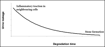
Potential and hypothetical relationship between urine leakage (graft permeability) and inflamatory reaction from one side and graft degradation time (water resistant) and stone formation within urinary tract.
Cell Source
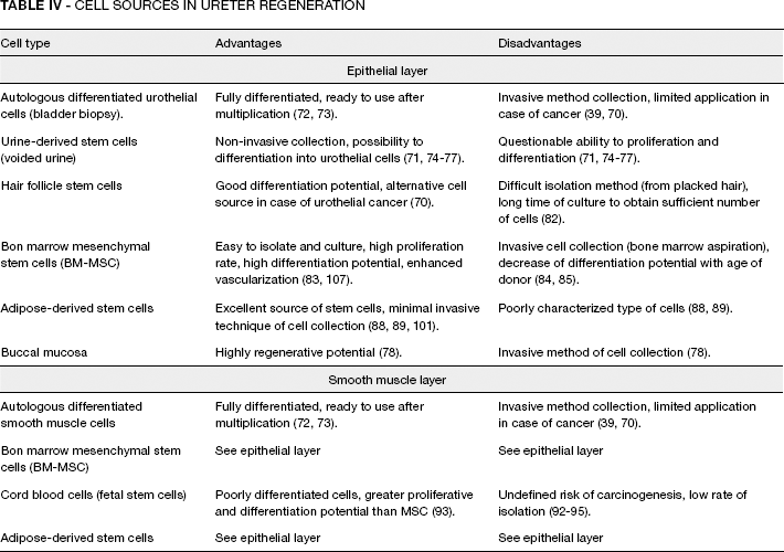
The construction of the graft for ureter regeneration requires two layers of cells: epithelium and muscle (Tab. IV). If urothelium alone were enough for this purpose, the ureter would be easy to regenerate due to its small dimensions. But regeneration does not occur for graft coated by epithelium (69). The reason of this phenomenon has not been explained yet. It may be caused by the toxic urine environment or by biofilm and urine stone formation on the scaffold surface.
Cell Sources in Ureter Regeneration
The outer layer of the ureter is formed of smooth muscle cells. Regeneration of this layer is very important because it provides correct peristaltic activity, causing physiologic urine transport. Proper muscle regeneration will result in adequate blood supply to the regenerated tissue. On the other hand, when improper fibrotic tissue with a poor blood vessel net is formed it does not maintain proper function of the graft.
Epithelial Cells
Transitional epithelium is one of the ureter cell layers. The main function of the ureter epithelium is to prevent resorption of urine ingredients (23).
Autologous urothelial bladder mucosa is excellent material for epithelium regeneration. It can be used to treat the main causes of ureter injury, which are mechanical, and especially iatrogenic, damages. Its value is reduced by limited application in case of urothelial cancer of the bladder, ureter or renal pelvis (70). Harvesting of urothelial cells usually requires an invasive procedure such as a bladder biopsy (71). Autologous epithelium cells were used in bladder and urethra regeneration (72, 73).
Urine-derived stem cells (USC) have been used as a cell source for urethral regeneration. USCs can easily differentiate into urothelial cells (UCs) because of their origin, and they can be easily harvested using non-invasive procedures (72, 74–77). Zhang et al and Wu et al have done very interesting research on isolating stem cells from excreted urine. They suggest that urine contains urothelial progenitors. This fact is controversial and was not proven by an independent laboratory. The main contradiction is that if progenitor cells are lost every day, as constituents of excreted urine, they should run out very quickly. Moreover, the urine environment is very toxic for cells, so we do not think that USCs will survive under these conditions. Localization of these stem cells (in the basal layer) also undermines this theory (23). The applications of USCs are limited by different diseases of the urinary system, similar to urothelial cells (71, 74). In the case of benign diseases, the best source of tissue for epithelium regeneration is bladder autologous urothelium. In the case of cancer, the cell source has not been defined yet, but autologous urothelium has been excluded. Cells from buccal mucosa can also serve as a potentially source for urothelium regeneration. It is a good cell source because of high regenerative potential. A disadvantage is invasive cell collection procedure.
Lu et al have transplanted porcine buccal mucosa into a defect created in the bladder. They showed that in the bladder microenvironment, cells from buccal mucosa trans-differentiated into bladder urothelium (78).
Skin stem cells from hair follicles can be used as a source of cells for ureter regeneration if urothelial cancer limits the use of more convenient types of cells. Hair follicles are highly promising sources of relatively easy accessible multipotent stem cells (79). Such cells have good differentiation potential. The use of conditioning culture media enables trans-differentiation of stem cells derived from hair follicles. (80). They can differentiate into neurons, glia, keratinocytes, smooth muscle cells, and melanocytes in vitro (81). The disadvantage of this type of cells is the difficult and time consuming method of culturing necessary to obtain the proper cell number (82).
Mesenchymal stem cells isolated from bone marrow (BM-MSC) can also be considered as a cell source for ureter regeneration. They are easy to isolate and culture. Their high proliferation rate makes it possible to appropriate the amount of cells in a relatively short time. BM-MSCs have a high differentiation potential similar to hair follicles. Studies conducted on scaffolds seeded with BM-MSCs indicated that these cells enhanced neovascularization and provided better growth of neo-tissue (38, 83). The main disadvantage of BM-MSC use are invasive cell collection and a potential decrease in differentiation with age of donor (84, 85). Tian et al have successfully differentiated human MSCs into urothelium using conditioning medium and co-culture with human urothelial cells (86). Anumanthan et al have differentiated MSCs from mouse bone marrow into urothelium using heterospecific recombinant xenografts created from embryonic rat bladder mesenchymal cells combined with MSCs. The graft was transplanted to the renal subcapsular space in mice (87).
Adipose-derived stem cells (ADSCs) may also be a convenient source of stem cells for ureter regeneration. The concept of their use is relatively new. The first mention of ADSCs appeared in 2001 (88). Fat tissue is an excellent source of mesenchymal stem cells. They can be obtained during minimally-invasive cosmetic liposuction (89). Liu et al have differentiated human ADSCs into urothelium-like cells using a co-culture of ADSCs with urothelial cells. They suggested that cell-to-cell contact induced differentiation (90). Embryonic stem cells and Induced Pluripotent Stem Cells (IPS) have had a marginal significance for potential clinical tissue regeneration until now.
Smooth Muscle Cells
There are two distinct muscle layers that compose the ureter wall: the internal circuit and the external longitudinal layers. Smooth muscle layers are very important for ureter function. Proper excretion of urine from the renal pelvis to the bladder is maintained by rhythmic peristaltic movements and the peristaltic wave of the ureter muscularis (23). Autologous smooth muscle cells have been used in bladder and urethra regeneration (72, 73).
BM-MSC can also be used in muscle layer regeneration. The advantages and disadvantages are the same as in the case of epithelium regeneration. Trans-differentiation of BM-MSC into smooth muscle will probably be easier than into urothelium. Joachimiak et al successfully trans-differentiated animal BM-MSC to smooth muscle cells using conditioned medium and medium with TGF-β1 as well (80). Cord Blood Stem Cells (CBSC) are good candidates for smooth muscle regeneration. Cord blood banking started in the 1930s, and has become more popular over time, so methods that use cord blood cells are very promising (91). Cells from cord blood are poorly differentiated and have greater proliferation and differentiation potential than mesenchymal stem cells due to donor age. Collection of these cells is completely safe for mother and child. There are two disadvantages that limit the use of CBSC. The first is the inefficiency in isolating stem cells from cord blood, which is limited to 30%. The second disadvantage is a possible, but not proven, risk of cancer development after transplantation of undifferentiated cells (92–95). This risk has been proven for embryonic stem cells (96).
Wu et al used urine-derived stem cells for smooth muscle cell regeneration. Trans-differentiation of urine-derived stem cells into smooth muscle is the most interesting and controversial issue of this study due to the endothelial origin of urothelial cells (74).
ADSCs are also able to differentiate into smooth muscle cells. Zheng et al have reported that ADSCs can undergo multilineage differentiation in vitro, but differentiation into muscles was limited (97). In another study Viera et al showed that ADSCs restored dystrophin expression in Duchenne muscular dystrophy (DMD) patients (98). ADSCs have a similar phenotype and gene expression profile to bone marrow MSCs (99, 100). It suggests that appropriate techniques of ADSC differentiation will provide successful trans-differentiation into muscle cells. Fraser et al have shown an increase of myosin and MyoD1 expression and transcriptions factors: myf5, myf6, and myogenin after dexamethasone and hydrocortisone ADSC treatment. The appearance of the obtained cells closely resembled myotubes (101). Jack et al have differentiated ASCs using inductive media. These cells expressed smooth muscle molecular markers (102). Zhu et al have demonstrated that ADSCs seeded on bladder acellular matrix grafts (BAMGs) were able to regenerate smooth muscle and nerve tissue in a rabbit model (103). Zhao et al were able to differentiate ADSCs into smooth muscle cells and used them for ureter regeneration in rabbits (104).
Based on the reviewed papers, the most appropriate cell sources for ureter smooth muscle layer regeneration seems to be bone marrow-derived mesenchymal stem cells and adipose-derived stem cells, because of their ability to differentiate into smooth muscle cells, the easy isolation method, and the minimally invasive procedure of the tissue collection. It should be noted that there are so many poorly examined different stem cell niches, e.g. amniotic membrane or amniotic fluid stem cells, which could be significant in future research of ureter regeneration.
Neo-Ureter Creation
Models Tested Outside the Urinary Tract
Baumert et al have used small intestinal submucosal (SIS) seeded with cells from bladder biopsy as a potential scaffold for ureter regeneration. Scaffolds were transferred to the omentum of 5 pigs. One year later the same group created a neo-ureter with a similar method, but tissue was taken laparoscopically, and placed in the omentum of 3 pigs also using the laparoscopic method. The results showed the creation of neo-ureter with regeneration of the urothelium and muscle layers (105, 106). The weakness of these studies was the casuistic character of both studies and the lack of anastomosis of the created neo-ureter with the native ureter. Shen at al used a polycaprolactone-lecithin construct (PCL-L) produced by the electrospinning method and seeded with cells to prepare a ureter graft in vitro. They showed that lecithin units provided good conditions for cell growth and adhesion in vitro, however the scaffold was implanted into the peritoneum and not into the ureter (107). It should be noted that the peritoneum environment which is different from the extraperitoneal environment in urine can induce fibrosis and ureter constriction. Wu et al have successfully used SIS modified with 5% peracetic acid in order to increase porosity of this material as a scaffold for ureter regeneration. Cells seeded on this type of scaffold formed a thicker layer of tissue in comparison to unmodified SIS but no in vivo experiment was performed (74). In another study, urothelial cells isolated from ureters of patients who had undergone nephroureterectomy were seeded on PLLA-collagen scaffold. Cells were seeded using a centrifugal seeding system which enhances the cell number loading to the scaffold which were then transplanted subcutaneously into nude rats for 14 days. The results indicated that the seeded cells remained in the scaffold for at least 2 weeks (108). Some attempts have been done to prepare matrix providing delivery of Growth Factors (GF). Shen et al concluded that the use of biomatrices loaded with growth factor increased cell to matrix attachment (109). In contrary to these results, Nuininga et al showed that scaffold surface overloaded with GF decreased its regenerative properties, but this experiment was done on rabbit urethra (110). Addition of growth factors may enhance the properties of matrices for tissue engineering, yet there is little experimental data for evaluation of the problem.
Models Tested Inside the Urinary Tract (Anastomosed with Native Ureter)
Living autologous tissues (vascularized flaps), like appendix, ileal fragments formed using the technique of Monti have been used in ureter regeneration as templates (111, 112). Additionally autologous grafts like buccal mucosa, bladder mucosa, and blood vessels were used (113–115). Natural collagen-based acellular matrices, such as small intestinal submucosa (SIS) or bladder submucosa was also used (116–119) for this purpose.
Some experiments have been performed on cell-free (unseeded) matrices to verify the urethral self-regeneration (120). Total regeneration of matrices appeared after 4 weeks when scaffolds of 0.5 cm length were used. Longer scaffolds (1, 2 and 3 cm) have regenerated only at the anastomotic edges.
In another study, acellular matrix created from canine decellularized ureter was used to regenerate 3 cm ureter segments in mongrel dogs. After 8 weeks massive hydroureteronephrosis, shrinkage of neoureter with marked narrowing, or complete occlusion and extensive subepithelial fibrosis were observed (121). In research performed on 5 dogs SIS was used to regenerate a 4 cm ureter segment. Muscle and urothelium regeneration on SIS was observed but coexistence of fibrosis and luminal obstruction were also observed. Coexistence of hydroureteronephrosis and ureter dilatation were detected. (122). This result proves that unseeded matrix is unsuitable for regeneration of clinically important ureter defects (i.e., re-anastomosis does not occur).
SIS was also used for regeneration of ureter segments in 5 pigs. The results were satisfying, and ureter continuity was preserved (123). Gajda et al used tunica albuginea as a graft for ureter regeneration in 20 mongrel dogs. One ureter was regenerated with preservation of ureter continuity and a second on the entire ureter length (2–3.5 cm). Ureter regeneration occurred only when continuity of ureter was preserved; substitution of complete ureteral loss with tube-graft resulted in necrosis and restenosis (124). Regeneration of the ureter segment is possible only when ureteral continuity is maintained. A small fragment of tissue is enough to stimulate regeneration of muscle tissue on collagen grafts. This explains difficulty in reconstruction of the muscle layer and restoration of peristaltic movements on the entire ureter length.
Matsunuma et al attempted to regenerate ureter using 2 cm piece of canine ureteral decellularized matrix (UDM) as a scaffold seeded with cells and transplanted into rats and mice. Regeneration of the uroepithelial layer was successful (2 weeks after transplantation), indicating that UDM is useful as scaffold material for ureter tissue engineering, but the disproportion between dogs and rodents must be emphasized (83). The latest study was conducted on 20 rabbits. The ureter was regenerated using vessel extracellular matrix seeded with smooth muscle cells differentiated from ADSCs. After 16 week followup, muscle layer regeneration and lack of ureteral stricture and hydroureteronephrosis were observed (104). In this study, lack of urography and photo documentation of the implanted scaffold during followup reduced the credibility of this work.
Conclusions
Ureter reconstruction in large animal models has not been reported. A high failure rate of ureter reconstruction in small animal models has been observed. Ureter regeneration based on tissue engineering techniques is new and being developed. The most appropriate materials for scaffolds are polyesters, because of their capacity for biodegradation and the ability to be combined with other compounds, thus controlling porosity, cell attachment, and biodegradation time. Polyesters do not introduce allogeneic tissues or proteins when implanted. The use of electrospun nanofibers as scaffolds makes it possible to control the direction of cell growth, to apply a minimal amount of foreign material, and to enable the scaffold to mimic extracellular collagen matrix. The most appropriate cell source for epithelium regeneration is autologous urothelium in cases of benign diseases. An alternative source of cells, especially in the case of urinary cancer, may be hair follicle stem cells. As a cell source for muscle layer reconstruction, the best candidate seems to be bone marrow mesenchymal stem cells and adipose-derived stem cells. Despite their invasive cell collection method, their good proliferation capacity, easy isolation method, and enhanced neovascularization make them the best choice. The choice of scaffold construction and cell seeding method are also important. Some studies indicate that urine is toxic for transplanted cells. These results showed that failure in ureter regeneration could be caused by urine influence on the cell seeded graft. That is why we suggested that scaffolding for ureter regeneration should minimize the negative influence of urine and prevent urine-induced inflammation along with consequent fibrosis.