






Abstract
Objectives
This study aims to examine biofilm formed on vascular prostheses by Staphylococcus epidermidis with different ica and aap genetic status, and to evaluate the effect of antibiotic-modified prostheses on bacterial colonization.
Methods
Biofilm formation was determined using fluorescence microscopy imaging. Quantitative analysis was conducted using the biofilm coverage ratio (BCR) calculations.
Results
Our investigations prove that the BCR method with fluorescent dye enabled an accurate assessment of biofilm coverage and comparison of the obtained results. The ica+ aap+ strains formed a biofilm on all of the examined vascular prostheses. Uni-Graft® modified with covalently immobilized amikacin was effective in preventing bacterial adherence.
Conclusions
Molecular biology techniques combined with phenotype studies give a broad insight into biofilm formation mechanisms. On the other hand, fluorescence microscopy imaging along with BCR calculations are reliable and simple tools to quantitatively estimate biofilm formation, as well as the effectiveness of antimicrobial prosthesis modification.
Keywords
Introduction
The coagulase-negative staphylococci (CNS) were originally known as the normal microflora of healthy human skin and mucous membranes, rarely causing diseases in immunocompromised patients. However, they have become serious pathogens hindering medical progress and have brought a new threat: biomaterial-associated infections (1, 2). Frequent use of biomaterials in modern medicine results in an increased number of infections related to implanted medical devices. Among CNS, Staphylococcus epidermidis is the most commonly isolated species responsible for the infections. The pathogenicity of CNS results from their ability to colonize the surface of medical devices by forming a biofilm (3-5). Biofilm is established by the accumulation of microbial cells which are irreversibly associated with the surface and embedded in a matrix of extracellular polysaccharides that they produce (6, 7). Several genes are involved in biofilm formation. For example, the ica operon controls the biosynthesis of polysaccharide intercellular adhesin (PIA), whereas the aap gene is responsible for the synthesis of accumulation-associated protein (Aap). The role of the ica and aap genes is emphasized in a series of literature data (8-13) as an important factor in the pathogenesis of biomaterial-associated S. epidermidis infections.
The adherence of bacteria to medical devices and their subsequent colonization and biofilm formation cause infections as well as device dysfunction. Additionally, the bacteria from biofilm can disperse from the original site of colonization and cause further local and systemic infections. Therefore devices colonized by the bacteria may become the source of infection for the whole organism (14, 15). The diagnosis and treatment of such diseases constitute one of the major problems engendered by bio-film-associated infections. Microbes in biofilms are notably resistant to antimicrobial chemotherapy and the host immune response. High resistance of microbes in biofilms arises from specific conditions in a biofilm community. The multilayer structure of a biofilm inhibits the diffusion of antibiotics, whereas the extracellular matrix protects the bacteria from elimination by the immune system. What is more, reduced growth rate and phenotypic changes make biofilm resistant to antimicrobials, while easy exchange of plasmids causes a rapid spread of antibiotic resistance (4, 16, 17).
Despite the fact that antibiotic therapy is less effective against biofilm populations, it is still the most common practice in preventing bacterial colonization of biomaterials. Antimicrobials which are administered systemically or orally often do not reach the site of infection and infections may occur on the surface of implanted devices even in the presence of antibiotics. In addition, the extensive use of antimicrobial agents and disinfectants in the hospital environment is an important factor causing the rapid spread of multi-drug bacterial resistance. Because antimicrobial treatment has little or no effect against biofilm populations, these bacterial infections are very difficult to eradicate. Therefore surgical removal and substitution of the device is often required (3, 14, 18, 19).
Since biomaterial-associated infections have become a serious threat to public health and have entailed social and economic costs, searching for new solutions to this problem remains important. New strategies of preventing biofilm formation seem to be significantly more efficient than combating a developed biofilm. A greater understanding of biofilm processes should lead to novel, effective treatment strategies for biofilm prevention. High hopes are associated with the development of new surfaces for medical devices which would prevent bacterial colonization (6, 14, 16, 17). The use of antibiotic-immobilized biomaterials appears to be a straightforward and appropriate strategy to reduce the chances of biofilm formation. Furthermore, a better understanding of the complexities of biofilm formation requires future investigations.
The objective of the present study was to examine the biofilm formed on vascular prostheses by S. epidermidis strains with different genetic and phenotypic profiles. Moreover, we evaluated the effect of antibiotic-modified prostheses on bacterial colonization and tested whether fluorescence microscopy imaging could be used as a simple tool for quantitative analysis of biofilm formation.
Materials and Methods
Bacterial strains
Two reference strains of S. epidermidis: ATCC 35984 (also known as RP62A) and ATCC 12228 as well as two clinical isolates of S. epidermidis: 37IINL64 and 19IINL35 were studied in this report. The clinical isolates were obtained from the nasopharynx of lung cancer patients and were identified using standard microbiological techniques. All the strains used in this investigation were genetically and phenotypically characterized as was described previously (20). Briefly: slime secretion was determined using the Congo red agar (CRA) test, while the microtiter plate (MtP) method was used as a test for determining biofilm formation. PCR of the icaA and aap genes was performed in the examined Staphylococcus strains. For the detection of icaA, the primers were as follows: 5′-AACAAGTTGAAGGCATCTCC (forward primer) and 5′-GATGCTTGTTTGATTCCCT (reverse primer). Two primers for the detection of aap were 5′-ATACAACTG-GTGCAGATGGTTG (forward primer) and 5′-GTAGCCGTC-CAAGTTTTACCAG (reverse primer), respectively. Cycling conditions were as follows: preheating for 4 min at 96°C, followed by 35 cycles of denaturation at 96°C for 30 s, annealing at 60°C for 30 s, primer extension at 70°C for 30 s and final extension at 70°C for 4 min. Amplified products were analyzed using agarose gel electrophoresis. Detailed strain information is available in Table I. The S. epidermidis 37IINL64 clinical isolate was confirmed as sensitive to amikacin using the disc-diffusion method.
CHARACTERISTICS OF THE STAPHYLOCOCCUS EPIDERMIDIS STRAINS USED IN THIS STUDY

Vascular prostheses
Four vascular prostheses were examined: Gelsoft™, a knitted polyester gelatin-impregnated graft (Vascutek, Scotland, UK); Uni-Graft®, a knitted double velour polyes-ter gelatin-impregnated graft (B. Braun Melsungen, Melsungen, Germany); Protegraft®, a knitted polyester graft (B. Braun Melsungen, Melsungen, Germany); and GORETEX® Vascular Graft, a graft made of expanded polytetrafluoroethylene (ePTFE) (W. L. Gore & Associates, Newark, DE, USA).
Biofilm formation on vascular prostheses
The vascular prostheses were cut into circles of 6.0 mm in diameter. Sterile graft pieces were placed in 15 mL of tryptic soy broth medium (TSB; Oxoid, Basingstoke, UK) containing 5 × 10⁵ CFU/ml of corresponding bacteria and were incubated at 37°C with gentle shaking for 24, 48, or 72 h. Growth medium was refreshed every 24 h. After the incubation period, the grafts were washed two times with 5 mL phosphate-buffered saline to remove nonadherent cells. The grafts were then examined using fluorescence microscopy. All examined strains were tested for biofilm formation on four types of vascular grafts. Control grafts were incubated in TSB without bacteria and then handled in the same manner as the materials incubated with bacteria. The experiments were performed in triplicate and repeated twice.
Testing the amikacin-modified vascular prosthesis
Uni-Graft® prostheses modified with covalently immobilized amikacin were compared to commercially available Uni-Graft® prostheses. Prosthesis modification with antibiotic was performed according to Polish Patent No. PL201383 (21). Covalent immobilization of amikacin was performed by prosthesis activation with glutaraldehyde and then antibiotic reaction followed by reduction of Schiff bases by NaBH4 (22). Sterile graft pieces (6.0 mm squares) were placed in 5 mL TSB containing 5 × 10⁵ CFU/ml of the S. epidermidis 37IINL64 clinical isolate. After 24 h of incubation at 37°C with gentle shaking, the grafts were washed twice with 5 mL phosphate-buffered saline to remove non-adherent cells. The grafts were then examined using fluorescence microscopy. The experiments were performed in triplicate and repeated twice.
Fluorescence microscopy imaging and BCR calculation
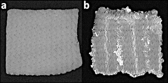
The biofilm coverage ratio (BCR) method was performed as described by Adachi et al (23) with modification. The modification concerned a substitution of crystal violet with the fluorescent dye Hoechst 33342 (Sigma-Aldrich, St Louis, MO, USA). The grafts were stained with Hoechst 33342 according to the manufacturer's instructions and examined on a fluorescence microscope (BX41; Olympus, Tokyo, Japan) equipped with wide band UV excitation filter cube (U-MWU2; Olympus, Tokyo, Japan; exciter filter 330-385, dichromatic mirror 400, barrier filter 420) and 100 W mercury lamp (U-RLF-T; Olympus, Tokyo, Japan). Color images were converted into gray-scale images. All quantifications were performed with 4x lens magnification. An example of a colonized vascular graft in comparison to the control graft is presented in Figure 1. BCR was calculated with cellSens Imaging Software (Olympus, Tokyo, Japan). All data are expressed as the mean ± standard deviation.
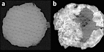
Comparison between a biofilm-covered graft and an uninfected graft, used as a control; (a) uninfected Gelsoft™ graft, (b) Gelsoft™ graft infected with S. epidermidis ATCC 35984 (BCR 62%) after 24 h of incubation.
Results
Differences in BCR between the examined strains
The experiment visualized biofilm forming on the following vascular graft types: Gelsoft™, Protegraft®, Uni-Graft®, and ePTFE. The obtained results indicated the differences in biofilm formation by the tested S. epidermidis strains. The examined ica+ aap+ strains formed a biofilm on all tested vascular grafts. The examined ica- aap+ strain formed a biofilm only on the ePTFE graft, whereas the examined ica- aap- strain did not form a biofilm on any of the tested grafts.
The results are presented in Figures 2 and 3 using the Gelsoft™ graft as an example. BCR for the ica+ aap+ clinical isolate of S. epidermidis 37IINL64 after 24 h of incubation was 11.17% ± 5.78% (Figs. 2c and 3a). Particularly high BCR (68.33% ± 12.64%) was observed after 24 h for the ica+ aap+ reference strain of S. epidermidis ATCC 35984 (Figs. 2d and 3a). The ica- aap- clinical isolate of S. epidermidis 19IINL35 did not demonstrate any biofilm production either after 24 h (BCR 0.13% ± 0.20%; Figs. 2a and 3a) or during the following days of incubation (BCR at 72 h 1.03% ± 1.03%; Fig. 3c). The ica- aap+ reference strain of S. epidermidis ATCC 12228 did not develop any biofilm structure on the Gelsoft™ graft, although small bacteria clusters were observed (BCR at 24 h 1.39% ± 0.73%; Figs. 2b and 3a).
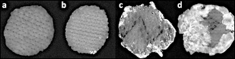
S. epidermidis biofilm formed after 24 h on the Gelsoft™ grafts; (a) S. epidermidis 19IINL35, (b) S. epidermidis ATCC 12228, (c) S. epidrmidis 37IINL64, (d) S. epidermidis ATCC 35984.
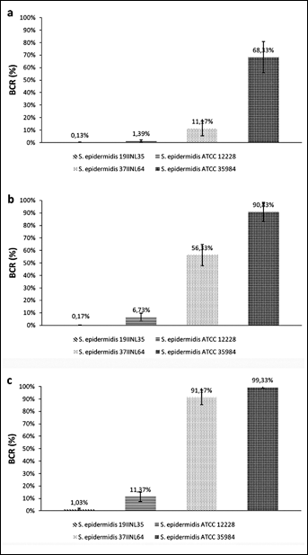
Differences in BCR between the examined strains on the Gelsoft™ grafts after three incubation times; (a) 24 h, (b) 48 h, (c) 72 h.
Comparison of BCR between the examined prostheses
Two types of bacterial growth depending on the graft structure were distinguished based on the tested materials. On the ePTFE graft, bacteria grew only on the surface, without penetrating deeper layers of the graft. Conversely, the bacteria grew both on the surface and deeper into the knitted structure of the other grafts – the Gelsoft™, Protegraft®, and Uni-Graft®. An example of bacterial growth with biofilm penetrating the structure of a knitted graft is shown in Figure 4. Moreover, no significant differences in BCR between the tested knitted grafts (Gelsoft™, Protegraft®, Uni-Graft®) were observed for each strain. Biofilm formed on all four types of the examined material is presented using the ica+ aap+ isolate of S. epidermidis 37IINL64 as an example (Fig. 5). BCR for this isolate on the ePTFE graft was lower than on the knitted grafts (Fig. 6). On the other hand, extensive biofilm formation (BCR at 72 h 58,73% ± 17.31%) was observed on the ePTFE graft infected with the ica- aap+ reference strain of S. epidermidis ATCC 12228, although this strain is considered biofilm negative and did not form a biofilm on other tested grafts.
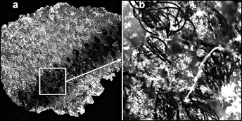
Biofilm formed on the fibers of Protegraft® by the S. epidermidis 37IINL64 clinical isolate after 24 h of incubation; lens magnifications: (a) 4x, (b) 20x.
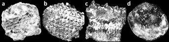
Coverage with biofilm formed by the S. epidermidis 37IINL64 clinical isolate after 72 h of incubation with different vascular graft types: (a) Gelsoft™, (b) Protegraft®, (c) Uni-Graft®, (d) ePTFE.
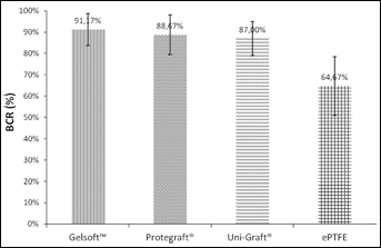
BCR on vascular grafts colonized by the S. epidermidis 37IINL64 clinical isolate after 72 h of incubation.
BCR over time
Differences in the rate of biofilm formation between the ica+ aap+ strains were observed. The data obtained for the S. epidermidis 37IINL64 clinical isolate and the S. epidermidis ATCC 35984 reference strain during the following three days of incubation on the Gelsoft™ grafts are shown in Figures 7 and 8. Biofilm coverage increased from day to day. It was observed that biofilm not only covered the surface but also grew vertically. The surface of the grafts infected with the S. epidermidis 37IINL64 clinical isolate was almost completely covered with biofilm on the third day of experiment (BCR 91.17% ± 7.47%; Fig. 7a.3). The S. epidermidis ATCC 35984 reference strain colonized a larger surface (BCR 68.33% ± 12.64%; Fig. 7b.1) compared to the S. epidermidis 37IINL64 clinical isolate (BCR 11.17% ± 5.78%; Fig. 7a.1) after 24 h of incubation. Additionally, the fastest vertical growth was observed already after 24 h in the case of S. epidermidis ATCC 35984.
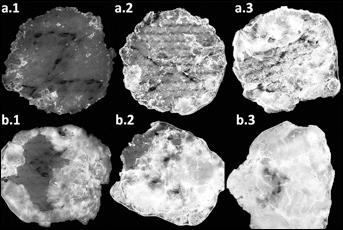
Bio film coverage of Gelsoft™ grafts after different incubation times: (a.1, b.1) 24 h, (a.2, b.2) 48 h, (a.3, b.3) 72 h; (a) S. epidermidis 37IINL64, (b) S. epidermidis ATCC 35984.
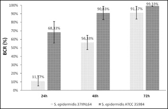
BCR over time for the biofilm-forming strains on the Gelsoft™ grafts.
Effect of amikacin-modified prosthesis on bacterial biofilm
The study data indicate differences in bacterial adhesion on the amikacin-modified grafts (Fig. 9). The biofilm-forming strain S. epidermidis 37IINL64 (ica+ aap+) did not exhibit any adherence to the Uni-Graft® surface with covalently attached antibiotic after 24 h (BCR 0.00%), however, the adherence was noticeable on the nonmodified Uni-Graft® surface (BCR 10.00% ± 6.43%).
Effect of graft modification after 24 h of incubation with the S. epidermidis 37IINL64 clinical isolate; (a) Uni-Graft® modified with covalently immobilized amikacin, (b) Uni-Graft® without antibiotic modification.
Discussion
Biomaterials are increasingly used in modern medicine, however, infections associated with implantation are a frequently occurring problem. Biomaterial-associated infections are the effect of bacterial ability to colonize implanted material surface and form a biofilm. The growing incidence of this type of infection requires reliable methods of biofilm examination. What is more, new strategies of preventing biofilm formation seem to be significantly more efficient than combating a developed biofilm. The application of fluorescence microscopy imaging enabled the observation of bacterial adhesion and biofilm formation on biomaterials. The use of Hoechst 33342 fluorescent dye allowed us to clearly distinguish the areas covered with biofilm from the graft surface. The calculated biofilm coverage ratio (BCR) was based on this distinction. Crystal violet used in the original BCR method (23) could not be applied in our examinations as it would stain not only the biofilm but also the knitted grafts. Our modification with Hoechst 33342 due to the specificity of the dye interaction with bacterial cells expands the BCR method by adding the possibility of applying it to various biomaterials. The BCR method with fluorescent dye enabled an accurate assessment of biofilm coverage and comparison of the results. However, the method is based on the biofilm coverage area and it is impossible to quantify vertical biofilm growth. Nevertheless, bacterial biofilm first grows two-dimensionally and subsequently maturates by vertical growth. Therefore two-dimensional measurement of the initial stages of biofilm formation is sufficiently reliable and may be used to estimate biofilm formation on biomaterials, which is in agreement with the data presented by Adachi et al (23) and Kajiyama et al (24). Additionally, fluorescence microscopy imaging combined with the BCR method could be used to evaluate the effectiveness of graft surface protection against bacterial colonization and constitute a reliable method for biofilm examination not only on vascular prostheses but also on other biomaterials, such as: venous catheters, urinary catheters, prosthetic heart valves, orthopedic devices, contact lenses, shunts, pacemakers, and others.
Our experiment visualized biofilm forming on four types of vascular grafts: Gelsoft™, Protegraft®, Uni-Graft®, and ePTFE. Furthermore, we compared the BCR of four S. epidermidis strains with different genetic status. The examined ica+ aap+ strains formed a biofilm on all tested vascular grafts. Among the examined ica-negative strains, the ica- aap- strain did not form a biofilm on any of the tested grafts, whereas the ica- aap+ strain formed a biofilm only on the ePTFE graft. According to the data presented by other authors (5, 25), a simultaneous pres-ence of the ica operon and the aap gene plays an important role in the biofilm-positive phenotype. Our study confirmed that the ica+ aap+ strains form a biofilm not only in standard conditions (as we indicated using the MtP method and the CRA test in the earlier investigation (20)) but also in our experiment with vascular prostheses. These results indicate that the ica+ aap+ strains may become a particularly serious threat for patients after implantations. However, the ica- aap+ reference strain of S. epidermidis ATCC 12228, which is considered to be biofilm-negative, can also form a biofilm on an ePTFE graft. Although in our experiment the ica- aap- clinical isolate did not form a biofilm, it is possible for some ica- aap- strains to form a biofilm, as was indicated by the MtP method (20). These data confirm that biofilm formation on medical devices is a complex process. Other genes besides ica and aap — such as atlE, bap, fbe, or embp—may also influence biofilm development (26-30). In addition, the surface and the environment also play a significant role in biofilm formation (13, 26, 31). These facts may explain why some ica-aap- strains are capable of forming a biofilm. However, the ica- and aap-independent biofilm formation mechanisms are still poorly understood.
Once a biofilm has been formed, treating an infection becomes extremely difficult. Therefore prevention of S. epidermidis adherence to biomaterials remains a priority. Prepared colonization-resistant surfaces of biomaterials may minimize the frequency of infections after implantation. Antibiotic-impregnated grafts are an example of successful implementation of biomaterial-associated infection prevention (17, 32, 33). The results of our study demonstrate that amikacin covalently immobilized to Uni-Graft® (according to the method described in Polish Patent No. PL201383) efficiently inhibits the bacterial attachment process. Thus, antibiotic-modified prostheses may contribute to enhancing the safety of biomaterial implantation.
Conclusions
Molecular biology techniques combined with phenotype studies give a broad insight into biofilm formation mechanisms. Our investigation proved that fluorescence microscopy imaging along with BCR calculations gave an accurate assessment of biofilm coverage, as well as measuring the effectiveness of antimicrobial prosthesis modification. The study demonstrated that the ica+ aap+ strains form a biofilm not only in standard conditions but also in experimental conditions on vascular prostheses. The results confirmed that the ica+ aap+ strains may become a particularly serious threat for patients after implantations. Moreover, the findings of this study indicate that amikacin covalently immobilized to Uni-Graft® efficiently inhibits the bacterial attachment process.