
Abstract
Purpose
Electrically active supports provide new horizons for bio-sensing and artificial organ design. Cell-based electrochemical biosensors can be used as bio-microactuators, applied to the biorobotics. Microchip-based bioassay systems can provide real-time cell analysis for preclinical drug design or for intelligent drug delivery devices. In regenerative medicine, electrically active supports can be used as bio-reactors to monitor cell activity, optimize the stem cell differentiation and control cell and tissue morphology. Biocompatibility and direct interaction of the electrically active surface with the cell surface is a critical aspect of this technology.
Methods
In this work embryonic stem cells (AK7 ES) have been cultivated on the surface of thin films achieved through the evaporation of two aromatic compounds (T6 and PDI-8CN2) of particular interest for the fabrication of organic field-effect transistors (OFET). One of the potential advantages offered by the application of OFETs as bio-electronic supports is that they represent a powerful tool for the detection of bio-signals because their electrically active surface is an organic film.
Results
The cell morphology on T6 and PDI-8CN 2 surface shows to be similar to the usual cell appearance, as obtained when standard culture support (petri dish) are employed. Moreover, our experimental results demonstrate that stem cells can be lead to differentiation up to “beating” cardiomyocytes even on these electrically-active organic films.
Conclusions
This investigation encourages the perspective to develop OFET-based biosensors in order to accurately characterize stem cells during the cardiac differentiation process and eventually increase their differentiation efficiency.
Introduction
Cell-based electrochemical biosensors are applied in medicinal chemistry and drug design (1), providing new horizons for bio-sensing and artificial organ design, micro-bio-robots (2) or artificial micro-devices, like micro-bio-actuators or micro-bio-pumps (3). Promising techniques for the realization of these devices are based on the isolation and in vitro cultivation of cardiomyocytes (cardiac cells) that show spontaneous contracting capability (4). In regenerative medicine electrically active supports can be used as bio-reactors to monitor cell activity, optimize the stem cell differentiation and control cell and tissue morphology, with particular application to electric active cells, like neurons and cardiomyocytes. Due to the limited proliferative capacity of adult heart cells, a promising therapy for heart diseases is the use of stem cells for large-scale production of cardiomyocytes in order to obtain engineered tissues to regenerate injured myocardium (5, 6). Alternative therapies try to transplant tissue-engineered three-dimensional heart grafts (7) which offers the advantage of inducing the desired cell morphology in the tissue before the implant. This can be achieved by culturing the cells on specific supports, where the signals present in the native heart, which can be electric, chemical or mechanical, can be mimicked to induce cardiomyocyte alignment, beating, and bioactivity. An external electrical signal can be applied to mimic those in the native heart (8) and induce synchronous contractions of cultured cardiac constructs, lead the organization and contractility of cardiomyocytes in vitro, increase cell density, and enhance tissue morphology (9). These may be essential aspects for the integration of the tissue construct to the native heart tissue upon implantation (10). On the other hand, the measurement of the cell electric activity can be used as an indicator of the ongoing differentiation.
To guide regeneration processes even within the patient's body, new or modified materials with specific properties are being continuously developed.
During the last two decades, organic semiconductors have been widely explored with the aim of developing innovative electronics based on low-cost fabrication techniques, low-temperature (T<200°C) processing and the use of flexible large-area and transparent substrates. Moreover, thanks to their chemical composition, these materials exhibit an inherent high degree of biocompatibility with living cells and tissues. This feature makes them particularly attractive for applications in the field of bio-electronics, where reliable devices suitable to interface biological and electronic signals are required (11–13).
In particular, Organic Field-Effect Transistors (OFETs), in which the active channel consists of a conjugated oligomer or polymer film, represent powerful tools for the detection of bio-signals. Indeed, in solution environments, the changes in charge and/or dipole (related to ions or molecules) concentrations (14) can be monitored by observing the related variations of specific OFET electrical parameters. Furthermore, OFETs could be employed for direct, real-time observation of signals generated by electrogenic cells and tissues. OFETs are inherently low-dimensional devices since the charge carrier movement is confined within a few molecular layers at the interface between the organic layer and the dielectric barrier (15). Consequently, their response sensitivity can be enhanced by using ultra-thin films to form the device active channel in such a way to assure close proximity between the cell membrane and the charge transport region.
In order to achieve these application aims, before setting up in vivo experiments, detailed experimental data are currently needed to demonstrate the real capability of these materials to support the adhesion and the proliferation of in vitro cell cultures. In general, a few hours after cells are seeded into a sterile culture dish containing a nutrient-rich cell culturing medium, they adhere to the surface and start to proliferate. This process may take days and the selection of a substratum suitable to cell growth is not trivial, since some materials can result toxic, inhibiting the proliferation or the adhesion of the cells. For this reason, in order to check the effective applicability of organic devices for bio-system activity monitoring, it is mandatory to investigate the effects of the organic films on cell viability. In this paper, OFET biocompatibility was tested by performing experiments on embryonic stem cells: pluripotent stem cells that have a unique capacity to renew themselves and to give rise to specialized cell type.
Embryonic stem cells are derived from the inner cell mass (ICM) of the mammalian blastocyst (16, 17). They can be grown in vitro in an undifferentiated state in presence of leukemia inhibitory factor (LIF) and feeder cells. In the absence of feeder cells and anti-differentiating agents, mouse ES cells spontaneously differentiate and, under appropriate conditions, generate progeny consisting of derivatives of the three embryonic germ layers: mesoderm, endoderm, and ectoderm (18, 19). Mesoderm-derived lineages include the hematopoietic, vascular, and cardiac. Endoderm derivatives include pancreatic β cell and hepatocytes. Ectoderm differentiation of mouse ES cells is well established, since numerous studies (20) have documented and characterized neuroectoderm commitment and neural differentiation.
Here, a preliminary experimental work devoted to the verification of the biocompatibility of OFET with mouse embryonic stem cells is reported. We show that, after 9 days, AK7 ES cells differentiate in cardiac myocytes on the surface of two different types of organic semiconductors, exhibiting rhythmic beating.
Materials and Methods
T6 and PDI-8CN2 transistor fabrication
Sexithiophene (T6) and N,N'-bis (n octyl)-dicyanoperylenediimide (PDI-8CN2) films, exhibiting hole (p-type) and electron (n-type) transporting properties, respectively, were grown on SiO2 layers and used as substrates for the cell cultivation. Figure 1a shows the chemical structures of the two semiconducting organic materials studied in this work. T6 is an archetypal small molecule compound displaying hole transporting properties, which has been widely investigated in the last 20 years (21).
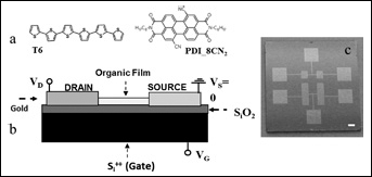
Chemical structures of the two semiconducting organic materials studied in this work. a) Chemical structures of T6 and PDI-8CN2 molecules; b) Transistor configuration; c) A photo of the electrode configuration.
PDI-8CN2, instead, belongs to the family of perylene diimide molecules which have been introduced very recently as organic semiconductors capable of exhibiting robust electron transport properties even under ambient conditions (22). In this work, both T6 and PDI-8CN2 films were Joule evaporated in a high vacuum (Pr∼ 10−7/10−8 mbar) system by using Knudsen cells on substrates (Fig. 1b) consisting of a highly doped 500 μm thick silicon (Si++) layer acting as Gate electrode, a thin (200 nm) silicon dioxide (SiO2) dielectric barrier and four pairs of interdigitated gold (Au) electrodes (Fig. 1c), working as source/drain electrodes. It is significant to remember that this same type of substrate has recently been used to perform impedance spectroscopy techniques to analyze the properties of human epithelial (HeLa) cells in the form of confluent monolayers (23). Before the film deposition, Si++/SiO2/Au substrates were cleaned by ultrasonic baths in acetone and isopropanol followed by a drying with Nitrogen.
After the organic layer evaporation, these structures represent complete OFETs in what is called the bottom-contact bottom-gate configuration. As Bao et al showed (14), the application of a voltage (VGS) between the Gate and the Source electrodes induces the accumulation of charges at the interface between the dielectric layer (SiO2, in our case) and the organic layer. In this way, the current IDS between the Drain and the Source electrodes as a function of the corresponding Drain-Source (VDS) voltage can be largely changed also by varying VGS. When using T6 films as active channels, the charge accumulation (and the related IDS increase) is achieved by applying negative VGS. This means that the free charge carriers are holes (positive charges) and the field-effect devices are classified as p-type. Conversely, with PDI-8CN2 active channels, positive VGS voltages provide the charge accumulation at the organic/dielectric interface and n-type devices, supporting electrons are free charge carriers. For the electrical response of a field-effect transistor, the charge carrier mobility μ, defined as the drift speed gained by a charge under the application of a unit electric field, is the main figure of merit. By considering OFETs with the same bottom-contact bottom-gate configuration used in this work, maximum mobility values of about 0.01 cm2/volt*sec can be measured for both T6 and PDI-8CN2 devices (24, 25).
During all the film depositions, the substrate temperature was fixed to about 100°C with an evaporation rate of 1 nm/min. For the biocompatibility tests reported below, T6 and PDI-8CN2 films with thickness of 30 nm were considered. The thickness was monitored during the evaporation by using a quartz micro-balance. The morphological properties of the film surface were characterized in air by an XE100 Park AFM microscope (non-contacting mode with amplitude regulation) (Park Systems, Suwon, Korea). Images were acquired using silicon-doped cantilevers provided by Nanosensors™ (Neuchatel, Switzerland).
ES Cell Lines
AK7 ES cell lines were maintained in an undifferentiated state by culture on a monolayer of mitomycin-C-inactivated fibroblast in the presence of leukemia-inhibiting factor (LIF) (26). Under these conditions, the cell population remained undifferentiated, as determined by visual inspection under phase-contrast microscopy. Two days before the initiation of differentiation, cells were seeded into gelatinized dishes. Undifferentiated cells were maintained on Dulbecco's modified Eagle's medium (DMEM) (Life Technologies, Carlsbad CA, US), 15% heat-inactivated fetal bovine serum (Euroclone, Milano, Italy) 100 U/ml penicillin/streptomycin, 2 mM glutamine, 1 mM sodium pyruvate (Life Technologies, Carlsbad CA, US), 1,000 U/ml leukemia inhibiting factor (LIF) (Millipore, Darnstadt, Germany), and 0.1 mM β-mercaptoethanol (Sigma, St. Louis MO, US).
In Vitro Differentiation
To induce differentiation, wt ES cell lines were dissociated by trypsinization and were suspended in Dulbecco modified Eagle medium (DMEM) containing 15% fetal bovine serum (FBS), 1 mM sodium piruvate, 2 mM glutamine, 100 U/ml penicillin/streptomycin, and 0.1 mM β-mercaptoethanol. Cells were cultured for 2 days by the hanging-drop method (3×10² ES cell for 30 μL in each drop). ES cells are allowed to aggregate and form three dimensional colonies known as embryoid bodies (EBs) (18, 27). After 2 days, the EBs in hanging-drops were transferred to suspension culture in 60 mm dishes and were cultured for another 3 days; the resultant EBs were plated onto chip. Cultures were maintained in a humidified environment with 5% CO2 in air at a temperature of 37°C. At day 9 or 11, rhythmic beating of EBs, indicating cardiac muscle differentiation, were monitored by inspection of the cultures using stereomicroscopy (Lieca MZ16FA; Leica Microsystems, Wetzelar, Germany) (26).
Immunocytochemistry
Cells were fixed in methanol/acetone (7:3) on ice for 30 min. Following fixation, samples were hydrated with 1X PBS for 30 min and then incubated with 5%BSA/0.1% triton in 1X PBS for 15 min at room temperature and after with 5% BSA in 1X PBS for 30 min. The cells were then incubated with primary antibodies monoclonal anti-MF20 (1:50 Ibridoma Bank, city, country) in 5% BSA/1X PBS. Following primary antibodies incubation, cells were rinsed 5 times in 0,1% tween/1X PBS and further incubated with secondary antibodies: goat anti-mouse IgG texas red-conjugated (1:400; Invitrogen, city, country) in 5%BSA/PBS 1X for 30 min at RT. Finally, samples were washed 4 times in 1X PBS and counterstained with DAPI (250 ng/ml; Sigma-Aldrich, St Louis, MO, USA). Labeling was visualized by fluorescent illumination using an inverted microscope (DMIRB; Leica Microsystems, Wetzelar, Germany); images were acquired on a DC 350 FX camera (Leica).
RNA Extraction and RT-PCR
Total RNA was isolated by acid-phenol extraction (Trizol; Gibco/BRL, Gaithersburg, MD, USA). Reverse transcription-PCR (RT-PCR) was performed with the Perkin-Elmer RT-PCR kit, as recommended by the manufacturer. cDNA was amplified by PCR. The number of cycles was chosen to select PCR conditions on the linear portion of the reaction curve to avoid the saturation effects of PCR. Sequences of specific primers, number of cycles, annealing temperature and the length of the amplified products were reported in the Table I.
Primers Used and Pcr Conditions
OFET Treatment
Before cell seeding, OFETs were sterilized with penicillin/streptomycin (25%) and amphotericin B (25%) in 1X PBS overnight under UV light. They were then washed 3 times with 1X PBS.
Results
Morphological Properties of T6 and PDI-8CN2 film
Images of T6 and PDI-8CN2 film surfaces analyzed in this work are reported in Figures 2a and b, respectively. As shown, similar to what is commonly achieved for evaporated small molecule layers (14), both T6 and PDI-8CN2 films display a polycrystalline structure, derived from the coalescence of crystalline islands grown with different structural orientations in the a-b plane (parallel to substrate surface). The shape of the crystalline domains is peculiar to each compound. Indeed, while for T6 films the crystallites are characterized by a basic disk shape with the possible presence of fractal boundary regions, PDI-8CN2 layers are composed of elongated islands with one preferential growth direction and rounded corners. Despite these differences, the root-mean-square (RMS) roughness, determined as the standard deviation of the film height distribution, was comparable for the two compounds, which were about 1.8 nm in the case of 30 nm thick films. As previously reported (15, 22), both T6 and PDI-8CN2 molecules grow with their longer molecular axis almost perpendicular to the SiO2 surface. In this way, the conjugation planes, where p molecular orbitals superimposed, are parallel to the substrate surface. This condition has been demonstrated to be the most favorable for the efficiency of π electron transport (14). Detailed investigations about the electrical responses in different environment conditions (vacuum and air) of the OFETs considered here have already been reported by some of the authors (24, 25). In particular, we want also to outline that low-voltage operation PDI-8CN2 devices have been realized and electrically characterized even when immersed in DMEM liquid. The stability analysis of the electrical response of these transistors when operated under these very challenging conditions has been reported elsewhere (28).
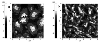
Atomic Force Microscopy (AFM) images (size 5 μm x 5 μm) of
Biocompatibility Test
As the first biocompatibility test, cell adhesion was tested and compared by considering both naked and T6 or PDI-8CN2-covered Si++/SiO2/Au substrates. In the first case, cells were basically in contact with the SiO2 surface. In Figure 3a we report a stereomicroscope image of an undifferentiated murine stem cell plated on one of the supports. In Figure 3b, we report a stereomicroscope image of cells at day 9 of cardiac differentiation, correctly seated on the SiO2 surface (uncovered substrates).
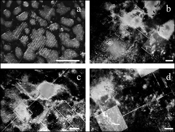
Stereomicroscope images of cells on substrates.
The same observation was repeated when analyzing the cells in contact with the surface of T6 and PDI-8CN2 films, reported in Figures 3c and d, respectively. The cell morphology appears to be comparable among the different supports, and similar to the usual cell appearance, in the presence of standard culture support (petri dish).
Cell Differentiation
To investigate further whether ES cells could differentiate on the previously described supports, we induced differentiation using the hanging-drop method. It is well known that this method preferentially induces differentiation of ES cells into the mesodermal fate and more particularly into cardiomyocytes. After 2 days in hanging-drop and another 3 days of culture in suspension, at day 5 the formed embryoid bodies (EBs) were transferred onto a chip. It was described that the development of the cardiac lineage in ES cell differentiation cultures is easily detected by the appearance of areas of contracting cells that display characteristics of cardiomyocytes (20). On day 9, we observed the first appearance of contracting cell foci on all chips, indicating the presence of cardiomyocytes. Moreover, as reported in Figures 4A and B, formation of cardiomyocytes was confirmed by immunofluorescence analysis using a specific antibody against sarcomeric myosin and by expression of specific cardiac markers analyzed on the EBs differentiated on tissue culture plates as controls for the ones differentiated on the chips. The beating phenomena on the chip was further confirmed at day 11 of differentiation. Growth and differentiation times are in agreement with previous observations in standard conditions (18, 29). The rhythmic contractions of the cellular layer (beating), typical of cardiomyocytes, was clearly visible at the stereomicroscope. In Figure 5 we report the measurement of the mean gray level of the images acquired at the stereomicroscope, as measured over a region that showed beating and is normalized with respect to its average value. The data show a clear periodic oscillation. Similar measurement was done on different beating areas on every chip; the periodic signal did not showed significant differences between the samples.
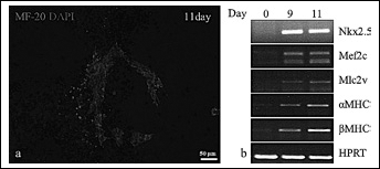
Immunofluorescence analysis of cardiomyocytes.
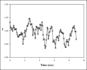
Normalized image intensity fluctuation as a function of time are reported as a quantification of cell periodic contraction (beating).
Discussion
The preliminary results reported here encourage further experimental investigations. The final goal of the proposed research is to investigate the stem cell response to a wide number of external stimuli during differentiation, in order to study how to induce and control an appropriate development of new tissues. A powerful and versatile technique, widely used to investigate the dynamic evolution of biological systems in vitro is Time Lapse Microscopy (30, 31), where the samples are periodically imaged while kept in a controlled environment. The methodology we plan to develop is focused on the direct imaging of stem cells plated on OFET-based sensors in order to visually investigate the dynamic evolution of tissue morphology during the differentiation, in the presence of electric stimuli. The cell electric activity during differentiation will be also monitored.
On top of this, the simultaneous effect of a chemical gradient mimicking a chemotactic signal will also be investigated through direct viewing chemotaxis chambers (32) where the cells are subject to a controlled diffusive flow (33, 34) in a three-dimensional collagen matrix. The presence of mechanical stress can also be investigated by superimposing a confined flow through specific microfluidic devices (35) or confined flow cells (36–38). Beyond qualitative observation of cell and tissue morphology, the motility of single and clustered cells will be also monitored thanks to image analysis algorithms able to segment the images for cell identification (39). In fact, during the differentiation, the ES cells make definite movements, thus modeling tissues and embryos. These movements may occur because the murine stem cells can migrate along extra-cellular signals towards the concentration gradient (chemotaxis). Moreover, it has already been demonstrated (40) that ES cells improve their ability to differentiate in cardiomyocytes if subjected to electric fields. Since cardiomyocytes are unable to regenerate following an injury in the adult heart, cell-based therapies provide a potential alternative approach to replace damaged myocardial tissue and restore cardiac function. OFET could help to set up a new protocol that will increase the efficiency of cardiomyocytes differentiation monitoring their functionality during differentiation.
Conclusions
In conclusion, we have demonstrated that embryonic stem cells (AK7 ES) can be differentiated in cardiomyocytes and produce rhythmic beating on the surface of both T6 and PDI-8CN2 films. These compounds are of interest for the fabrication of organic filed-effect transistors which could be utilized in bio-sensing applications for the detection of bio-signals and their transduction in electrical information, or in the field of bio-actuators.
The results of this work encourage new experimental activities aimed at optimizing the electrical response of organic transistors when operated in biological liquids with strong ionic concentrations. The objective is to investigate the stem cell response to a wide number of external stimuli during differentiation in order to study how to induce and control appropriate development of new tissues. The final goal is to define bioreactor-based approaches for the manufacturing of engineered tissues (41) to be used in regenerative medicine.
This study is related to the activity of the European network action COST MP1106 “Smart and green interfaces - from single bubbles and drops to industrial, environmental and biomedical applications”.
Footnotes
G.M. was supported by Medical Research in Italy (MERIT: RBNE-08HM7T_003) “Innovative models of repair and regeneration of tissues in orthopedic trauma” - MIUR.