
Abstract
We conducted experiments to determine the most effective calcium chelating agents for use in enhancing adhesion of human bone marrow mesenchymal stem cells (BM-MSCs) on nano-hydroxyapatite (nHAp)-coated titanium substrates by covalently immobilizing bone morphogenetic protein-2 (BMP-2). The quantity of amine groups on the chitosan chelated surface was 7 μg/surface area, and it was 1.4 μg/surface area on the alendronate chelated surface. The quantity of BMP-2 on the BMP-2 immobilized surface chelated with chitosan (4 ng/surface area) was higher than that on BMP-2 immobilized surface chelated with alendronate (2.2 ng/surface area). Contact angles of the nHAp-coated titanium, alendronate chelated, chitosan chelated, and BMP-2 immobilized surfaces chelated with alendronate were 68.8 ± 3.6°, 78.2 ± 1.9°, 74.8 ± 5.2°, and 76.0 ± 2.5°, respectively. The contact angle of the BMP-2 immobilized surface chelated with chitosan was significantly lower (56.2 ± 2.0°) than that of any of the other groups. BM-MSCs on the chitosan surface and BMP-2 immobilized on the surface chelated with chitosan appeared to be healthy and showed a spindle-like fibroblastic morphology. In addition, BM-MSCs on these surfaces appeared to have the ability to differentiate into bone-forming cells. We suggest that chitosan can be used as an effective calcium chelating agent for implants.
Introduction
It is essential to maintain a stable bone-biomaterial interface to ensure long-term success of an endosseous dental implant. Early osseointegration and biocompatibility in the location of the implant are associated with long-term success (1). Metal prostheses have excellent mechanical properties (2); however, osseointegration and biocompatibility are dependent on biomolecules that enhance bone regeneration. In addition, it has been revealed that the use of bone cement for permanent bone implantation presents several problems (3).
Interest in surface modification methods to stimulate cell function at the bone-implant interface has increased (4). The key part of a clinical implant application is to immobilize biomolecules on biomaterials, and this has been a widely researched approach to modify metal surfaces to control cell and tissue responses (5). Hence, delivering growth factors to the bone-implant interface using implant coating techniques has recently become a popular method to control healing and fixation of implants (6). Adsorption of bone morphogenetic protein-2 (BMP-2) on the surface of titanium or hydroxyapatite (HAp) ceramics results in intense acceleration of implant osseointegration (7–9).
Osteoconductive calcium phosphate, which is mainly composed of HAp, is an attractive and typical biocompatible ceramic material used in prosthetic devices and has received much attention for its application as an endosseous dental implant (10–12). HAp, Ca10(PO4)6(OH)2, was first established as the mineral component of bone in 1926 by DeJong, and synthetic HAp was approved as a biomaterial for use in orthopedics, bone grafts, and dentistry about 35 years ago (13). HAp can enhance osseointegration, but it is brittle, which restricts its use in endosseous dental applications (14–15). The technique of HAp coating by methods such as plasma spray, sputtering, electrolysis, and sol-gel have been studied to overcome this defect. Effective coating of a load-bearing substrate with HAp can overcome the physical weakness of HAp (13).
However, despite the enormous benefit of biomimetic coatings on a titanium surface, proteins such as BMP-2, which is pre-adsorbed onto the material surface, may be insufficiently immobilized and release may be uncontrolled due to a lack of functional groups. Covalent immobilization on the material surface achieves prolonged retention of BMP-2 and growth factors at their site of action. A two-step, zero length, cross-linking strategy can be applied to covalently immobilize BMP-2 by exposing the amino-functionalized ceramic discs to a cross-linker (16). A solution to this problem may be the use of calcium chelating techniques that provide amine groups needed for covalent attachment of proteins with the cross-linker (17).
Many calcium chelating agents have been researched for calcium chelating technique applications. Pamidronate (C3H11NO7P2) and alendronate (C4H18NNaO10P2) have been used widely by researchers to evaluate the suitability of this approach (16). In addition, EDTA (C10H16N2O8) is an effective calcium chelating agent as it is a single hexadentate chelon. Due to the lone pairs of electrons, the four oxygen atoms in the four carboxyl groups and two nitrogen atoms chelate with the metal and calcium ions (18). However, due to their cytotoxic properties, bisphosphonates such as pamidronate and alendronate can cause apoptosis in a variety of cell types in vitro (19).
In this study, we investigated the effectiveness of calcium chelating agents for providing amine groups to immobilize protein on nano-hydroxyapatite (nHAp) surfaces. We adopted natural chelating material that is non-cytotoxic, has excellent biocompatibility, and immobilizes BMP-2. Chitosan, which is widely used in various forms of biomaterials, shows good biocompatibility and no cytotoxicity in either human or mouse fibroblasts in tissue cell culture (20). Chitosan (C6H13NO5) has chelating ability toward metal and calcium ions, since the calcium chelating effect occurs with the amine group of the chitosan molecule (21). In our study, we evaluated the possibility of using chitosan as a novel calcium chelating agent for immobilizing BMP-2 by culturing bone marrow mesenchymal stem cells (BM-MSCs) and Jurkat cells on a modified titanium surface to test biocompatibility.
Materials and Methods
Materials
Grade 4 titanium discs were used. The triethyl phosphate (P(OC2H5)3) solution, propionic acid, and calcium ethoxide (Ca(OC2H5)2) powder were obtained from the Electronic Functional Materials Laboratory of Seoul National University. 4-amino-1-hydroxy-1-phosphonobutyl phosphonic acid monosodium (alendronate sodium trihydrate) was purchased from Sigma-Aldrich Chemical (St Louis, MO, USA). Chitosan and Protosan UP CL 213 were purchased from Novametrix (Brakeroya, Drammen, Norway). Human BMP-2 was a gift from Daewoong Research and Development, Daewoong Pharmaceutical Co., Ltd (Seoul, Korea).
Preparation of titanium discs
The diameters of the titanium discs used in this experiment were 8 and 14 mm (Fig. 1), and the size of titanium foil was 5 cm × 5 cm. The titanium discs and foil were coated with nHAp using a spin-coater. The nHAp solution was prepared inside a glove box that was purged with argon gas (99.999%). P(OC2H5)3 liquid was diluted with propionic acid to produce the nHAp solution, then Ca(OC2H5)2 powder was dissolved with propionic acid as a solvent. Two bottles of each solution were stored separately in the glove box and stirred until absolutely dissolved. The two bottles of each solution were then mixed in a 1:1 ratio in the glove box for 10 min. Afterwards, the mixed solution was stirred at a temperature of 60°C in a water bath for 6 h. The nHAp solution was spin-coated onto the titanium discs with a spinning velocity of 4000 rpm and a 20 s spin coating time. nHAp spin-coated titanium discs were then sintered in a sintering oven at a temperature of 500°C for 2 h.
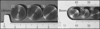
8 mm and 14 mm nano-hydroxyapatite (nHAp)-coated titanium discs. Sample
Calcium chelating agent treatment
The nHAp coated 8 mm and 14 mm discs and the 5 cm × 5 cm titanium foil were placed in 12-well plates containing 2 mL of a 1 mg/ml alendronate or chitosan aqueous solution. The titanium discs were then totally immersed in solution. The well plates were shaken at room temperature for 4 h on an orbital shaker set at 20 rpm in the dark. The titanium samples were washed three times with distilled water, dried, and stored under vacuum until further use.
Immobilization of BMP-2
The concentration of human BMP-2 stock solution was 1 μg/ml in phosphate-buffered saline (PBS). BMP-2 was dispersed on the nHAp coated titanium surfaces, which had been treated with each of the calcium chelating agents. Before BMP-2 treatment, human BMP-2 stock solution and cross-linking solution were mixed for 5 h at a 1:1 ratio to achieve efficient covalent immobilization of BMP-2 on the calcium chelated nHAp coating surface. The cross-linking solution was composed of 20 mL 40% (v/v) ethanol and 50 mM MES (pH 5.5), 24 mM 1-ethyl-3-(3-dimethyl aminopropyl)carbodiimide (Fluka Chemic AG, Milwaukee, WI, USA) and 5 mM N-hydroxysuccinimide (Fluka Chemic AG). The diluted human BMP-2 solution was 0.5 μg/ml. A 10 μL aliquot of the diluted human BMP-2 solution was loaded onto the 8 mm titanium disc surfaces, 30 μL was loaded onto the 14 mm titanium disc surfaces, and 500 μL was added to the 5 cm × 5 cm titanium foil surfaces. All samples were placed overnight at room temperature, washed five times with distilled water, and dried at room temperature. We compared the nHAp-coated titanium surface (cont nHAp group), the alendronate chelated cont nHAp group (alen group), the BMP-2 immobilized alen group (alen + BMP group), the chitosan chelated cont nHAp group (chito group), and the BMP-2 immobilized chito group (chito + BMP group).
Amine assay
The surface density of the amine groups introduced onto the 8 mm calcium chelated titanium discs was quantified by reaction with 2,4,6-trinitrobenzenesulfonic acid (TNBS), which interacts with primary amine groups to form trinitrophenyl derivatives (22). The amine assay was performed according to the method of Puleo (5). Titanium samples were incubated with 0.1% TNBS in 3% sodium borate at 70°C for 5 min, followed by washing with triple distilled water, then hydrolyzed with 1 N NaOH at 70°C for 10 min. These reactions provided a yellow color that was proportional to the number of trinitrophenyl groups, which was proportional to the number of amine groups. Absorbance of hydrolysate was detected at 410 nm. Standard curves were prepared by adding L-DOPA (Sigma-Aldrich) in 0.1% TNBS in 3% sodium borate, followed by a serial dilution. The standard was then hydrolyzed with 1 N NaOH at 70°C for 10 min.
BMP-2 assay
The BMP-2 assay was performed using the Human BMP-2 Super X-ELISA kit (Antigenix America, Huntington Station, NY, USA). BMP-2 immobilized on the 8 mm nHAp-coated titanium discs and other titanium discs were placed in 48-well plates. These titanium discs were then immersed in 1 mL of 0.1 mg/ml bovine serum albumin (BSA) solution and incubated at room temperature for 2 h in the dark. Then, the samples were washed four times with the wash buffer provided in the kit. A 20 μL aliquot of the 0.5 μg/ml biotin-labeled tracer (tracer antibody) stock solution was loaded onto each titanium disc surface, and the titanium discs were incubated at room temperature for 2 h in the dark. Samples were then washed four times again using the wash buffer. The streptavidin-HRP conjugate solution was diluted with diluent, 0.05% Tween-20 (Uniqema, Wilmington, DE, USA), and 0.1% BSA in PBS, at a ratio of 1:500. A 20 μL aliquot of the diluted streptavidin-HRP conjugate solution was loaded onto each titanium disc surface, and the titanium discs were incubated at room temperature for 30 min in the dark. The samples were then washed four times again using the wash buffer provided in the kit. Titanium discs were placed upside down in another 48-well plate. TMB substrate solutions A and B were mixed at a 1:1 ratio. A 100 μL aliquot of TMB substrate mixed solution was added to the 48-well plates, followed by a 30-min incubation at room temperature in the dark. After completing the incubation, 100 μL of stop solution (2 N sulphuric acid) was added, and absorbance was measured at 450 nm.
Contact angle measurement
A contact angle measurement device (Tensiometer/Pendant Drop, Model DSA100; KRUSS Advancing Surface Science, Hamburg, Germany) was used to measure water contact angles. The Drop Shape Analysis software was used to estimate the contact angle of various nHAp-coated titanium surfaces. Water contact angles were measured on dry surfaces.
BM-MSC culture on surface-modified titanium discs
BM-MSCs were purchased from Lonza (Basel, Switzerland). Human BM-MSCs were cultured on 14 mm nHAp coated titanium discs and 5 cm × 5 cm titanium foil surfaces in high glucose DMEM medium (Invitrogen, Carlsbad, CA, USA) containing 10% foetal bovine serum (FBS; Cambrex, East Rutherford, NJ, USA), 1% penicillin streptomycin (10,000 units/ml penicillin, 10,000 μg/ml streptomycin, WelGENE Inc., Daejeon, Korea), and 25 μM ascorbic acid (Sigma-Aldrich) at a density of 2.5 × 10⁴ cells/disc and 1 × 10⁵ cells/foil. The 14 mm nHAp-coated titanium discs were placed in 24-well plates with the medium, and the cells were incubated at 37°C in a humidified atmosphere of 5% CO2 for 3 days. The 5 cm × 5 cm titanium foil sheets were placed in 100 mm diameter Petri dishes with medium, and the cells were incubated at 37°C in a humidified atmosphere of 5% CO2 for 10 days. Then, the culture medium, which was low glucose DMEM medium containing 10% FBS, 1% penicillin streptomycin, 50 μM ascorbic acid, 10 mM β-glycerophosphate (TCI, Seoul, Korea), and 10−7 M dexamethasone (Sigma-Aldrich) was used during 2 weeks.
Scanning electron microscopy (SEM)
SEM samples were fixed overnight at room temperature using a fixing reagent (2.5% glutaraldehyde in PBS); they were fixed in 2% OsO4:0.2 M phosphate buffer (1:1) reagent for 2 h in the dark. The samples were then rinsed with PBS twice for 10 min. The BM-MSC samples were washed with an ethanol: water mixture (30%, 50%, 70%, 90%, and 100% ethanol) twice for 10 min, in sequence. Samples were then treated with 98% 1,1,1,3,3,3-hexamethyldisilazane (HMDS):100% ethanol (1:1) solution for 5 min. They were then treated with 98% HMDS twice for 10 min and allowed to dry overnight.
The morphology of the BM-MSCs on nHAp-coated titanium disc surfaces was examined under SEM (Model HITACHI S-3000N; Hitachi Instruments, Tokyo, Japan).
MTT assay of BM-MSCs
Cell number was determined using a 3-(4,5-dimethylthiazol-2-yl)-2,5-diphenyl tetrazolium bromide (MTT; Sigma-Aldrich) assay (18). This assay identifies metabolically active cells through the action of a mitochondrial dehydrogenase that is changed into an insoluble formazan pigment. BM-MSCs in 24-well plates were incubated for the designated times in 0.33 mg/ml MTT supplemented cell culture medium at 37°C and 5% CO2 for 2 h. The intense purple-colored formazan derivative formed during active cell metabolism was eluted and dissolved in 1 mL dimethyl sulfoxide and absorbance was measured at 540 nm.
T-lymphocyte culture
T-cells were cultured for the biocompatibility analysis. The Jurkat cell line (T-lymphocyte, human leukemia, suspension cell line) was purchased from the ATCC (Manassas, VA, USA). Jurkat cells were cultured in 12-well plates containing 8 mm nHAp-coated titanium discs with 1 mL of RPMI1640 (Invitrogen) containing 10% FBS and 1% penicillin streptomycin (10,000 units/ml of penicillin, 10,000 μg/ml streptomycin) at a density of 5 × 10⁵ cells/well. Cells were incubated at 37°C in a humidified atmosphere of 5% CO2 for 3 days.
The BrdU assay was performed using the Cell Proliferation ELISA Assay, BrdU (colorimetric) kit (Roche Diagnostics, Mannheim, Germany) to evaluate proliferation of the Jurkat cell line. All 8 mm nHAp-coated titanium discs were removed during this assay. One microliter of the BrdU labeling solution provided in the kit was added to the Jurkat cell line cultured in 12-well plates, followed by a 2-h incubation at 37°C in the dark. The final concentration of BrdU labeling solution was 10 μM. The solution was centrifuged at 300 × g for 10 minutes using a 1.5 mL tube and the labeling medium was removed. The 1.2 mL of FixDenat provided in the kit was then added to the cells, followed by a 30-min room temperature incubation. The solution was centrifuged again at 300 × g for 10 minutes, followed by removal of the FixDenat solution. Then, 600 μL of anti-BrdU-POD working solution (antibody conjugate), which was diluted 1:100 with antibody dilution solution, was added to the cells, followed by a 90-min room temperature incubation. The antibody conjugate was removed by aspiration of the centrifuged samples, and the wells were rinsed three times with 1.2 mL PBS. A 600 μL aliquot of substrate solution was added to the cells, followed by 15-min room temperature incubation. Absorbance was measured immediately at 370 nm.
Tumor necrosis factor-α (TNF-α) assay
The quantity of secreted TNF-α, an inflammatory cytokine, was estimated after the T-cell culture. The TNF-α assay was performed using the TNF-α ELISA kit (Biosource, Nivelles, Belgium). Two hundred microliters of standards and samples were added to anti-TNF-α-coated wells of the microtiter plate with 50 μL of incubation buffer provided in the kit. The 96-well microtiter plates were incubated for 2 h at room temperature on a horizontal shaker set at 700 ± 100 rpm. The liquid was aspirated from each well, and the plates were washed three times by dispensing 0.4 mL of wash solution into each well, and aspirating the contents. One hundred microliters of standard 0 and 50 μL of anti-TNF-α conjugate, which were diluted 1:10 with conjugate buffer, were added to all wells. The 96-well plates were incubated for 2 h at room temperature on a horizontal shaker set at 700 ± 100 rpm. The liquid was then aspirated from each well. The plates were washed three times by dispensing of 0.4 mL of wash solution into each well and aspirating the contents. Following the washing step, 200 μL of freshly prepared chromogenic solution was added to each well within 15 min and the plates were incubated for 30 min at room temperature on a horizontal shaker set at 700 ± 100 rpm, avoiding direct sunlight. Finally, 50 μL of Stop Solution was added to each well, and absorbance was read at 450 nm within 3 h.
Reverse transcription polymerase chain reaction (RT-PCR) of BM-MSCs
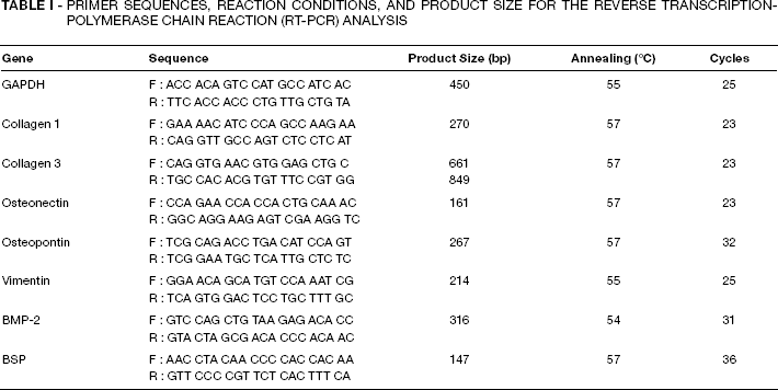
The RT-PCR analysis was performed to compare the expression of bone-inducing markers in BM-MSCs. Total cellular RNA was isolated using 1 mL of Trizol reagent (Invitrogen). cDNA was synthesised by reverse transcription using 1 gg of total RNA. PCR was conducted by subjecting the samples to 23 to 35 cycles (within the linear range of amplification) of denaturation (94°C, 1 min), annealing (53-57°C, 1 min), and extension (72°C, 1 min). The products were then analyzed on 2% agarose gels and visualized by SYBR Safe DNA Gel Staining (Invitrogen). The relative abundance of type I collagen, type III collagen, osteonectin, osteopontin, vimentin, BMP-2, bone sialoprotein (BSP), and GAPDH (an internal control) transcripts was measured using RT-PCR. Primers used for RT-PCR were purchased from Bioneer, and their sequences, reaction conditions, and product size (bp) are listed in Table I. Image J software (Wayne Rasband, National Institutes of Health, Bethesda, MD, USA) was used to quantitatively analyze the RT-PCR amplicons on digitized gel images.
Primer Sequences, Reaction Conditions, And Product Size For The Reverse Transcription-Polymerase Chain Reaction (Rt-Pcr) Analysis
Statistical analysis
Student's t-test was used to evaluate the artificial titanium sample data. Data are given as means ± standard deviations. A p<0.05 was considered significant.
Results
Quantification of amine groups
Amine groups were quantified using the amine assay with the TNBS reaction. The quantity of amine groups on the chito group surface was approximately 7 μg/surface area (Fig. 2). However, the alen group surface contained approximately 1.4 μg/surface area of amine groups. Thus, the chitosan chelated surface appeared to have a large number of amine groups, compared with the alendronate chelated surface. As expected, the cont nHAp group surface had scarcely any amine groups on its surface.
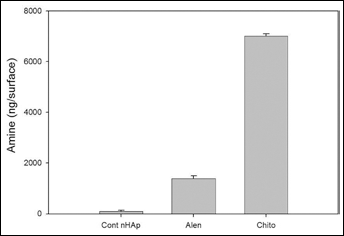
Quantification of amine groups on the calcium chelated surface. The nano-hydroxyapatite (nHAp)-coated titanium surface (cont nHAp) graph indicates quantification of amine groups on the nHAp-coated titanium surface. The Alen graph indicates quantification of amine groups on the alendronate chelated surface. The Chito graph indicates quantification of amine groups on the chitosan chelated surface.
BMP-2 estimation
The BMP-2 assay was performed when all samples were dried perfectly. We compared the quantity of BMP-2 immobilized on the cont nHAp, alen, alen + BMP, chito, and chito + BMP groups (Fig. 3). The quantity of BMP-2 on the chito + BMP (about 4 ng/surface area) group was higher than that on the alen + BMP (about 2.2 ng/surface area). The chitosan agent appeared to hold many BMP-2 growth factors due to the large quantity of amine functional groups. We also detected some BMP-2 on the surface of the sample groups that were not immobilized with BMP-2, which may have been caused by a reaction with the calcium chelating agent.
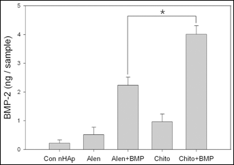
Estimation of bone morphogenetic protein-2 (BMP-2) on the calcium chelated surface. The Cont nHAp graph indicates quantification of BMP-2 on the nano-hydroxyapatite (nHAp) coated titanium surface. The Alen graph indicates quantification of BMP-2 on the alendronate chelated surface. The Alen + BMP graph indicates quantification of BMP-2 on the BMP-2 immobilized surface chelated with alendronate. The Chito graph indicates quantification of BMP-2 on the chitosan chelated surface. The Chito + BMP graph indicates quantification of BMP-2 on the BMP-2 immobilized surface chelated with chitosan. Results are expressed as means ± standard deviation (n = 3) (∗p<0.0001).
Contact angle goniometry
Contact angles of the alen and chito groups were significantly higher (more hydrophobic) than those of the cont nHAp group. In addition, the contact angle of the alen + BMP group was also significantly higher than that of the Cont nHAp group. Angles of the cont nHAp, alen, chito, and alen + BMP groups were 68.8 ± 3.6°, 78.2 ± 1.9°, 74.8 ± 5.2°, and 76.0 ± 2.5°, respectively. However, the contact angle of the chito + BMP group was significantly lower (56.2 ± 2.0°) than that of any other group (Fig. 4). Consequently, the chitosan chelated surface was modified by immobilizing BMP-2 to improve the hydrophilicity of its surface.
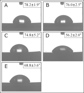
Results for the calcium chelated surface contact angles.
BM-MSC morphology and spreading
The morphology of BM-MSCs on the nHAp-coated titanium discs was assessed by SEM. High-magnification images (x 1.0 K) of these samples show cell adhesion. BM-MSCs were more adherent on the alen + BMP (Fig. 5B) group than those on the cont nHAp and alen groups (Figs. 5A and E). No differences were observed between the cont nHAp and alen groups. BM-MSCs on the chito and chito + BMP (Figs. 5C and D) groups were more adherent than those on the cont nHAp group.
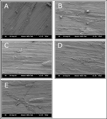
Scanning electron microscopy (SEM) images of bone marrow mesenchymal stem cells (BM-MSCs) on a calcium chelated surface (magnification: x2.0 k).
In particular, adhesion of the chito + BMP group was higher than that of the chito group, which may have occurred due to the presence of abundant amine functional groups, suggesting that chitosan has the ability to increase adhesion of BM-MSCs. Thus, chitosan is an effective biomolecule with an adhesion role in cells. In addition, BMP-2 on chitosan increased cell adhesion.
Normally, cells attached on the surface, and the adhesion area was not large. But if the cells spread their cytoplasm on the ECM or growth factor-coated surface, then the surface increased adhesion and the spreading area increased. In Figure 6, when the cells adhered on the HAp-coated surface, the adhesion area of the cytoplasm was not large. But, when MSCs attached to the BMP linked surface, the spreading area increased because BMP increased cytoplasmic spreading. Thus, even though the cell number was similar, the spreading area was wider. The spreading cell area was measured by Image J software and is shown in Figure 6.
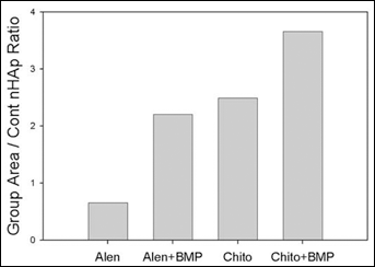
Analysis of spreading cell area measured by Image J software. Group area was divided by the nHAp-coated titanium surface (cont nHAp) area. (Group area = spreading cell area for each experiment group; cont nHAp = spreading cell area of the control group).
BM-MSC proliferation
BM-MSC proliferation on nHAp coated titanium discs was evaluated by the MTT assay (Fig. 7) 3 days after cell seeding. The number of BM-MSCs was measured with a standard MTT assay.
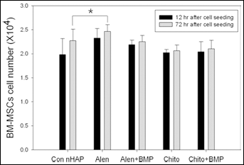
Proliferation of bone marrow mesenchymal stem cells (BM-MSCs) on the calcium chelated surface. The Cont nHAp graph represents cell proliferation on the nano-hydroxyapatite (nHAp) coated titanium surface. The Alen graph represents cell proliferation on the alendronate chelated surface. The Alen + BMP graph represents cell proliferation on the bone morphogenetic protein-2 (BMP-2) immobilized surface chelated with alendronate. The Chito graph represents cell proliferation on the chitosan chelated surface. The Chito + BMP graph represents cell proliferation on the BMP-2 immobilized surface chelated with chitosan. Each cell proliferation graph is divided into 12 and 72 h after cell seeding. Results are expressed as means ± standard deviations (n = 3) (∗p<0.5).
The number of BM-MSCs on the cont nHAp group surface after 12 h and 72 h was approximately 2.01 ± 0.33 × 10⁴ cells/disc and 2.27 ± 0.23 × 10³ cells/disc, respectively. The cell number appeared to increase slightly over 3 days. However, the other groups did not show an outstanding growth effect. The numbers of BM-MSCs on the alen group surface after 12 h and 72 h were approximately 2.32 ± 0.20 × 10⁴ cells disc and 2.43 ± 0.54 × 10³ cells/disc. The numbers on the alen + BMP group surface after 12 h and 72 h were approximately 2.18 ± 0.09 × 10⁴ cells/disc and 2.25 ± 0.13 × 10³ cells/disc. The numbers on the chito group surface after 12 h and 72 h were approximately 2.02 ± 0.07 × 10⁴ cells/disc and 2.06 ± 0.12 × 10³ cells/disc. The numbers on the chito + BMP group surface after 12 h and 72 h were approximately 2.04 ± 0.21 × 10⁴ cells/disc and 2.10 ± 0.17 × 10³ cells/disc, respectively. We found no differences in cell proliferation. Despite what appeared to be enhanced cell attachment on the alen group, no differences were observed among the samples.
Number of Jurkat cells
The BrdU assay was performed to measure the number of Jurkat cells 3 days after cell seeding (Fig. 8). The number of Jurkat cells on the alen + BMP group (approximately 33.3 ± 4.94 × 10⁵ cells/well) was higher than that of cells on the alen group (approximately 15 ± 5.06 × 10⁵ cells/well). We also confirmed that the number of Jurkat cells on the chito + BMP group (approximately 41.4 ± 4.23 × 10⁵ cells/well) was higher than that of cells on the chito group (approximately 34.7 ± 2.04 × 10⁵ cells/well). In other reported research, exposure to toxic molecules results in a concentration-dependent decrease in Jurkat T-cell proliferation (23). Our results suggest that alendronate is more cytotoxic than chitosan; however, BMP-2 had the ability to reduce cytotoxicity of alendronate by increasing the number of Jurkat cells.
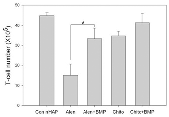
The number of Jurkat cells was measured by the BrdU assay. The Con nHAp graph represents Jurkat cell number on the nano-hydroxyapatite (nHAp) coated titanium surface. The Alen graph represents Jurkat cell number on the alendronate chelated surface. The Alen + BMP graph represents Jurkat cell number on the bone morphogenetic protein-2 (BMP-2) immobilized surface chelated with alendronate. The Chito graph represents Jurkat cell number on the chitosan chelated surface. The Chito + BMP graph represents Jurkat cell number on the BMP-2 immobilized surface chelated with chitosan. Results are means ± standard deviations (n = 3) (∗p<0.0001).
Quantification of TNF-α
The TNF-α assay was performed with RPMI1640 medium from Jurkat cells, which was obtained 3 days after cell seeding (Fig. 9). This result showed a large amount of TNF-α secreted in the alen group. TNF-α decreased significantly in the alen + BMP group, when compared with that of the alen group due to immobilization of BMP-2 on the surface. In addition, the chito and chito + BMP groups showed decreased TNF-α. Thus, chitosan was less cytotoxic than alendronate in Jurkat cells.
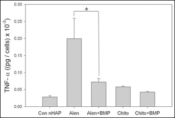
Tumor necrosis factor-α (TNF-α) assay results for Jurkat cells on the calcium chelated surface. The Con nHAp graph represents the quantity of TNF-α on the nHAp coated titanium surface. The Alen graph represents the quantity of TNF-α on the alendronate chelated surface. The Alen + BMP graph represents the quantity of TNF-α on the bone morphogenetic protein-2 (BMP-2) immobilized surface chelated with alendronate. The Chito graph represents the quantity of TNF-α on the chitosan chelated surface. The Chito + BMP graph represents the quantity of TNF-α on the BMP-2 immobilized surface chelated with chitosan. Results are means ± standard deviations (n = 3) (∗p<0.005).
RT-PCR analysis
We investigated whether preconditioning BM-MSCs results in an increase in their bone-inducing activity using RT-PCR analysis (Fig. 10). Although type I collagen mRNA expression appeared to increase in the chito group when compared with that in the control group, no significant difference was observed among any of the groups.
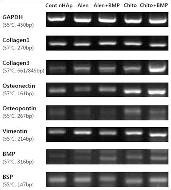
Reverse transcription polymerase chain reaction (RT-PCR) analysis of GAPDH, collagen type I, collagen type III, osteonectin, osteopontin, vimentin, bone morphogenetic protein (BMP), and bone sialoprotein (BSP).
In contrast, type III collagen expression increased in the chito + BMP group when compared with that in the other groups. Levels of osteonectin and osteopontin expression were higher in the chito and chito + BMP groups, compared with the other groups. Osteonectin expression in the chito + BMP group increased more than that in the chito group. In addition, the chito + BMP group showed greater BMP expression than that in any of the other groups. These combined results suggest that chitosan may have a significant autonomous, osteoconductive effect. Thus, bone-inducing activity was more enhanced when BMP-2 was immobilized on the chitosan chelated surface. Additionally, we analyzed BMP release from the BMP-2 immobilized surface and found that 10% of the BMP was released into the media every 24 h during the 4 days. As a result, this osteogenic effect was due to immobilized BMP and BMP released into the media. Alendronate may also have an osteoconductive effect. However, the levels of type III collagen, osteopontin, vimentin, and BSP mRNA expression decreased when BMP-2 was immobilized on the alendronate chelated surface. The quantitative analysis of the RT-PCR amplicons on digitized gel images is shown in Figure 11. We suggest that chitosan can be used as a ligand to bind BMP-2 and can work as a bioactive agent for biomaterials that promote osteogenic differentiation.
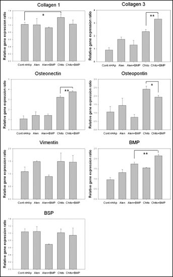
Quantitative analysis of reverse transcription-polymerase chain reaction (RT-PCR) amplicons on digitized gel images measured by Image J software (∗p<0.1, ∗∗p<0.05).
Discussion
The key point of our study was immobilization of BMP biomolecules on biomaterials, which is a widely researched approach to modify metal surfaces to control cell and tissue responses (5). We used alendronate and chitosan as calcium chelating agents to improve immobilization of BMP-2 on an nHAp-coated surface. We hoped to find the most effective calcium chelating agent to enhance adhesion of BM-MSCs on nHAp-coated titanium substrates by covalently immobilizing BMP-2. The results suggest that chitosan, a biomolecule that contains amine functional groups, increased the ability to retain BMP-2, increased the adhesion of BM-MSCs, and reduced cytotoxicity. In addition, BMP-2 immobilized on a chitosan chelated surface was increasingly hydrophilic, which can enhance differentiation to bone compared with a chitosan-only chelated surface. Chitosan is a non-toxic, non-immunogenic, and biodegradable natural biopolymer that enhances bone healing in various animal models (24).
We found that the chitosan chelated surface had a larger number of amine groups compared to that of the alendronate chelated surface. Alendronate has a primary amine on its R2 side chain (25), whereas chitosan has many amine groups that have a chelating effect (26). BMP-2 tends to adhere to cross-linker activated surfaces. Other researchers have revealed that titanium surface hydroxyl groups can be activated with carbonyldiimidazole and that the carboxyl groups are activated with N-hydroxysuccinimide to bind amine-containing molecules. In addition, BMP-2 covalently attaches to activated titanium surfaces (27). Therefore, chitosan has a large number of amine functional groups that enhanced immobilization of BMP-2 with the cross-linker. We also found that the quantity of BMP-2 on the chito + BMP group was higher than that on the alen + BMP group. Thus, we demonstrated an association between the number of amine groups on the surface and that of BMP-2 immobilized onto an amine-grafted surface.
We found that the hydrophilicity of the BMP-2 immobilized surface chelated with chitosan could be improved. Bone-inducing activity was enhanced when BMP-2 was immobilized on the chitosan chelated surface. The correlation between surface hydrophilicity and osteogenic activity of cells has been demonstrated in other studies. Our results suggest that hydrophilic titanium can lead to an alterations in osteogenic activity (28). However, we found no significant difference in cell proliferation among the samples. Other researchers have already shown that cell proliferation on both titanium grafted with carboxymethyl chitosan and BMP-2 functionalized substrates does not increase compared with that on pristine titanium (29). We propose that although we did not detect a significant increase in cell proliferation, enhanced cell adhesion due to the presence of chitosan and BMP-2 would lead to more complete osseointegration.
Research has shown that chitosan has a wide range of applications, including antibacterial activity (30, 31). Analogous studies of immobilizing BMP-2 to enhance the osteoconductive effect of a titanium surface using chitosan have also been reported. Shi et al immobilized BMP-2 on a titanium surface, which was functionalized by covalent grafting with carboxymethyl chitosan, and showed an antibacterial effect (29). Shi et al grafted chitosan on titanium by immobilizing L-DOPA on its surface. Their results showed that bacterial adhesion on both the carboxymethyl chitosan-grafted (CMCS) surface and BMP-2 immobilized CMSC surfaces decreased significantly, compared with that on pristine substrates. In addition, BMP-2 immobilized CMSC surfaces promote significant attachment, alkaline phosphatase activity, and calcium mineral deposition in both osteoblasts and human BM-MSCs. However, we immobilized BMP-2 on chitosan, which was chelated on an nHAp-coated titanium surface. We adopted an nHAp-coated titanium surface, as it contains a large number of calcium ions for chelation with chitosan.
Chitosan enhanced binding ability and cell adhesion of BMP-2 and also reduced cytotoxicity. We also demonstrated that chitosan had an excellent autonomous osseointegration effect. In addition, BMP-2 immobilization on a chitosan chelated surface increased hydrophilicity of the surface. BM-MSC differentiation to bone on a BMP-2 immobilized surface chelated with chitosan was better than that on a chitosan chelated surface.
Conclusions
More amine groups were found on the chitosan chelated surface than on the alendronate chelated surface. The quantity of BMP-2 on the BMP-2 immobilized surface chelated with chitosan was higher than that on the BMP-2 immobilized surface chelated with alendronate. Hydrophilicity of the BMP-2 immobilized surface chelated with chitosan showed a significantly greater increase than that of any other group. BM-MSCs on a BMP-2 immobilized surface chelated with chitosan appeared to have a significant ability to differentiate into bone-forming cells. Based on these results, we suggest that chitosan is an effective calcium chelating agent for implants. Our future work will focus on methods to immobilize various bioactive molecules on surfaces grafted with bioinert material. In addition, we will investigate various analytical tests.