
Abstract
The aim of the present study was to develop and investigate nanoporous activated carbon materials for their ability to adsorb inflammatory cytokines directly from blood, for a range of therapeutic applications, including: systemic inflammatory response syndrome (SIRS) related to sepsis, cardio-pulmonary by-pass surgery, or ischemic reperfusion injury. Building on the previously established relationship between the porous structure of beaded polymer-derived activated carbon and its capacity to adsorb inflammatory molecules, we have developed and characterized monolithic porous carbon columns produced from the same polymer precursor matrix as carbon microbeads. The monolithic columns developed were assessed for their ability to adsorb inflammatory molecules from blood in a circulating system. Preliminary findings demonstrated good removal of the inflammatory cytokines IL-8 (100% removal), IL-6 (80% removal), and TNF (51% removal) from blood. The efficiency of cleansing is dependent on the size of the adsorbed molecule and the porous structure of the monolith, highlighting their potential for use as a hemoadsorption device.
Introduction
Sepsis is one of the most widespread conditions resulting in critically ill patients being admitted to the Intensive Care Unit (ICU) and it is associated with a high mortality rate. A study conducted in the United States estimated the occurrence as 3.0 cases occurring per 1000 population per year, which calculates to ∼20 million cases per year (1). In a recent outcome from the Surviving Sepsis Campaign, which collected data from 200 sites in Europe and the US, ICU mortality for patients with severe sepsis and septic shock was 31% in the US and 32% for patients in Europe (2). Although recent studies are encouraging, demonstrating decreased mortality as a result of new guidelines, interventions, and therapeutics, due to the increasing incidence of sepsis the number of people who die each year continues to increase (3–6).
The use of adsorbents incorporated into an extracorporeal device has been considered either for plasma adsorption (7) or direct hemoperfusion as a therapy to treat sepsis and other conditions related to systemic inflammatory response syndrome (SIRS). A number of clinical trials have been conducted or are currently ongoing to evaluate different extra-corporeal devices, with varying degrees of success (7–10). We have shown that a novel polymer/pyrolyzed activated carbon (AC), with a finely controlled pore size can be used for the direct and effective removal of cytokines and other biologically active molecules, including bacterial exotoxin, from blood without prior separation of the blood into cells and plasma (11). This process occurs with no detectable hemolysis, while other adverse effects such as cytotoxic-ity or activation of white blood cells have not been noted (12–14). It is important for good hemocompatibility of this polymer derived carbon that it allows direct contact with blood without an additional coating. Coating hampers the adsorption properties of AC and has restricted the use of hemoperfusion in comparison to other blood purification techniques such as hemodialysis and hemofiltration. During hemoperfusion with a device containing uncoated AC, the diffusion of molecules from blood to the outer surface (40 m2) or to the inner porous surface (up to 3000 m2/g−1) is the rate limiting step. Whereas, in coated AC devices, diffusion of the molecule through the biopolymer coating is the rate limiting step, therefore substances of low molecular mass are only slightly delayed by the coating, but diffusion of molecules of 3500 Da in size or larger is significantly delayed or restricted. The use of uncoated AC enhances its adsorption capacity towards middle and large molecular weight biomolecules and increases the rate of adsorption compared to coated carbons currently used in direct hemoperfusion (15). Inflammatory cytokines fall within this size range; for example, TNF has a molecular weight ranging from 17 kDa to 51 kDa depending on whether it is found in the monomer, dimer or trimeric form, whereas IL-8 is much smaller, being 8 kDa in size.
The porous structure of carbon material can be comprized of micropores (≤2 nm), mesopores (2-50 nm), and macropores (≥50 nm) according to the nomenclature recommended by the International Union of Pure and Applied Chemistry, which is used in this paper. Of particular importance for adsorption of cytokines are small macropores (50-100 nm in diameter) as shown previously (11). In this study we investigated two types of carbon materials: spherical carbon beads (250-500 μm) and three-dimensional (3 D) monolithic columns produced from the same phenol-formaldehyde resin precursor used to prepare the carbon microbeads (Fig. 1). Within the beads, the mesopore and small macropores (∼100 nm) are created by pore-forming substances at the resin-precursor preparation stage (16). This porous structure was also introduced into the monolith by incorporation of AC powder produced from the beads themselves.
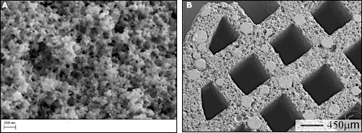
A) SEM image showing meso-macroporous domains within the carbon beads (Zeiss NTS, Sigma FEG-SEM).
At the outset, carbon beads were tested for their adsorption efficiency for a range of pro-inflammatory cytokines relevant to the clinical condition of sepsis (11). However, although achieving good cytokine clearance when used in an extracorporeal device, because of their small particle size they would suffer from the disadvantage of an excessive pressure drop across the packed bed. Current packed bed hemoperfusion systems such as the Prismaflex® system HP containing Adsorba® C cartridges (Gambro) contain much larger coated granules (1mm by 2-5 mm) and thus offer low resistance to blood flow through the system although, they are limited to removal of small molecules, mainly in poisoning applications.
The challenge was to develop a monolithic carbon material with the required porous structure to enable removal of inflammatory cytokines directly from blood. The advantage of using a monolithic structure for blood flow is the low pressure drop, however, it may be less effective in the adsorption of macromolecules due to the hydrodynamic flow characteristics of the channel structure which influence the movement of molecules into the pores. The removal capacity of the monolith was therefore investigated in a continuous blood recycle system, consistent with current extracorporeal therapies.
Materials and Methods
Synthesis: carbon beads
The meso/macroporous carbon beads were produced using a novolac phenol-formaldehyde resin dissolved in a pore-forming solvent (ethylene glycol) together with a cross-linking agent, hexamethylenetetramine, by being dispersed in hot mineral oil containing a dispersing agent. Novolac concentration ranged from 33% (TE3), 24% (TE7) and 20% (TE9) and hexamethylenetetramine from 7%, 5% and 4% respectively (16). The pre-polymer solution formed spherical droplets which solidified as curing of the resin proceeded. The average bead particle size was controlled by the rate of stirring and the ratio of reagents. The beads were produced in the size range between 5 μm and 2000 μm. The resin beads (TE3, TE5, TE7 and TE9) were then carbonized by heating them in inert atmosphere at 600°C to 900°C and optionally activated to varying degrees of burn-off in carbon dioxide at 800°C to 900°C and sieved to isolate the required particle size fraction. Carbons thus obtained were coded as TE3, TE5, TE7 or TE9, followed by a two-digit number corresponding to the degree of activation (burn-off), and the letter C to indicate activation by CO2. The code 00 means no activation; it was assigned to the carbonized-only material.
Synthesis: carbon monoliths
Carbonized and optionally activated monoliths containing large transport channels were also formed from the same phenolic resin precursors as the carbon beads. The findings from the carbon bead adsorption study (see section below on Cytokine adsorption by carbon beads) determined that the TE7 resin provided the optimum size of small macropores in the 80 nm range for maximal adsorption capacity. Thus two combinations of this resin, TE7/16 and TE7/20, were used with the aim of producing monoliths with inherent porosity in this size range. Two types of carbon monoliths, A and B, were produced; monolith A was then carbonized at 800°C for 1 hour, and monolith B at 850°C for 2 hours. These monoliths were compared with a purely microporous carbon monolith C, prepared from microporous resin and carbonized at 800°C for 3 hours. Manufacturing process of porous resin monoliths followed several steps: (i) partial curing a resin into a solid, (ii) comminution of the partially cured resin, (iii) mixing with activated carbon powder obtained from carbon beads to give the required meso- and small macroporous structure, (iv) forming of the comminuted particles into a dough by the addition of water and an extrusion agent, and (v) extruding the dough to form a resin monolith. The resulting monolith was carbonized by heating in an inert atmosphere to 600°C to 900°C. The carbonized monolith has a bimodal distribution of pores, and a BET surface area, SBET of 250-800 m2/g without activation (Fig. 3). The carbon is further activated by heating at 800°C to 900°C, in the presence of carbon dioxide, resulting in a SBET of 750-1500 m2/g (18). The carbon monoliths used in this study had a diameter of 7 mm, length of 100 mm with 0.6 mm square channels (24 in total) running the length of the monolith, and they weighed 2 g. Each monolith was joined to stainless steel connectors at each end that connected to 3 mm diameter silicon tubing. The prototype device was tested for adsorption in a re-circulating system.
Material analysis
The porous structure of carbon materials was investigated using low temperature nitrogen adsorption porosimetry (Autosorb-1; Quantachrome Instruments, Hook, UK), mercury porosimetry (PoreMaster; Quantachrome Instruments) and scanning electron microscopy (Sigma FEG-SEM microscope, Carl Zeiss NTS, Cambridge, UK). Carbon samples (0.08-0.1 g) were degassed in the Autosorb-1 at 200°C for 3 hours prior to analysis. Nitrogen adsorption was carried out at 77 K using 40 adsorption and 40 desorption points; data was collected during the analysis and after completion the sample was transferred for analysis by mercury po-rosimetry. Porosimetry data analysis was carried out using Quantachrome data reduction software. For SEM analysis samples were analyzed without sputter coating, using a high resolution, high vacuum FEG-SEM.
Cytokine adsorption by carbon beads
Fresh frozen plasma (National Blood Service, London, UK) was defrosted at 37°C and then separated into a non-spiked and spiked sample that was supplemented with the human recombinant cytokines (BD Biosciences, Oxford, UK) TNF and IL-6 at a final concentration of 300 pg/ml and 1000 pg/ml, respectively. The cytokine levels chosen were maximum values reported in the literature for sepsis patients. The carbon beads were weighed into Eppendorf tubes (0.02 g) and pre-equilibrated with 1 mL of phosphate buffered saline (PBS) in a shaking incubator at 37°C for 2 hours, before centrifugation at 2000 g and removal of the supernatant. 800 μl of spiked plasma was added to each carbon and the samples were placed in a shaking incubator at 37°C. A positive control with no carbon beads and a negative control of non-spiked plasma were also included. At timed intervals (5, 45, and 90 min) the samples were cen-trifuged at 2000 g and 150 μL aliquots were removed and stored at -20°C before analysis by ELISA assay to determine cytokine concentration (BD Biosciences, Oxford, UK) according to manufacturer's instructions.
Cytokine adsorption using a monolith filtration/ adsorption system
Fresh blood was collected from healthy volunteers following institutionally approved ethical guidelines, put into citrated vacuette tubes, and separated into a non-spiked and spiked sample that was supplemented with the human recombinant cytokines TNF, IL-6 and IL-8 (BD Biosciences, Oxford, UK) at a final concentration of 500, 1000, and 500 pg/ml, respectively. The carbon monoliths were attached to silicone tubing and fed from a reservoir through a peristaltic pump set at a flow rate of 5 ml/min, and back through the monolith to the reservoir. The linear velocity of the experimental setup was calculated to be similar to that used during hemodialysis. The monoliths were primed with 20 mL PBS for 60 min, which was then replaced by 20 mL of spiked blood pumped through the monolith at a flow rate of 5 ml/min. Samples were collected at time points of up to 60 min. Controls consisted of non-spiked and spiked blood flowed through empty silicone tubing or non-spiked blood flowed through monoliths. Collected blood aliquots were centrifuged at 2500 g for 15 min at 4°C. The plasma was stored at -20°C before analysis of the cytokine concentration by ELISA assay (BD Biosciences, Oxford, UK) according to the manufacturer's instructions.
Results
Material synthesis and analysis
Carbon beads were prepared with a range of meso/ macropores and degrees of activation and examined by scanning electron microscopy (SEM), porosimetry and assessment of adsorption capacity for inflammatory cyto-kines. The porous internal structure of the carbon beads is clearly noticeable in the scanning electron micrograph shown as Figure 1a, while the monoliths internal channel structure can be seen in Figure 1b. The porous structure of the beads visible on the micrograph supports the porosim-etry data obtained for these carbons, although the smallest micropores are below SEM resolution (Fig. 1a).
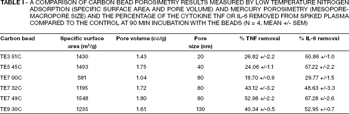
The porosity of the carbon beads was measured by both low temperature nitrogen adsorption to measure the specific surface area SBET; and pore volume data, and mercury porosimetry to determine meso/macropore pore size distribution using the software provided by Quantachrome. The carbon beads had SBET ranging from 580 m2/g to 1550 m2/g, a pore volume from 1.04 cm3/g to 1.80 cm3/g, and a mean meso/macropore diameter ranging from 20 nm to 130 nm (Fig. 2), depending on the type of polymer precursor (TE3, TE5, TE7 or TE9) used to prepare the beads (Tab. I). The TE7 series of beads demonstrate the influence of activation on the porosity: the non-activated beads (TE7 00C) have the lowest specific surface area and pore volume; while the most activated beads (TE7 49C, which means 49% activation burn-off of carbon in carbon dioxide) have the highest values for both, although the mean small macropore diameter remains constant at 80 nm.
A Comparison Of Carbon Bead Porosimetry Results Measured By Low Temperature Nitrogen Adsorption (Specific Surface Area And Pore Volume) And Mercury Porosimetry (Mesopore-Macropore Size) And The Percentage Of The Cytokine Tnf Or Il-6 Removed From Spiked Plasma Compared To The Control At 90 Min Incubation With The Beads (N = 4, Mean +/- Sem)
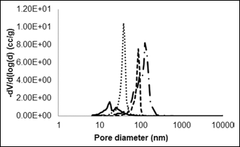
Calculated pore size distribution of carbon beads TE3-9 measured by nitrogen adsorption, displaying the increase in mean meso-macropore diameter across the series from TE3-9 (–- TE3 51C, TE5 45C, – – TE7 49C, –- . TE9 30C).
The pore size distribution for three types of monoliths (A-C) measured by mercury porosimetry (Fig. 3) demonstrate that use of the TE7 resin pre-cursor allowed successful tailoring of the pore structure to include pores in the small macropore size range similar to the TE7 beads. This offers the greatest potential for adsorption of inflammatory cytokines. Monoliths A and B had SBET of 760 m2/g and 850 m2/g and a pore volume of 0.72 cm3/g and 1.15 cm3/g, respectively, similar to the non-activated TE7 00C beads. Monolith C made from microporous resin precursor displayed very few pores in the meso/macro-pore range, but a relatively high surface area of 930 m2/g. The large macropores apparent in Figure 3 derive from the voids between the milled particles.
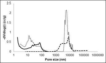
Calculated pore size distribution of Monoliths A-C measured by mercury porosimetry, displaying mesopores (10-50 nm) small macropores (50-150 nm) contained within Monoliths A and B (–- A, B, – – C).
Cytokine adsorption by carbon beads
Experiments to determine the adsorption capacity of the range of prepared carbon beads with varying porosity characteristics revealed the percentage adsorption of both TNF and IL-6 from plasma after 90 min incubation, and displayed a similar pattern related not only to the meso/ small macropore size but also to the specific surface area and pore volume (Tab. I). Although the TE7 carbon series (TE7 00C-TE7 49C) all possessed small macropores with a mean diameter of 80 nm, they displayed increasing specific (per g of carbon) surface area and pore volume in line with the degree of activation, which resulted in significantly increased efficiency of cytokine removal. The differing resin precursors resulted in the production of beads with an array of meso/macropore diameters, with the smallest at 20 nm (TE3 51C) to the largest at 130 nm (TE9 30C). The TE3 and TE5 beads with the smallest mesopores removed the lowest amount of TNF, in line with the non-activated TE7-00 beads, but more efficiently adsorbed the smaller cytokine IL-6. The TE9 beads with the largest diameter of pores (130 nm) demonstrated good removal of both cyto-kines, however the best performing beads were the highest activated TE7 49C, with the greatest surface area and pore volume and pores of 80 nm mean diameter. The carbon bead results suggest that size exclusion is playing a part in the adsorption profiles, with the largest TNF molecule (51 kDa) being excluded to some extent from the smaller mesopores, whereas adsorption of IL-6 molecules (∼26 kDa) is less restricted. Adsorption kinetics is significantly faster with the TE7 beads than either the TE3 or TE9 beads, for both TNF (Fig. 4) and IL-6 (data not shown) (p≤0.01, t-test).
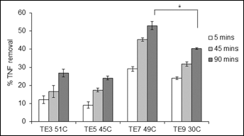
Adsorption of TNF by the series of beads, % removal of TNF compared to the control from plasma after 5, 45, and 90 min incubation (n = 4, mean +/- sem) ∗p≤0.01 T-test.
Monolith filtration/adsorption system
Monolith filtration/adsorption experiments to determine the adsorption capacity of the three monolith types A to C, revealed the increasing adsorption of the cytokines TNF, IL-6, and IL-8 from blood over time up to 60 min circulation (Figs. 5a-c). The concentration of TNF, IL-8, and IL-6 in the spiked blood after continuous circulation through the control tubing only remained constant over time (data not shown).
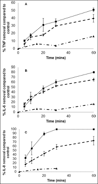
A) Adsorption of TNF by Monoliths A-C, % removal compared to the control from blood after 0-60 min continuous circulation through the monoliths (Monoliths A-B, n = 4, mean +/- sem, Monolith C, n = 2, mean) (– – A, B, –- · C).
The greatest removal was measured for the smallest cyto-kine IL-8 (8 kDa), with total removal achieved by Monolith B at the 30-minute time point. Monolith B also removed the most IL-6 with up to 80% removal by 60 min, whereas removal of the larger TNF molecule was less successful, reaching a maximum of 40% and 51% removal for monoliths A and B, respectively. In line with the bead adsorption findings, differences in the adsorption profiles for the two monoliths can be attributed to the porous characteristics of the materials. Monolith A has a surface area of 760 m2/g and pore volume of 0.72 cm3/g, much lower than those obtained for the activated carbon beads, and also lower than monolith B which has a surface area of 850 m2/g and pore volume of 1.14 cm3/g. In contrast, monolith C, which lacked the meso/small macroporous domains, demonstrated poor removal capacity for all three cytokines studied.
Discussion
Removal of the inflammatory cytokine TNF by adsorption appears to provide the greatest challenge not only for the carbon adsorbents investigated in this study but for other adsorbent materials studied in the literature (8, 17). TNF is the largest cytokine molecule studied in this investigation with a molecular weight of 51 kDa in its homotrimeric form (9.4 × 9.4x 11.7 nm) (18), whereas both IL-6 at 26 kDa (4.97 × 4.97 × 12.2 nm) and IL-8 at 8 kDa (4 × 4 × 9 nm) are smaller in size (19, 20). In our previous work, the best performing carbonaceous adsorbent (PGF 60C AM) with the largest mesopores (∼40 nm) and surface area (∼1800 m2/g) displayed the greatest uptake of TNF at 2.47 ng/1 g adsorbent (11). The study concluded that this was due to the larger pores allowing for more rapid intraparticle diffusion and orientation of the TNF molecule within the pores.
In this study, the highest activated TE7 49C beads performed significantly better than carbon PGF60C AM and the other carbon beads previously studied, removing the greatest amount of TNF and IL-6 (53 and 67%, respectively), reaching a value of 7.75 ng TNF and 14.3 ng IL-6 per 1 g of adsorbent. Similarly, in flow studies with the carbon beads packed into 2 mL columns with continuous circulation of plasma containing inflammatory cytokines at a rate of 5 ml/min, the TE7 49C and TE9 30C beads performed best compared to the microporous beads and commercially available, biocompatible, coated AC Adsorba® C (Gambro), which demonstrated negligible adsorption (data not shown). In comparison to other adsorbent materials that have been investigated for their potential cytokine removal, such as cellulose microspheres with ligands bound to the surface, used in the CFX (17), and CTR columns (21), and DHP-1 activated charcoal and Amberlite XAD-7 resin (1.4 and 7.25 ng TNF and IL-6/1g adsorbent respectively) (22), meso/macroporous carbon beads studied here perform considerably better. A study of coupled hemofiltration and adsorption using a carbon containing hemofilter (Detoxyl 3; Bellco, Mirandola, Italy) demonstrated the feasibility of efficiently removing cytokines under ex vivo conditions, although removal of TNF by the system was poor perhaps due to the porosity being suitable for molecules of up to 20 kDa in size (7). The TE7 49C carbon beads also appear to outperform the polymer resin CytoSorb™ beads (Medasorb Technologies, Malvern, PA, USA) which in similar studies removed ∼4.0 ng of TNF and 12.5 ng of IL-6 per 1 g adsorbent (23). These findings suggest that pores in the 80 nm range are most efficient at removing the cytokines TNF and IL-6, whereas increasing pore size to 130 nm as in the case of the TE9 30C carbon, does not additionally improve adsorption capacity.
The cytokine adsorption data for the TE7 carbon beads and monoliths A and B suggest that the meso/macropo-rous structures are retained when incorporated into the 3D monolith matrix, which is supported by the monolith pore size distributions. The results showed that monoliths A and B, with inherent meso/macroporosity have the greatest removal capacity for TNF, IL-6, and IL-8 from blood. In contrast, the purely microporous monolith C demonstrated poor removal capability for all three cytokines. The percentage removal of the cytokines by the best performing monolith B was similar to the removal for the best performing TE7 carbon beads, although actual values of adsorption in ng removal per 1 g monolith were lower (2.47ng of TNF and 7.78 ng of IL-6 for monolith B). This may be explained by slower adsorption kinetics from blood compared to plasma, and lower surface area and pore volume of the monolith, which was comparable to the TE7 00 non-activated beads.
However, using a monolith as an extracorporeal therapy, the kinetics of adsorption is not the rate limiting factor, as the blood is re-circulated through the device over a more extended time frame of 2 h to 4 h, and this would provide sufficient time to remove clinically relevant amounts of cy-tokines. This is supported by the hemoadsorption study using the CytoSorb™ column (Medasorb Technologies, Malvern, PA, USA), which has been effective in removing both pro- and anti-inflammatory cytokines, and has resulted in improvement in the short-term survival in a rat model of sepsis (24). The device depleted middle molecular weight cytokines from a circulating solution, although the authors attempted to increase the removal of TNF from 55% to 69% with covalently linked anti-TNF antibodies (25). This may allow more specific adsorption of one particular cytokine, but the increased cost to the device may be significant and other cytokines remain unaffected.
Over recent years both experimental and clinical studies have demonstrated that hemoadsorption therapy to adsorb cytokines is beneficial during endotoxemia and sepsis. Studies such as the use of the Lixelle β2 microglobulin column (Kaneka Corporation, Osaka, Japan) in patients with sepsis found that even after a 3-h session there was still some removal of cytokines (26). The MPCF-X column which consists of cellulose beads cross-linked with hexa-methylene-di-isocyanate and coated with 2-methacryloy-loxyethyl phosphorylcholine (MPC) was tested for cytokine removal in vitro with patient plasma. MPCF-X was shown to have an advantage over other adsorbents such as Lix-elle, by adsorbing considerable amounts of cytokines with decreased affinity for normal IgG (17). In an animal model of endotoxin-induced sepsis, the CTR column was found to improve mortality rates by adsorbing both cytokines and exotoxin TSST-1 (9). The CYT-860-DHP adsorbing device has demonstrated low in vitro adsorption rates for TNF and in animal sepsis model studies has significantly improved survival rates (27–29). This column has also undergone a small scale pilot study in critically ill patients with persistent or severe hypercytokinemia, and showed significant reductions of cytokine levels in blood (8).
Conclusions
The monoliths developed and investigated in this study have a potential to develop new and efficient hemoadsorption therapy for the removal of molecules involved in the pathogenesis of excessive and dysregulated inflammation, and compare favorably to current adsorbent materials undergoing animal or clinical investigations. Our findings support previous work in identifying the relationship between pore size and adsorption capacity towards large biomol-ecules. This study has also shown that the adsorbent properties of the beads can be retained when the resin material is used to manufacture a 3D monolith. This monolith has a large surface area, the required meso/small macroporous structure, is free from fines and can be used uncoated, with the added benefit over polymer bead-based materials in avoiding potential leachate and pressure drop complications. These solid monolithic columns have been specially designed and engineered for maximum efficiency, and warrant further investigation as a potential therapy for the treatment of hypercytokinemia, experienced in conditions such as multiple organ failure. We are currently working towards a clinically relevant scaled up version of the monolith for further evaluation of hemocompatibility, adsorption performance and axial pressure drop across the device. At the present time, monoliths with a diameter up to 10 cm can be produced in varying lengths, with an identical channel structure and internal pore structure of the small monoliths used in this study.