
Abstract
Objectives
This study investigates the molecular mechanisms by which Alpiniae oxyphyllae fructus (AOF) promotes neuron regeneration.
Methods
A piece of silicone rubber was guided across a 15 mm gap in the sciatic nerve of a rat. This nerve gap was then filled with different concentrations of AOF extract (0-200 mg/ml). We investigated the role of MAPK (ERK1/2, JNK and p38) pathways for AOF-induced matrix-degrading proteolytic enzyme (PAs and MMP2/9) production in RSC96 Schwann cells.
Results
The results showed that AOF increased the expressions of uPA, tPA, MMP-9, and MAPKs in vivo. In vitro, our results show that treatment with AOF extract induces ERK1/2, JNK, and p38 phosphorylation to activate the downstream PAs and MMPs signaling expression. AOF-stimulated ERK1/2, JNK, and p38 phosphorylation attenuated by individual pretreatment with siRNAs or inhibitors (U0126, SP600125 and SB203580), resulting in migration and uPA-related signal pathway inhibition.
Conclusions
Taken together our data suggests the MAPKs (ERK1/2, JNK and p38), PAs (uPA, tPA), MMP (MMP2, MMP9) regenerative and migration signaling pathway of Schwann cells regulated by AOF extract might play a major role in Schwann cell migration and damaged peripheral nerve regeneration.
Keywords
Introduction
Nerve regeneration is a complex phenomenon that has interested scientists for many years. Neurons can be separated into central and peripheral nervous systems (PNS), with different anatomical structures and regenerative ability. In mammals, the central neurons without a myelin sheath are difficult to regenerate. In contrast to the central nervous system, the PNS with a myelin sheath exhibit easier re-growth (1). Schwann cells are the PNS support cells and can differentiate into the PNS myelin sheath and proliferate and migrate to the distal end of the injured nerve area (2). Since Schwann cell migration is critical for axonal elongation and remyelination of injured nerves (3, 4), peripheral nerve injury locally activates Schwann cells and macrophages to synthesize a cocktail of neurotrophic factors, adhesion molecules, cytokines and growth-promoting surface molecules (5, 6). However, the action mechanisms that regulate Schwann cell migration, proliferation and signal transformation remain unclear.
The mitogen-activated protein kinase (MAPK) family plays an essential role in inducing cell migration (7). JNK, p38, and ERK1/2, the members of the MAPKs family, play crucial roles in nerve cell migration (8). Many experiments have determined that after injury, a rapid increase in tissue plasminogen activator (tPA) and urokinase plasminogen activator (uPA) expression has been observed in neurons (9, 10). uPA and tPA cleave plasminogen to plasmin to promote extracellular matrix (ECM) degradation for cell migration during recovery from injury (11). Therefore, the Schwann cell's ability to promote regeneration in peripheral neurons has led to an increasing interest in using Schwann cells for peripheral nervous system repair.
Biomaterials combined with Chinese herbal medicine have been previously applied to study nerve regeneration. For example, a filled silicon rubber chamber has been used with Schwann cells to bridge a 15 mm defect in rat sciatic nerves. Several Chinese medicines have been identified as enhancing neuron regeneration (12). Therefore, targeting Schwann cells with herbal medicines to induce neuron re-growth may be a possible therapeutic approach for treating injured nerves.
Alpiniae oxyphyllae fructus (AOF) (botanical name Alpinia oxyphylla Miq.), commonly known in English as sharp-leaf galangal berry or bitter cardamom, is one of the most important traditional Chinese medicines. According to the Chinese Pharmacopoeia, it is used for treating diarrhea, polyuria, ulceration, dementia, tumors, and gastralgia (13, 14). Several reports show that water and ethanol extracts of AOF have potential neuro-protective effects (13, 15–18). However, the detailed molecular mechanisms of how AOF acts as a nerve migration-enhancer on Schwann cells are still unknown. We therefore investigated the molecular events associated with AOF treatment in rat sciatic nerves and verified the deduced mechanism using RSC 96 cells by silencing appropriate protein expressions.
Materials and Methods
AOF extraction
Raw, fragmented Alpinia oxyphylla Miquel was purchased from Shin-Long Pharmaceutical Company (Taichung, Taiwan). One hundred and fifty grams of AOF was extracted with 600 mL of boiling water for 2 h. After filtration, the filtrate was concentrated at reduced pressure for convenience. The extract solution was stored at 4°C. This extract was spray-dried to produce a powdered extract.
Animal model
The surgery was performed as described in a previous study (19). Thirty-six adult Sprague-Dawley rats weighing 220 ± 20 gm underwent silicone tube (1.96 mm OD; Helix Medical, Carpinteria, CA, USA) placement. The animals were divided equally into six groups. In each animal the right legs received experimental treatment. In the first group the chambers were filled with only saline. The chambers in groups 2~6 were filled with different concentrations of AOF extract at 30, 60, 100, 150, and 200 mg/ml, respectively. The chamber lumen volume was 25.5 μL. AOF extract was injected through a precooled micropipette into the lumen by passing the tip of the needle into the silicone rubber chamber. Loading was performed as slowly as possible to prevent the formation of air bubbles. The distal stump was then secured at the other end of the chamber. Both the proximal and distal stumps were secured to a depth of 1 mm into the chamber, leaving a 15 mm gap between the stumps. The muscle layer was approximated using 4–0 chromic gut sutures and the skin was closed with 2–0 silk sutures. All animals were housed at 22°C and 45% humidity with a 12 h light/dark cycle. They had free access to rodent chow and water at libitum. After four weeks the animals were anesthetized using isoflurane. The nerves were re-exposed and the chambers across the 15 mm gap were examined for nerve regeneration. All animals were maintained in facilities approved by China Medical University for Accredited Laboratory Animal Care according to the regulations and standards of the National Science Council of Health of the Republic of China.
Cell Culture
RSC96 Schwann cells were purchased from American Type Culture Collection (ATCC; Manassas, VA, USA) and cultured in Dulbecco's modified Eagle's medium (DMEM) supplemented with 10% fetal bovine serum (FBS), 4 mM L-glutamate, 1.5 g/l sodium bicarbonate and 4.5 g/l glucose in humidified atmosphere of 5% CO2 and 95% air. After 4 h in serum-free culture the cells were treated with AOF at different concentrations, incubated for 16~24 h and then harvested for further analysis.
Cell viability assay
Cell viability was estimated using a colorimetric assay based on tetrazolium dye (MTT) conversion into a blue formazan product. Briefly, RSC96 Schwann cells were plated at a density of 2 × 10 4 cells/ well in 12-well plates. AOF at different concentrations (20-200 μg/ml) was added to the wells for a 24 h treatment. The culture medium was then replaced with 500 μL of MTT solution (5 mg/ml stock solution in PBS, diluted with serum-free essential medium to the final concentration 0.5 mg/ml). After 4 h incubation at 37°C the solution was removed and the produced formazan was solubilized in 200 μL dimethyl sulfoxide (DMSO). The absorbance was measured at 570 nm using an automated microplate reader.
Inhibitors
RSC 96 Schwann cells were treated with several inhibitors, including SB203580 (p38 MAP kinase inhibitor), U0126 (MEK1 and MEK2 inhibitor), and SP600125 (JNK inhibitor; all purchased from Promega, Madison, WI, USA).
Migration assay
We used a Boyden chamber and polyvinyl-pyrrolidone-free polycarbonate membranes with 8 μm pores (Neuro Probe, Gaithersburg, MD, USA) for the migration assay. The bottom wells of the chamber were filled with 10% FBS DMEM medium. The wells were covered with a serum-free membrane sheet added onto the top chamber. The membranes were stained with Giemsa stain (Sigma-Aldrich, St Louis, MO, USA). Cells that migrated through the membrane were counted using a counting grid fitted into the eyepiece of a phase contrast microscope.
Wound healing
Cells were initially seeded in 60 mm culture plates with an artificial “wound” carefully created at 0 h using a sterile P-200 pipette tip to scratch the sub-confluent, mono-layered cells to make an approximate 1.0 mm gap. After 24 h of culture with different AOF concentrations, the cell migration was calculated by counting the number of cells that had advanced into the cell-free space selected randomly from the initial wound border area. Photographs were taken of the wounded regions using an inverted Olympus microscope (CKX41; Olympus, Tokyo, Japan).
Western blotting
Cultured RSC96 cells were scraped and washed. All procedures were described in our previous study (20). Proteins were then separated in 12% gradient SDS-PAGE and transferred to nitrocellulose membranes. Non-specific protein binding was treated in blocking buffer (5% milk, 20 mM Tris-HCl, pH 7.6, 150 mM NaCl, and 0.1% Tween 20) and blotted with specific antibodies in the blocking buffer at 4°C overnight. Nitrocellulose membranes were stripped with Restore Western blot stripping buffer for repeated blotting (Pierce Biotechnology, Rockford, IL, USA) at 37°C for 15 min.
Zymography assay
MMP-2 and MMP-9 activity was determined using gelatin zymography. RSC96 cells were treated with different AOF concentrations. Cell medium was collected after incubation for 24 h. Sample medium was electrophoresed on 8% polyacrylamide gel containing 0.1% gelatin. After electrophoresis the gel was washed for 30 min 2 times in washing buffer (2.5% Triton X-100). The gel was then incubated in incubation buffer (1% NaN3; 2M Tris-HCl, pH 8.0; 1M CaCl2) at 37°C for 24 h with shaking and subsequently stained with Coomassie blue. The presence of MMP-2 and MMP-9 gelatinolytic activity was identified as clear bands on a blue background after destaining.
siRNA application
Double-strand siRNA sequences targeting MEK, p38, and JNK mRNAs were obtained from Dharmacon. A non-specific duplex (Dharmacon) was used as a control. RSC96 cells were cultured in 100 mm well plates in DMEM without FBS and transfected with double-stranded siRNA using the DharmaFECT Duo Transfection Reagent (Dharmacon/Fisher Scientific, Pittsburgh, PA, USA) according to the manufacturer's instructions. The ERK1/2, p38 and JNK protein level was detected by Western blot to assess gene silencing.
Statistical analysis
Each experiment was done in triplicate. Statistical differences were assessed using one way-ANOVA. P<0.05 was considered statistically significant. Data are expressed as the mean ± SEM.
Results
AOF Promotes Regeneration of Damaged Peripheral Nerves
To investigate whether AOF can promote regeneration of damaged nerves in rats, various concentrations of AOF were injected into a silicone tube connecting the distal and proximal stumps of sciatic nerves; after four weeks, corresponding protein expressions were examined. To identify the role of MAPK signaling in AOF-induced nerve cell migration, we examined the MAPK signaling activities in regenerated nerves and found that the levels of phosphorylated JNK, P38, and ERK were increased (Fig. 1A). The sciatic nerves were surgically removed from rats and the PA signaling proteins were examined using Western blotting analysis. AOF increased the expressions of uPA, tPA, and MMP-9 (Fig. 1B). These observations indicate that AOF promotes nerve cell regenerative and migratory markers; MAPKs, Pas, and MMP9-signaling pathways.
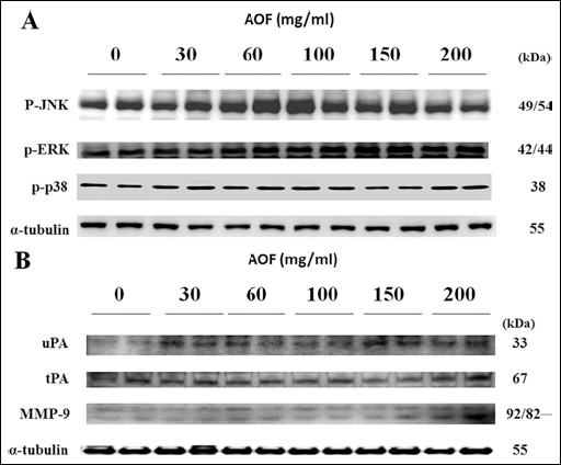
The regeneration of dissected sciatic nerves in chambers filled with AOF. The sciatic nerves from the chamber in rats with surgery were taken and the MAPK signaling activities (A) and the uPA, tPA, MMP-9 activities (B) were examined using Western blotting analysis. The chambers in the right legs were filled with various concentrations of AOF as indicated. α-tubulin was used as a loading control.
Effects of AOF on RSC96 Schwann cell viability
We evaluated the proliferative effect of AOF extract on the regenerative ability of RSC96 Schwann cells. During these experiments we first observed the effect of AOF at various concentrations (0, 20, 40, 60, 80, 100, 150 and 200 μg/ml) on cell viability for a 24 h period. We found that cellular viability was significantly elevated at the 20, 60 and 200 μg/ml concentrations at 24 h (Fig. 2). Therefore, these results may elucidate that treatment with 20 μg/ml to 200 μg/ml AOF for 24 hours appears to induce cell proliferation.
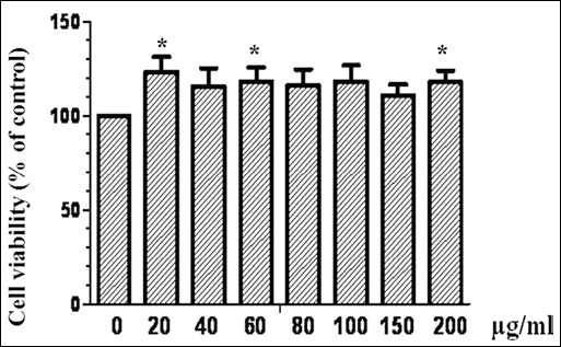
Effect of AOF extract on RSC96 cell viability. Schwann cells were treated with 0-200 μg/ml AOF extract for 24 h. Cell viability measured by MTT assay was described under materials and methods section. Data are shown as the mean of three independent experiments ± SE. *denotes significant differences from control values with p<0.05.
AOF Promotes the Migration of RSC96 Schwann Cells
The property of RSC96 Schwann cells to migrate along the growth direction is important in helping damaged peripheral nerve regeneration (21). We therefore performed an in vitro wound-healing experiment to evaluate the migration potential of RSC96 Schwann cells. As shown in Figure 3, treatment with AOF extract concentrations (0, 20, 40, 60, 80, 100, 150 and 200 μg/ml) for 24 h significantly enhanced the mobility of RSC96 Schwann cells. Using the Boyden chamber system to observe RSC96 Schwann cell migration, we found that cell migration increased after AOF treatment in a dose-dependent manner (Fig. 4). These results provide evidence that AOF has proliferative and migratory effects on RSC96 Schwann cells.
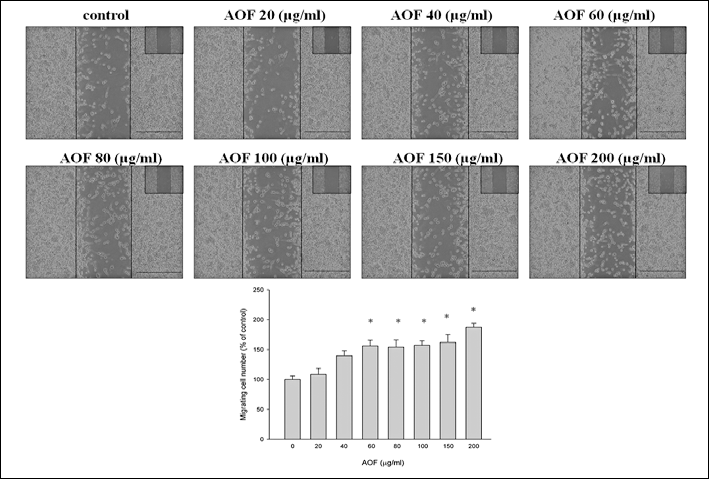
The migration effect of AOF extract on RSC96 cells. Schwann cells were incubated with different doses of AOF as indicated. Data are shown as the mean of three independent experiments ± SE. *denotes significant differences from control values with p<0.05.
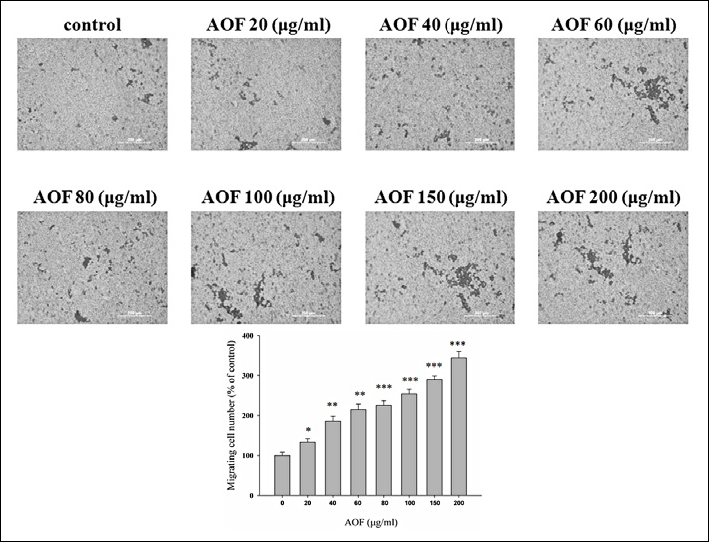
The migration effect of AOF extract on RSC96 cell. Schwann cells were treated with different doses of AOF as indicated. Cell migration as measured by Boyden chambers is described under materials and methods. Data are shown as the mean of three independent experiments ± SE. *denotes significant differences from control values with p<0.05.
Role of MAPKs in AOF-induced RSC96 Schwann cell migration
We further examined the mechanism of AOF extract-induced RSC96 Schwann cell migration. Intracellular MAPK signaling components, MEK1/2, ERK1/2, JNK1/2, p38, and their phosphorylate proteins were measured using Western blotting. Our data showed that the phosphorylation of MEK1/2, ERK1/2, JNK1/2, and p38 increased after AOF treatments (Figs. 5A and B). For the non-phosphorylated protein, MEK1/2, ERK1/2, JNK, and p38, protein was up regulated. We suggest that AOF extract could induce the phosphorylation of MEK1/2, ERK1/2, JNK1/2, and the p38 signal pathway to promote migration in RSC96 Schwann cells. Moreover, uPA and tPA proteins also increased and conversely the PAI-1 protein level decreased (Fig. 5C). RSC96 Schwann cells exposed to AOF extract also induced the expression of MMP-9 and MMP-2 proteins and decreased TIMP-1 and TIMP-2 levels (Fig. 5C). The zymography results further demonstrated that MMP9 and MMP2 activity increased after AOF extract treatments (Fig. 5D). These results indicate that AOF might be mediated through the activation of ERK1/2, JNK1/2, and p38 pathways to induce PAs and MMP2/9, resulting in RSC96 Schwann cell migration.
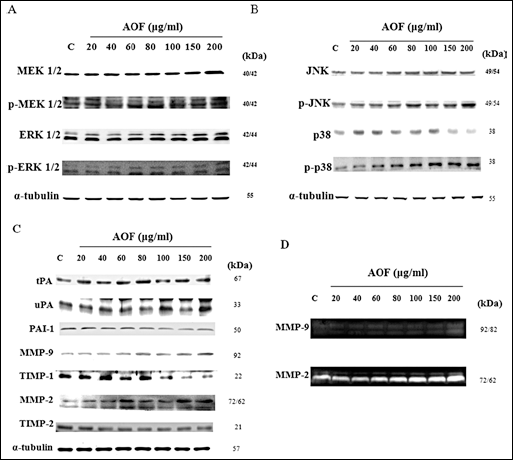
The migratory effect of AOF on RSC96 cell viability mediated by MAPK signaling. RSC96 cells were treated with various doses of AOF. The MAPK-signaling activities were measured using Western blot (A-C). α-tubulin was used as a load control. Further to confirm the MMP-9 and MMP-2 activity by gelatin zymopraphy (D).
RSC96 Schwann cell migration enhanced by AOF is ERK1/2, JNK, and p38 signaling-dependent in vitro
It has been demonstrated that AOF extract can significantly activate ERK1/2, JNK, and p38. We then examined whether AOF-induced cell migration indeed mediated through ERK1/2, JNK, and p38 MAPK. We used specific MAPK cascade inhibitors: U0126, SP600125, and SB203580. Schwann cells were pretreated with U0126, SP600125, and SB203580 pharmacological inhibitors, followed by incubation with AOF extract at 100 μg/ml concentration for 24 h. Our data revealed that the downstream signaling substrates of ERK1/2, JNK, and p38 that were activated by AOF extract were significantly blocked by the MAPK inhibitors (Fig. 6). These results were confirmed further by siRNA, which was applied to knock down ERK, JNK, and p38 proteins (Fig. 7). Western blots showed that there was a significant reduction in the p-ERK, p-JNK, and p-p38 protein levels as well as the downstream signaling substrates in AOF-treated Schwann cells after being transfected with MEK, JNK, or p38 siRNA. Our results demonstrate that AOF extract-induced Schwann cell migration occurs by activating the PAs and MMPs, which are dependent on the ERK1/2, JNK, and p38 MAPK pathways.
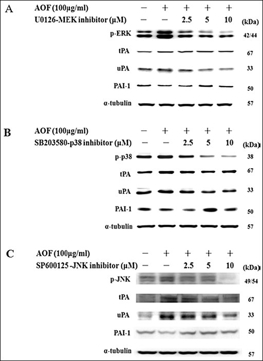
AOF extract effects on Schwann cell migration was ERK1/2, JNK and p38 signaling dependent. RSC96 cells were pretreated with U0126 (A), SB203580 (B) or SP600125 (C) for 1 h, then treated with 100 μg/ml AOF extract for 24 h. Western blot analysis of many protein levels, as indicated, in RSC96 cells treated with AOF extract. α-tubulin was used as a load control.
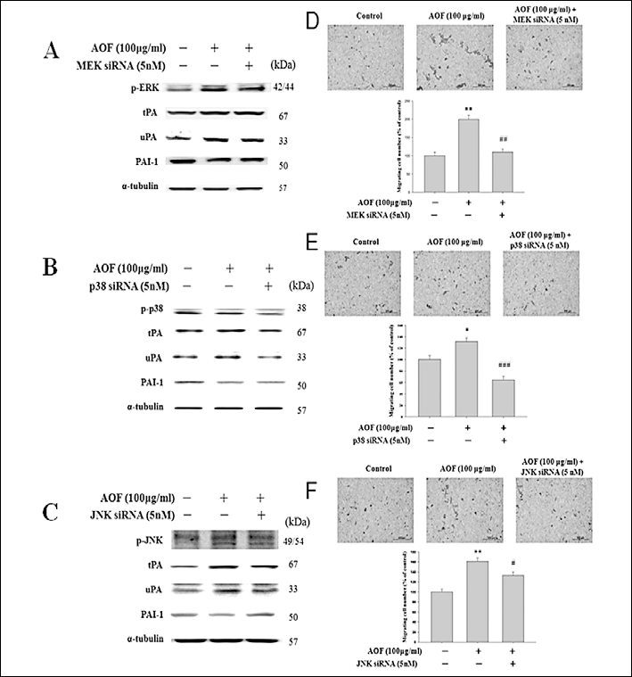
ERK1/2, p38 and JNK signaling required for AOF-induced cell migration. RSC cells were transiently transfected with ERK1/2 siRNA (5 nM), p38 siRNA (5 nM) or JNK siRNA (5 nM) for 8 h, then treated with 100 μg/ml AOF extract for 24 h (A-C). Western blot analysis of many protein levels, as indicated, in RSC96 cells treated with AOF extract. α-tubulin was used as a load control. After incubation with AOF extract, migration was assayed using Boyden chambers (D-F). Data are shown as the mean of three independent experiments ± SE. *denotes significant differences from untreated control values with p<0.05. #denotes significant differences from treated with AOF extract only values with p<0.05.
Discussion
The combination of herbal medicine and biomedical material science in promoting the functional recovery of damaged peripheral neurons has received much attention in recent years. Based on our previous study (19, 22), using a silicone rubber chamber filled with a mixture of herbal product and Schwann cells in bridging a 15 mm gap in rat sciatic nerves is a good model for evaluating the capacity for regenerating neurons. Schwann cells produce and release adhesion molecules and trophic factors that are vital requirements for successful nerve regeneration following injury (23). Chinese herbal medicines have attracted a great deal of attention as alternative and supplemental medicines (24, 25). AOF is one of the most important traditional Chinese medicines, reported to have potential as a neuro-protective agent (13, 15–18). Schwann cells migrating to the injured nerve area form a Bungner band that supports axonal regrowth (2). While the effect of AOF extract on nerve regeneration is unknown, the mechanism of AOF-induced Schwann cell migration is totally obscure.
This study investigated the mechanism in which AOF extract regulates RSC96 Schwann cell migration. We demonstrated a specific signaling migration pathway in AOF-stimulated RSC96 Schwann cells, inducing the ERK, JNK, and p38 mediated activation of uPA and tPA (Fig. 8). Cells treated with AOF extract resulted in ERK1/2, JNK, and p38 phosphorylation (Figs. 5A and 5B), inducing the expression of uPA and tPA in a dose-dependent manner (Fig. 5C), and leading to elevated MMP9 and MMP2 levels and activity (Fig. 5B). Using chemical inhibitors (Fig. 6) and SiRNA (Fig. 7), the effects of AOF extract on inducing RSC96 cell migration were further identified to be dependent on ERK1/2, JNK, and p38 signaling. In addition, wound-healing assay (Fig. 3) and Boyden chamber system experiments (Fig. 4) showed that the ECM enzyme activity regulating protein (MMP9 and MMP2) and Schwann cell migration were increased after AOF treatment. In the sciatic nerve-injured rat model the results showed that AOF increased uPA, tPA, MMP-9, and MAPKs expression (Fig. 1). These results provide evidence that AOF extract promotes the migration of Schwann cells and might enhance the reconstruction of peripheral nerve defects.

Schematic model of AOF extract migratory effects on RSC96 Schwann cells. Stimulation of Schwann cells with AOF activates ERK1/2, JNK and p38 signaling, leading to up-regulating uPA and tPA, and contributing to activating MMP9 and MMP2, enhancing the Schwann RSC96 cell migration.
The mitogen-activated protein kinases (MAPKs) that mediate critical signaling pathways are relevant to cell proliferation and differentiation (26). There are three sub-families of MAPKs: extracellular signal-regulated kinases (ERK), NH2-terminal kinases (JNK), and p38 kinase. They play important roles in regulating nerve cell migration (8). In our study the results from Western blotting analysis revealed that JNK and P38 mediated mechanism is involved in the AOF induced migration that aided in vivo neuron regeneration (Fig. 2A). This further demonstrated that AOF extract stimulated ERK1/2, JNK1/2, and p38 in a dose-dependent manner (Figs. 5A and 5B), leading to Schwann cell migration in vitro. AOF-induced Schwann cell motility and the phosphorylation of ERK1/2, JNK, and p38 were attenuated by pretreatment with MEK1/2 (U0126), JNK (SP600125), and p38 (SB203580) inhibitors (Fig. 6). Transfection with siRNA of MEK1/2, JNK, and p38 significantly reduced migration in response to AOF extract in Schwann cells as well (Fig. 7). These assays allow us to examine the individual steps in the complex signaling cascades and clearly illustrate direct AOF extract effects on Schwann cell migration. We further identified that AOF extract enhances uPA and tPA expression directly through the ERK, JNK, and p38 signaling pathway. To promote migration, cells secrete proteases that are thought to degrade matrix molecules and cell adhesion. These proteases include tPA and uPA (9). In contrast to PAs, PAI-1 is thought to be the major inhibitor. Pittman and Dibenedetto reported that overexpressing tPA regenerates neurites to a greater extent and causes faster migration than control cells in complex extracellular matrix (27). Our data clearly shows that the phosphorylation of ERK1/2, JNK, and p38 accompanies the increased expression of uPA and tPA. Conversely, PAI-1 expression is gradually decreased (Fig. 5C).
The development and regeneration of PNS is highly dependent on the migration of Schwann cells and the extension of axons toward their distant targets. PAs are associated with several neural cell types where they are believed to mediate localized degradation of the ECM, thus facilitating cell motility (28). ECM degradation is associated with neuron tissue growth. One of the key regulators of this process is the serine protease, uPA, acting on a wide variety of ECM components (29). The cell proliferation and angiogenesis processes are events involving uPA catalytic ECM degradation (30, 31). We suggest that ERK1/2, JNK, and p38 phosphorylation could promote uPA and tPA expression in AOF extract-treated cells.
Another family of proteases, the matrix metallo-proteases (MMPs), are also implicated in peripheral nerve regeneration and involved in many cell migration phenomena produced by many cell types, including neurons (32, 33). MMPs are secreted as inactive molecules and require activation via other proteases (34). Plasmin, activated by tPA or uPA, can activate MMP-9 and MMP-2 (35). Our results also show the elevated protein expression and activity of MMP2 and MMP9 (Fig. 5D).
Based on all of these findings, we suggest that AOF extract or some of its components could promote nerve cell migration and/or regeneration potential. Our results demonstrate that AOF extract can stimulate Schwann cell migration and upregulate PAs and MMP2/9 expression mediated through the MAPK pathways, ERK1/2, JNK, and p38. Further analyses are needed to determine the presence of bioactive compounds that promote cell migration in AOF extract. The findings of our study provide another novel neuron regeneration function. Therefore, an appropriate dose of AOF extract should be carefully selected to reach the highest potential for enhanced Schwann cell migration. AOF extract might serve as a promising migration inducing and/or therapeutic drug for nerve regeneration.